Clinical Cases in Gerodontology
Здесь есть возможность читать онлайн «Clinical Cases in Gerodontology» — ознакомительный отрывок электронной книги совершенно бесплатно, а после прочтения отрывка купить полную версию. В некоторых случаях можно слушать аудио, скачать через торрент в формате fb2 и присутствует краткое содержание. Жанр: unrecognised, на английском языке. Описание произведения, (предисловие) а так же отзывы посетителей доступны на портале библиотеки ЛибКат.
- Название:Clinical Cases in Gerodontology
- Автор:
- Жанр:
- Год:неизвестен
- ISBN:нет данных
- Рейтинг книги:3 / 5. Голосов: 1
-
Избранное:Добавить в избранное
- Отзывы:
-
Ваша оценка:
Clinical Cases in Gerodontology: краткое содержание, описание и аннотация
Предлагаем к чтению аннотацию, описание, краткое содержание или предисловие (зависит от того, что написал сам автор книги «Clinical Cases in Gerodontology»). Если вы не нашли необходимую информацию о книге — напишите в комментариях, мы постараемся отыскать её.
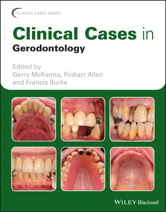
Gerodontology
Clinical Cases in
Gerodontology
Offers a case-based guide to geriatric dental care Includes the thinking behind clinical decision making Fosters independent learning and prepares for case-based examinations Contains review questions and relevant literature citations Written for graduate and undergraduate dental students and professionals,
offers an instructive case-based guide to the oral health of older adults.
Clinical Cases in Gerodontology — читать онлайн ознакомительный отрывок
Ниже представлен текст книги, разбитый по страницам. Система сохранения места последней прочитанной страницы, позволяет с удобством читать онлайн бесплатно книгу «Clinical Cases in Gerodontology», без необходимости каждый раз заново искать на чём Вы остановились. Поставьте закладку, и сможете в любой момент перейти на страницу, на которой закончили чтение.
Интервал:
Закладка:
3 Chapter 3Figure 3.16.1 Pre‐operative radiograph of fractured canine.Figure 3.16.2 Post‐obturation radiograph.Figure 3.16.3 Post‐operative clinical scenario with amalgam restoration in s...Figure 3.17.1 Pre‐treatment anterior view.Figure 3.17.2 Pre‐operative maxillary occlusal view.Figure 3.17.3 Pre‐operative mandibular occlusal view.Figure 3.17.4 (a) and (b) Pre‐operative periapical radiographs of 11, 12, 21...Figure 3.17.5 (a) and (b) Extracoronal restorations removed for restorabilit...Figure 3.17.6 Upper anterior teeth prepared for indirect restorations (22 ha...Figure 3.17.7 Final indirect restorations placed on upper anterior teeth.Figure 3.17.8 Tooth 36 prepared for cuspal coverage gold onlay.Figure 3.17.9 Gold onlay cemented onto 36.Figure 3.18.1 Patient at initial presentation.Figure 3.18.2 (a)–(d) Periapical radiographs of upper anterior teeth.Figure 3.18.3 Abutment teeth after bridge removal.Figure 3.18.4 Immediate denture fitted following extractions.Figure 3.18.5 Healing of soft tissues after 12 weeks.Figure 3.18.6 Aesthetics of definitive removable partial denture in situ.Figure 3.19.1 Patient at initial presentation; note the poor aesthetics of t...Figure 3.19.2 Palatal view demonstrating the extensive nature of the existin...Figure 3.19.3 Crowns constructed to facilitate the design for the upper remo...Figure 3.19.4 Fit of upper removable partial denture with components engagin...Figure 3.19.5 Final result 6 months after fit.Figure 3.20.1 Patient at initial presentation; note the poor aesthetics of t...Figure 3.20.2 Working model created after implant‐level impression.Figure 3.20.3 Wax try‐in of new bridge.Figure 3.20.4 Bridge on working model prior to fit.Figure 3.20.5 Final result after bridge fit.
4 Chapter 4Figure 4.21.1 Patient after surgical resection.Figure 4.21.2 (a) and (b) Construction of the surgical obturator.Figure 4.21.3 Patient wearing surgical obturator two weeks post surgery.Figure 4.21.4 (a) and (b) Silicone impression for definitive prosthesis.Figure 4.21.5 Insertion of definitive acrylic prosthesis.Figure 4.22.1 Patient at initial presentation.Figure 4.22.2 Pre‐operative maxillary view.Figure 4.22.3 Pre‐operative mandibular view.Figure 4.22.4 Roots prior to cementation of root copings.Figure 4.22.5 Post‐operative mandibular view with root canal–treated teeth w...Figure 4.22.6 Post‐operative view with final denture in situ.Figure 4.23.1 Patient’s remaining upper teeth at initial presentation.Figure 4.23.2 Radiographic assessment of patient.Figure 4.23.3 Clinical appearance of the patient’s tongue.Figure 4.24.1a c Gingival overgrowth in the lower arch at initial presentati...Figure 4.25.1 Patient at initial presentation; note the use of shade tabs in...Figure 4.25.2 Patient’s teeth recorded as shade C3 after a course of tooth w...Figure 4.25.3 Final result six months after tooth whitening.
Guide
1 Cover
2 Table of Contents
3 Begin Reading
Pages
1 ii
2 iii
3 iv
4 ix
5 xi
6 xii
7 xiii
8 1
9 2
10 3
11 4
12 5
13 6
14 7
15 8
16 9
17 10
18 11
19 12
20 13
21 14
22 15
23 16
24 17
25 18
26 19
27 20
28 21
29 22
30 23
31 24
32 25
33 26
34 27
35 28
36 29
37 30
38 31
39 32
40 33
41 34
42 35
43 36
44 37
45 38
46 39
47 40
48 41
49 42
50 43
51 44
52 45
53 46
54 47
55 48
56 49
57 50
58 51
59 52
60 53
61 54
62 55
63 56
64 57
65 58
66 59
67 60
68 61
69 62
70 63
71 64
72 65
73 66
74 67
75 68
76 69
77 70
78 71
79 72
80 73
81 74
82 75
83 76
84 77
85 78
86 79
87 80
88 81
89 82
90 83
91 84
92 85
93 86
94 87
95 88
96 89
97 90
98 91
99 92
100 93
101 94
102 95
103 96
104 97
105 98
106 99
107 100
108 101
109 102
110 103
111 104
112 105
113 106
114 107
115 108
116 109
117 110
118 111
119 112
120 113
121 114
122 115
123 116
124 117
125 118
126 119
127 120
128 121
129 122
130 123
131 124
132 125
133 126
134 127
135 128
136 129
137 130
138 131
139 132
140 133
141 134
142 135
143 136
144 137
145 138
146 139
147 140
148 141
149 142
150 143
151 144
152 145
153 146
154 147
155 148
156 149
157 150
158 151
159 152
160 153
161 154
162 155
163 156
164 157
Clinical Cases Series
Wiley‐Blackwell’s Clinical Cases series is designed to recognize the centrality of clinical cases to the dental profession by providing actual cases with an academic backbone. This unique approach supports the new trend in case‐based and problem‐based learning. Highly illustrated in full color, the Clinical Cases series utilizes a format that fosters independent learning and prepares the reader for case‐based examinations.
Clinical Cases in Pediatric Dentistry (2nd edition)by Amr M. Moursi (Editor) and Amy L. Truesdale (Associate Editor) December 2019
Clinical Cases in Dental Hygieneby Cheryl M. Westphal Theile, Mea A. Weinberg, and Stuart L. Segelnick January 2019
Clinical Cases in Endodonticsby Takashi Komabayashi November 2017
Clinical Cases in Orofacial Painby Malin Ernberg, Per Alstergren March 2017
Clinical Cases in Implant Dentistryby Nadeem Karimbux (Editor), Hans‐Peter Weber (Editor) December 2016
Clinical Cases in Orthodonticsby Martyn T. Cobourne, Padhraig S. Fleming, Andrew T. DiBiase, Sofia Ahmad June 2012
Clinical Cases in Periodonticsby Nadeem Karimbux December 2011
Clinical Cases in Prosthodonticsby Leila Jahangiri, Marjan Moghadam, Mijin Choi, Michael Ferguson October 2010
Clinical Cases in Restorative and Reconstructive Dentistryby Gregory J. Tarantola September 2010

Clinical Cases in Gerodontology
FIRST EDITION
Edited by
Gerry McKenna
BDS, MFDS RCSEd, PhD, FDS (Rest Dent) RCSEd, FHEA, FDTFEd
Clinical Reader and Consultant in Restorative Dentistry,
Centre for Public Health,
Queen’s University Belfast,
United Kingdom
Finbarr Allen
BDS, MSc, PhD, FDS RCPS, FDS (Rest Dent) RCPS, FFD RCSI
Professor and Dean,
Faculty of Dentistry,
National University of Singapore,
Singapore
Francis Burke
BDentSc, MSc, PhD, FDS RCSEd, FFD RCSI, DipTLHE
Senior Lecturer and Consultant in Restorative Dentistry,
Deputy Head (Academic Affairs),
College of Medicine and Health,
University College Cork,
Ireland

This edition first published 2021
Читать дальшеИнтервал:
Закладка:
Похожие книги на «Clinical Cases in Gerodontology»
Представляем Вашему вниманию похожие книги на «Clinical Cases in Gerodontology» списком для выбора. Мы отобрали схожую по названию и смыслу литературу в надежде предоставить читателям больше вариантов отыскать новые, интересные, ещё непрочитанные произведения.


Обсуждение, отзывы о книге «Clinical Cases in Gerodontology» и просто собственные мнения читателей. Оставьте ваши комментарии, напишите, что Вы думаете о произведении, его смысле или главных героях. Укажите что конкретно понравилось, а что нет, и почему Вы так считаете.