Clinical Atlas of Retreatment in Endodontics
Здесь есть возможность читать онлайн «Clinical Atlas of Retreatment in Endodontics» — ознакомительный отрывок электронной книги совершенно бесплатно, а после прочтения отрывка купить полную версию. В некоторых случаях можно слушать аудио, скачать через торрент в формате fb2 и присутствует краткое содержание. Жанр: unrecognised, на английском языке. Описание произведения, (предисловие) а так же отзывы посетителей доступны на портале библиотеки ЛибКат.
- Название:Clinical Atlas of Retreatment in Endodontics
- Автор:
- Жанр:
- Год:неизвестен
- ISBN:нет данных
- Рейтинг книги:3 / 5. Голосов: 1
-
Избранное:Добавить в избранное
- Отзывы:
-
Ваша оценка:
Clinical Atlas of Retreatment in Endodontics: краткое содержание, описание и аннотация
Предлагаем к чтению аннотацию, описание, краткое содержание или предисловие (зависит от того, что написал сам автор книги «Clinical Atlas of Retreatment in Endodontics»). Если вы не нашли необходимую информацию о книге — напишите в комментариях, мы постараемся отыскать её.
Clinical Atlas of Retreatment in Endodontics — читать онлайн ознакомительный отрывок
Ниже представлен текст книги, разбитый по страницам. Система сохранения места последней прочитанной страницы, позволяет с удобством читать онлайн бесплатно книгу «Clinical Atlas of Retreatment in Endodontics», без необходимости каждый раз заново искать на чём Вы остановились. Поставьте закладку, и сможете в любой момент перейти на страницу, на которой закончили чтение.
Интервал:
Закладка:
Dental history: discomfort due to impingement of food inside her molar. Previous treatment done on this tooth 1 year ago.
Clinical examination findings: deep decay, tooth was filled with food remnants, no mobility, no pain to percussion. After cleaning the tooth, big perforation was noted and bleeding also.
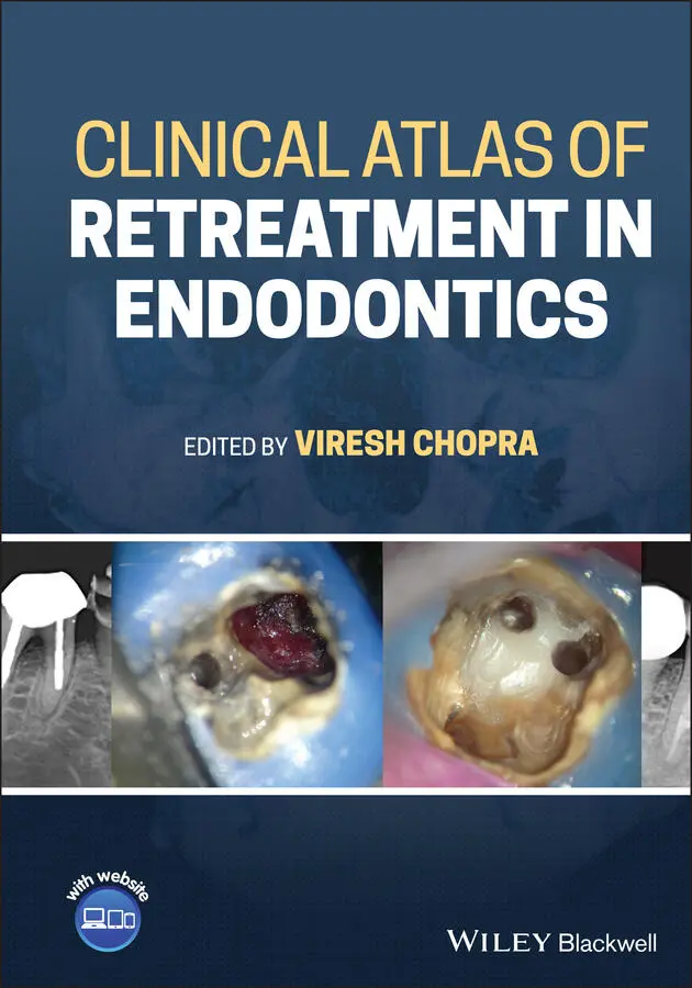
Preoperative radiological assessment: deep decay and lesion at furcation area due to perforation ( Figure 1.1).
Diagnosis (pulpal and periapical): previously initiated root canal therapy with asymptomatic apical periodontitis.
1.3 Treatment plan
First visit: local anaesthesia, rubber dam isolation, magnification (dental operative microscope), conventional access cavity, identification of orifices of the canals, placing cotton pellets inside them, stopping the bleeding physically with cotton pellet ( Figure 1.2).
Treatment plan for management of the endodontic mishap: applying MTA at the furcation area, then inserting a wet cotton pellet over MTA, temporary filling ( Figure 1.3). Figure 1.1 Preoperative radiograph showing radiolucency in the furcation area. Figure 1.2 Clinical picture showing the pulpal floor perforation. Figure 1.3 Radiograph showing MTA placed on the pulpal floor.
Second visit: removing temporary filling and cotton pellets, Check the condition of MTA (hardness), canal preparation with rotary files.
Irrigation protocol (solution and technique): 5.25% NaOCl; passive sonic irrigation.
Final irrigation protocol: 17% EDTA (syringe irrigation) for 1 minute.
Obturation (materials and technique): zinc oxide‐based sealer (SealiteTM Ultra) and gutta‐percha; warm vertical compaction.
Permanent filling ( Figures 1.4and 1.5).
1.4 Technical aspects
Key points to be taken care of while managing the endodontic mishap.
Stop bleeding before applying MTA.
Place wet cotton pellet over MTA and wait at least 4 hours to let it set.
1.5 Follow‐up
Follow for 2.5 years. The follow‐up radiograph shows formation of a bony trabecular pattern. Clinical and radiographic healing is evident on follow‐up visits ( Figure 1.6).
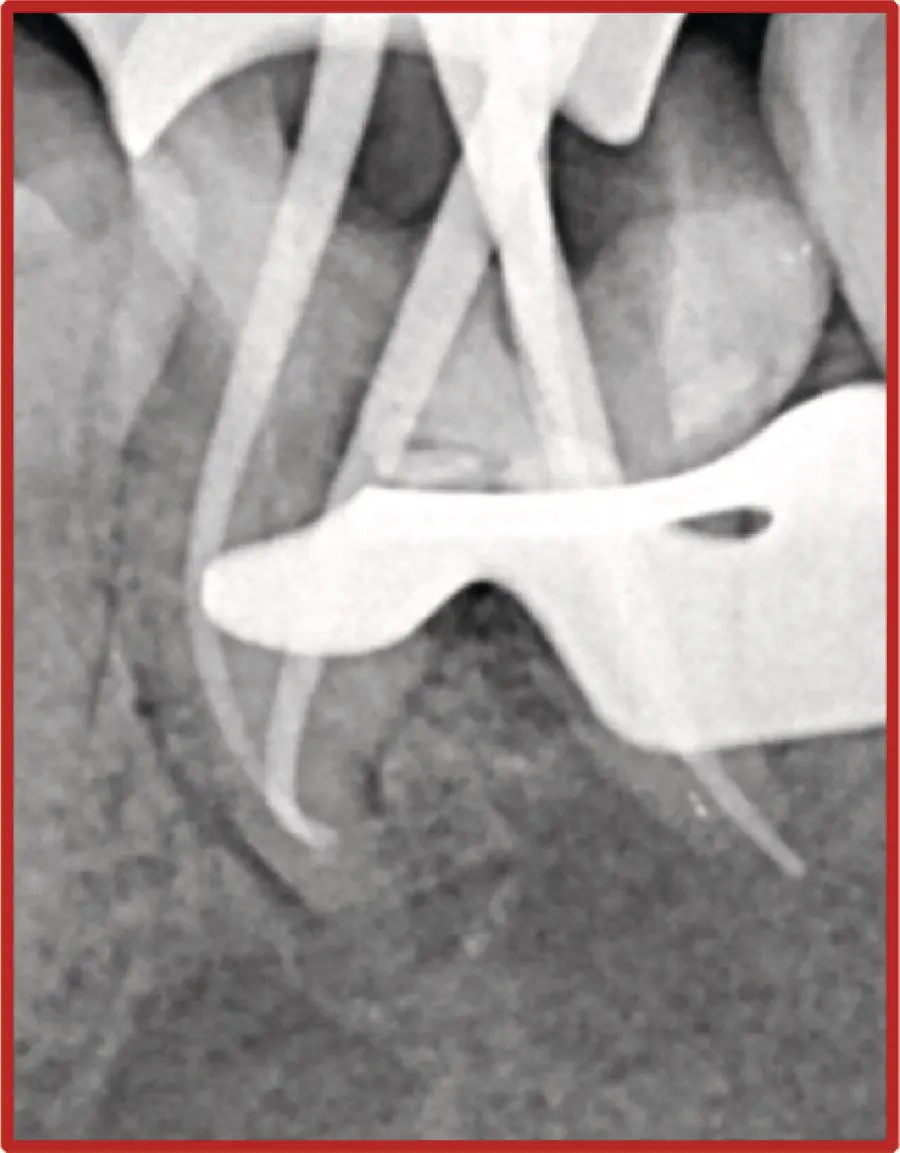
Figure 1.4 Radiograph showing master cone verification after biomechanical preparation of root canals.
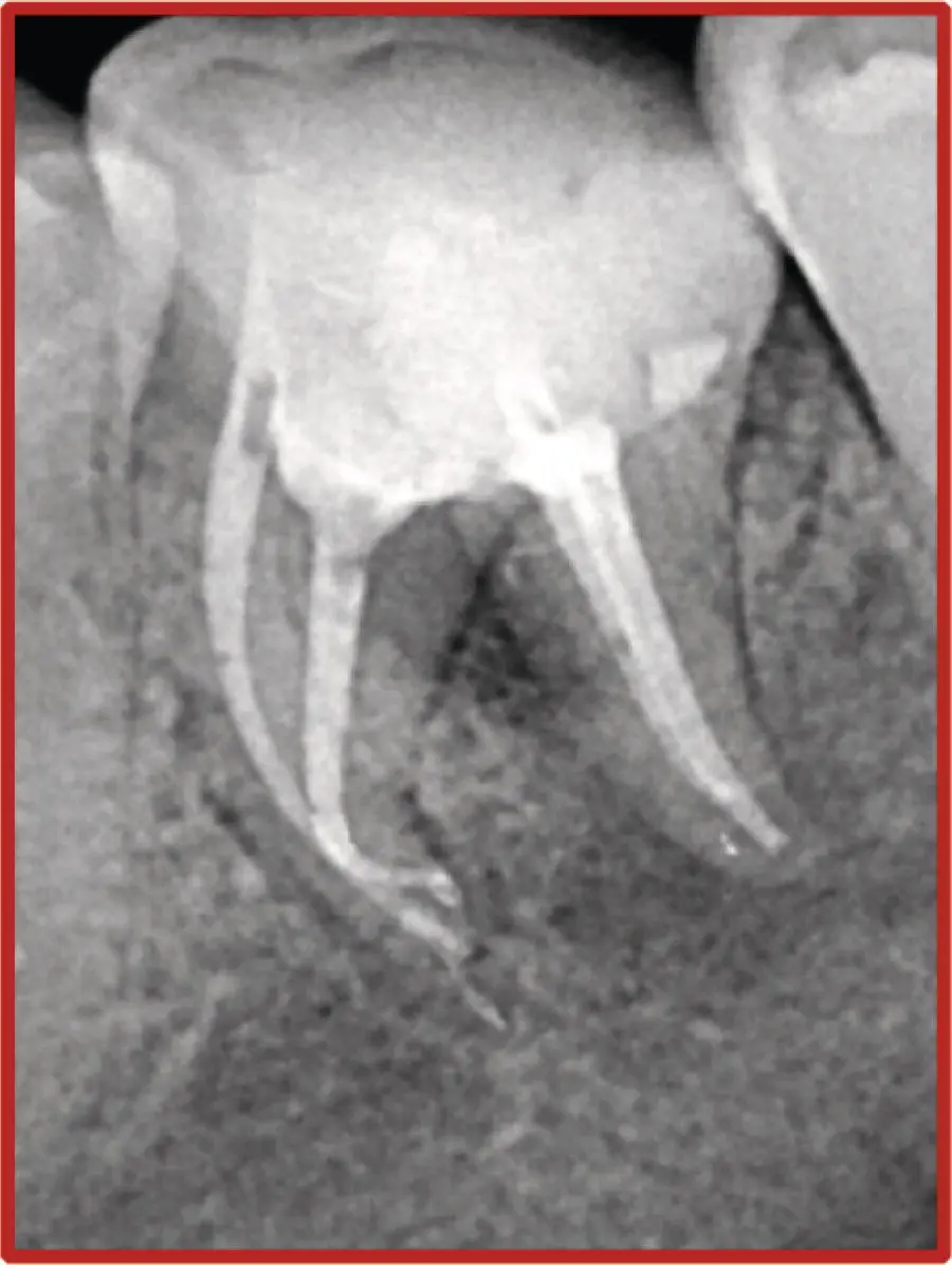
Figure 1.5 Radiograph showing obturation along with intact MTA.
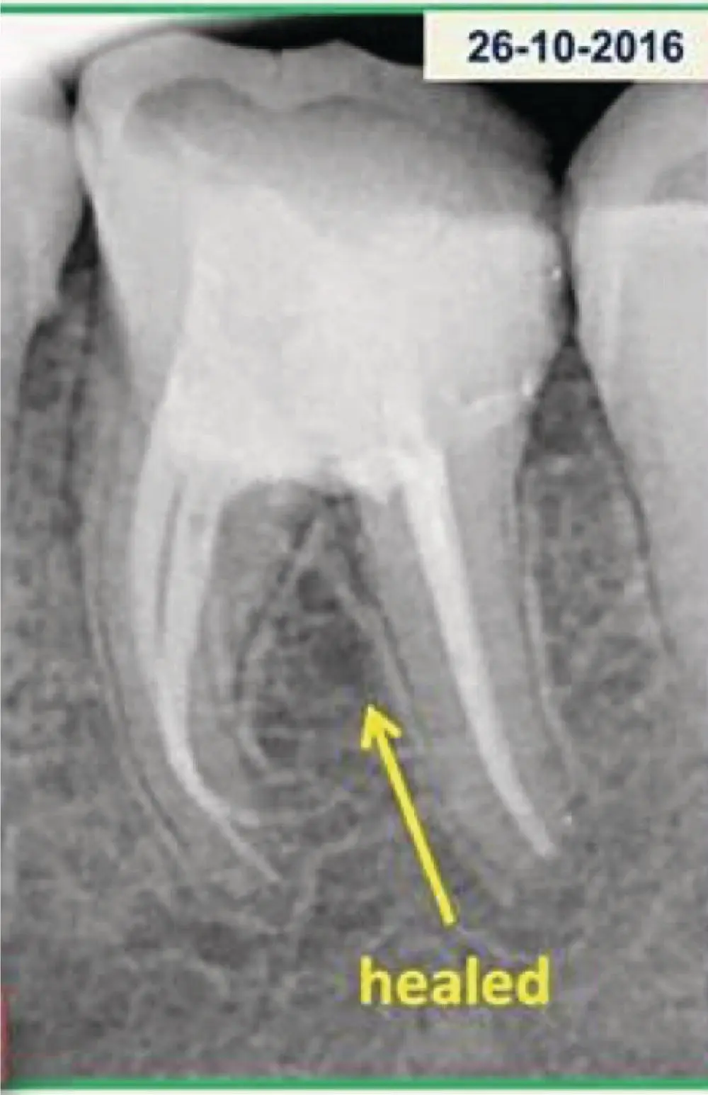
Figure 1.6 Follow‐up radiograph showing healing in the furcation area.
1.6 Learning objectives
How to approach a tooth with pulpal floor perforation.
The size and time of perforation do not justify extraction.
The priority is always for perforation repair, so do it as soon as possible.
1.7 How can this endodontic mishap be avoided?
Overdrilling should be avoided.
Location of canal orifices should be done with an endodontic explorer.
Once the operator feels a drop in the pulp chamber, no more vertical cutting should be done.
Use of safe‐ended, non‐cutting burs is recommended (e.g. Endo‐Z burs).
Конец ознакомительного фрагмента.
Текст предоставлен ООО «ЛитРес».
Прочитайте эту книгу целиком, купив полную легальную версию на ЛитРес.
Безопасно оплатить книгу можно банковской картой Visa, MasterCard, Maestro, со счета мобильного телефона, с платежного терминала, в салоне МТС или Связной, через PayPal, WebMoney, Яндекс.Деньги, QIWI Кошелек, бонусными картами или другим удобным Вам способом.
Интервал:
Закладка:
Похожие книги на «Clinical Atlas of Retreatment in Endodontics»
Представляем Вашему вниманию похожие книги на «Clinical Atlas of Retreatment in Endodontics» списком для выбора. Мы отобрали схожую по названию и смыслу литературу в надежде предоставить читателям больше вариантов отыскать новые, интересные, ещё непрочитанные произведения.


Обсуждение, отзывы о книге «Clinical Atlas of Retreatment in Endodontics» и просто собственные мнения читателей. Оставьте ваши комментарии, напишите, что Вы думаете о произведении, его смысле или главных героях. Укажите что конкретно понравилось, а что нет, и почему Вы так считаете.