Barbara J. Bain - Haematology
Здесь есть возможность читать онлайн «Barbara J. Bain - Haematology» — ознакомительный отрывок электронной книги совершенно бесплатно, а после прочтения отрывка купить полную версию. В некоторых случаях можно слушать аудио, скачать через торрент в формате fb2 и присутствует краткое содержание. Жанр: unrecognised, на английском языке. Описание произведения, (предисловие) а так же отзывы посетителей доступны на портале библиотеки ЛибКат.

- Название:Haematology
- Автор:
- Жанр:
- Год:неизвестен
- ISBN:нет данных
- Рейтинг книги:5 / 5. Голосов: 1
-
Избранное:Добавить в избранное
- Отзывы:
-
Ваша оценка:
Haematology: краткое содержание, описание и аннотация
Предлагаем к чтению аннотацию, описание, краткое содержание или предисловие (зависит от того, что написал сам автор книги «Haematology»). Если вы не нашли необходимую информацию о книге — напишите в комментариях, мы постараемся отыскать её.
Haematology: From the Image to the Diagnosis Haematology: From the Image to the Diagnosis
Haematology — читать онлайн ознакомительный отрывок
Ниже представлен текст книги, разбитый по страницам. Система сохранения места последней прочитанной страницы, позволяет с удобством читать онлайн бесплатно книгу «Haematology», без необходимости каждый раз заново искать на чём Вы остановились. Поставьте закладку, и сможете в любой момент перейти на страницу, на которой закончили чтение.
Интервал:
Закладка:
PICALM‐MLLT10 is most often found in T‐acute lymphoblastic leukaemia but also occurs in acute myeloid leukaemia and mixed phenotype acute leukaemia.
MCQ
1 Myeloid sarcoma:Can be the first sign of relapse of acute myeloid leukaemiaCan have a green colourCan be associated with t(8;21)(q22;q22.1)Is common in acute promyelocytic leukaemiaOccurs only at a single siteFor answers and discussion, see page 206.
13 Plasmablastic myeloma
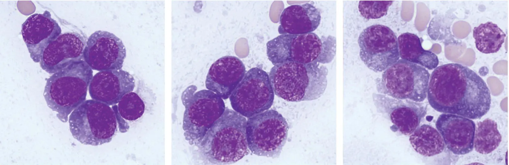
A 76‐year‐old woman treated with multiple lines of therapy for multiple myeloma presented to clinic with generalised debility and recent heavy nose bleeds. Her paraprotein levels had been noted to be on the rise despite lenalidomide and dexamethasone therapy. The full blood count showed Hb 95 g/l, WBC 3.5 × 10 9/l, neutrophils 1.5 × 10 9/l and platelets 18 × 10 9/l. An IgG paraprotein quantitation was 69 g/l. It was clear that the disease was becoming refractory to therapy but a bone marrow aspirate was taken in view of the problematic thrombocytopenia and bleeding. The aspirate was markedly hypercellular and well over 90% of cells were large pleomorphic plasma cells, some showing prominent nucleoli (images above ×100 objective). Normal haemopoiesis was markedly reduced and megakaryocytes in particular were scarce.
The malignant plasma cells of multiple myeloma are usually easy to recognise allowing accurate quantitation using a manual differential count. The morphology of plasma cells in refractory myeloma, however, often changes with increasing pleomorphism, increasing cell size and multinuclearity. The cells can sometimes resemble those of a high‐grade lymphoma. In addition many of these patients start to shed plasma cells into the peripheral blood (also noted in this case, not shown). Despite their morphological abnormality, the lineage is indicated in this patient by the strongly basophilic cytoplasm and the paler paranuclear Golgi zone. These features are important in helping recognise plasma cells when the nuclear morphology is atypical (images below ×100). Note the variation in nuclear morphology with bilobed and even binucleated forms but the Golgi zone and intense blue cytoplasm are prominent; all of these cells are plasma cells. As an adjunct, note the cytoplasmic fragments and particles due to the intense fragility of plasma cells on handling.
It may be worth reassessing the marrow in patients developing severe cytopenias since, in addition to the above features, some patients sadly develop a treatment‐related myelodysplastic syndrome or acute leukaemia.
MCQ
1 Plasmablastic myeloma:Can represent disease evolutionHas a high proliferation index on Ki‐67 stainingHas a worse prognosis than other cases of myelomaIs associated with worse renal function than other cases of myelomaIs associated with a higher serum calcium than other cases of myelomaFor answers and discussion, see page 206.
14 Septicaemia
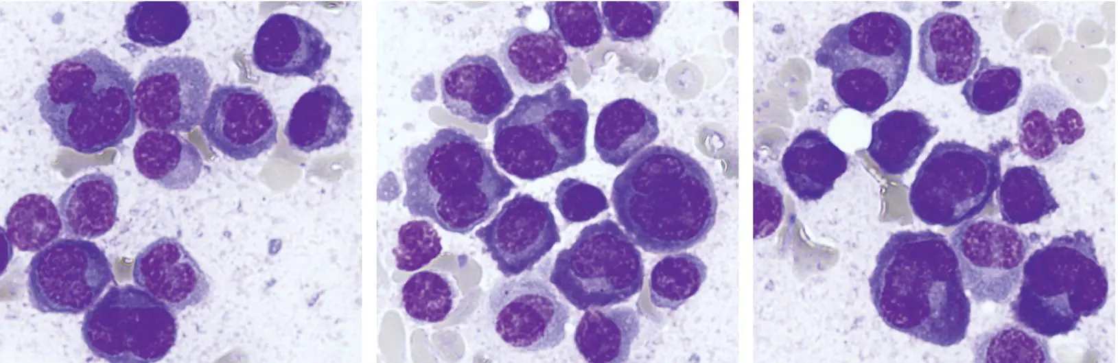
A 5‐year‐old girl was admitted as an emergency after a collapse following a fever at home. On assessment she was acutely unwell and hypotensive and had generalised petechiae. The clinical picture was of severe sepsis and after blood cultures had been taken she was commenced on broad‐spectrum antibiotics. The full blood count showed Hb 120 g/l, WBC 20 × 10 9/l, neutrophils 17 × 10 9/l and platelets 32 × 10 9/l. The coagulation profile was indicative of disseminated intravascular coagulation (DIC). The blood film (left images ×100 objective) showed neutrophilia with left shift; in particular there was prominent neutrophil vacuolation and there were cytoplasmic inclusions consistent with the appearance of diplococci. Note also the prominent Döhle bodies in the neutrophils (bottom left). Meningococcal septicaemia now seemed likely and was confirmed on blood cultures and PCR studies.
A second patient, a 62‐year‐old man, was admitted following a collapse after a 2‐day prodrome of feeling non‐specifically unwell. He had been bitten on the hand by a dog some days previously. He was febrile and hypotensive and had widespread petechiae. His condition deteriorated and he required intubation, ventilation and inotropic support. His full blood count showed Hb 137 g/l, WBC 24.2 × 10 9/l, neutrophils 22.7 × 10 9/l and platelets 9 × 10 9/l. The coagulation screen was again indicative of DIC (PT 25 s, APTT 92 s, TT 17 s, fibrinogen 1.5 g/l and D dimer 88 726 ng/ml). The blood film showed neutrophilia with left shift and some neutrophils had rod‐shaped bacterial inclusions (centre images). Blood cultures grew the Gram‐negative bacillus, Capnocytophaga canimorsus , which is a serious human pathogen that often forms part of normal canine and feline flora.
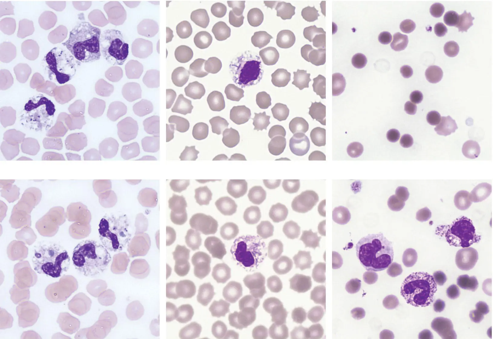
A third patient, a 66‐year‐old woman was in hospital with obstructive jaundice due to liver metastases from ovarian carcinoma and had external biliary stents in situ . She was receiving weekly paclitaxel chemotherapy. She became acutely unwell, developing hypotension and jaundice. The full blood count showed Hb 57 g/l, WBC 7.3 × 10 9/l and platelets 177 × 10 9/l. As in the previous cases, the blood film was informative; it showed profound red cell spherocytosis (right images) with red cell ghosts (top right image) and toxic granulation of neutrophils (bottom right image) but no bacterial inclusions were seen. Despite resuscitation efforts the patient died shortly afterwards. Blood cultures subsequently grew Clostridium perfringens . This bacterium can be a commensal of the normal hepatobiliary and gastrointestinal tracts but becomes a serious and life‐threatening pathogen when it gains access to the blood, most likely in this case in relation to the stent. It produces an alpha lecithinase enzyme which directly lyses the red cell membrane, inducing an acute haemolytic anaemia that further compounds the septicaemia. The blood film features are typical of this condition; the spherocytes are due to membrane perforation, as are the ghost cells where all red cell haemoglobin has been leaked. By the time the blood film has been assessed, most affected patients have succumbed to this form of septicaemia but in some cases the morphological features may indicate the need for rapid active management and save a life.
MCQ
1 Neutrophil vacuolation can be a feature of:Bacterial infectionChédiak–Higashi syndromeEthanol toxicityNeonatal necrotising enterocolitisNeutral lipid storage diseaseFor answers and discussion, see page 206.
15 An unstable haemoglobin and a myeloproliferative neoplasm
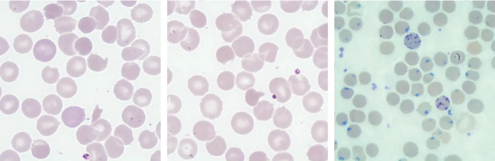
A 26‐year‐old man was well known to the Haematology clinic and had a chronic compensated haemolytic anaemia due to the unstable haemoglobin, haemoglobin Köln. He suffered exacerbations of haemolysis with intercurrent infection but his condition would settle thereafter. A typical full blood count showed Hb 114 g/l, WBC 4.7 × 10 9/l, neutrophils 2.4 × 10 9/l and platelets 154 × 10 9/l. His blood film is illustrated above; bite cells and irregularly contracted cells are evident (left and centre ×100 objective) and Heinz bodies were prominent (right, methyl violet ×100). Somewhat unexpectedly, he developed progressive and ultimately massive splenomegaly though his haemolytic disorder appeared clinically unchanged. CT scanning showed a 25 cm spleen with no focal abnormality; there was no evidence of chronic liver disease or portal hypertension and no lymphadenopathy was apparent. His full blood count now showed Hb 125 g/l, WBC 12.7 × 10 9/l, neutrophils 8.9 × 10 9/l and platelets 1830 × 10 9/l. He underwent a splenectomy in view of progressive splenic symptoms. The spleen weighed 2700 g (normal 200 g). Histological examination showed massive expansion of the red pulp with congested sinuses and extramedullary haemopoiesis. There was no evidence of lymphoma.
Читать дальшеИнтервал:
Закладка:
Похожие книги на «Haematology»
Представляем Вашему вниманию похожие книги на «Haematology» списком для выбора. Мы отобрали схожую по названию и смыслу литературу в надежде предоставить читателям больше вариантов отыскать новые, интересные, ещё непрочитанные произведения.


Обсуждение, отзывы о книге «Haematology» и просто собственные мнения читателей. Оставьте ваши комментарии, напишите, что Вы думаете о произведении, его смысле или главных героях. Укажите что конкретно понравилось, а что нет, и почему Вы так считаете.