Barbara J. Bain - Haematology
Здесь есть возможность читать онлайн «Barbara J. Bain - Haematology» — ознакомительный отрывок электронной книги совершенно бесплатно, а после прочтения отрывка купить полную версию. В некоторых случаях можно слушать аудио, скачать через торрент в формате fb2 и присутствует краткое содержание. Жанр: unrecognised, на английском языке. Описание произведения, (предисловие) а так же отзывы посетителей доступны на портале библиотеки ЛибКат.

- Название:Haematology
- Автор:
- Жанр:
- Год:неизвестен
- ISBN:нет данных
- Рейтинг книги:5 / 5. Голосов: 1
-
Избранное:Добавить в избранное
- Отзывы:
-
Ваша оценка:
Haematology: краткое содержание, описание и аннотация
Предлагаем к чтению аннотацию, описание, краткое содержание или предисловие (зависит от того, что написал сам автор книги «Haematology»). Если вы не нашли необходимую информацию о книге — напишите в комментариях, мы постараемся отыскать её.
Haematology: From the Image to the Diagnosis Haematology: From the Image to the Diagnosis
Haematology — читать онлайн ознакомительный отрывок
Ниже представлен текст книги, разбитый по страницам. Система сохранения места последней прочитанной страницы, позволяет с удобством читать онлайн бесплатно книгу «Haematology», без необходимости каждый раз заново искать на чём Вы остановились. Поставьте закладку, и сможете в любой момент перейти на страницу, на которой закончили чтение.
Интервал:
Закладка:
25 Reactive lymphocytosis due to viral infection
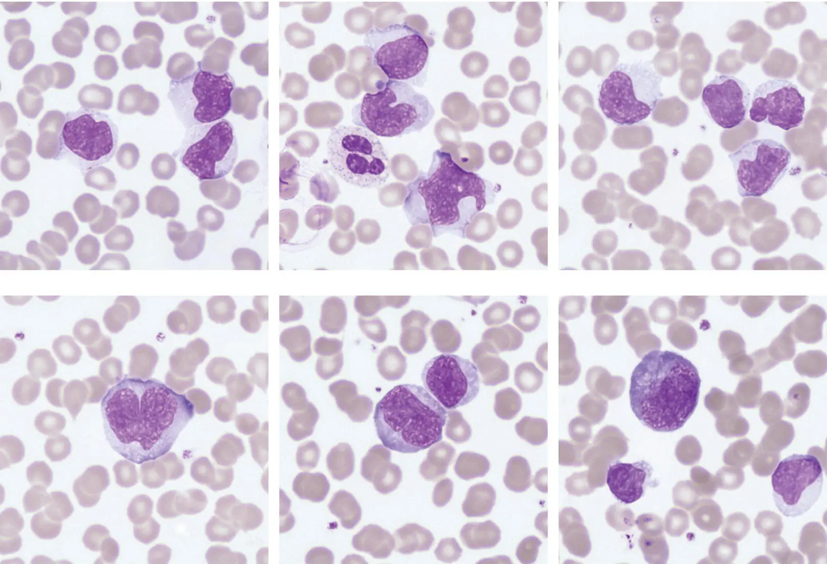
An 18‐year‐old ‘fresher’ medical student was referred to the university medical service with a fever, sore throat and cervical lymphadenopathy. His full blood count showed Hb 144 g/l, WBC 74 × 10 9/l, neutrophils 7.4 × 10 9/l, lymphocytes 66 × 10 9/l and platelets 203 × 10 9/l. The liver enzyme profile showed increased transaminases (AST 300 u/l, ALT 279 u/l) and a Monospot® test (which later became available) was positive. Epstein–Barr virus (EBV) was subsequently detected in his blood at 5606 copies/ml. The blood film caused some initial concern amongst the trainees due to the magnitude of lymphocytosis, but further careful morphological review from more senior members of the department was reassuring. Notably, the blood film shows a population of medium to large pleomorphic lymphocytes (all images ×100 objective), with some showing fine cytoplasmic granules. The cells have voluminous cytoplasm and tend to be indented by adjacent red cells (top images). Importantly, some cells have blastoid morphology, being larger with a prominent nucleolus and having more pronounced cytoplasmic basophilia. Flow cytometry identified a large population of CD8+, CD5+, CD2+, HLA‐DR+, CD7+/− cells which were not expressing precursor antigens, CD30 or CD25. The morphological diagnosis is a reactive lymphocytosis due to viral infection, in this case EBV infection. The flow cytometric studies are in keeping with this as expression of HLA‐DR is an activation marker of T cells and such cells frequently show some loss of CD7 expression. The activated cells are cytotoxic CD8+ T lymphocytes even though EBV targets the B‐lymphoid population.
Reactive lymphocytosis is relatively common in clinical practice and the morphological features described above are typical. Neoplastic disorders tend to have more monomorphic appearances, but of course a careful assessment of the clinical circumstances is always necessary. T‐cell neoplasms are remarkably variable in their presentation but the clinical history, morphology, laboratory investigations and flow cytometric findings were all consistent with a reactive process. Importantly, a wide range of viruses and other organisms can induce such a reaction; these include EBV, cytomegalovirus, human immunodeficiency virus (HIV), toxoplasma and pertussis. Most often the responsible organism is benign but if HIV is implicated it is important that this is identified. A positive test for a heterophile antibody is a pointer to EBV infection but not all cases are positive. Immunophenotyping is not generally indicated but was performed in this case because of initial concern about the possibility of a lymphoma.
MCQ
1 Infectious mononucleosis due to the Epstein–Barr virus:Can be associated with pure red cell aplasiaCan be complicated by haemophagocytic lymphohistiocytosisCan be followed by aplastic anaemiaIs associated with a subsequent increased incidence of Hodgkin lymphomaPredisposes to B‐lineage acute lymphoblastic leukaemiaFor answers and discussion, see page 206.
26 Therapy‐related acute myeloid leukaemia with eosinophilia
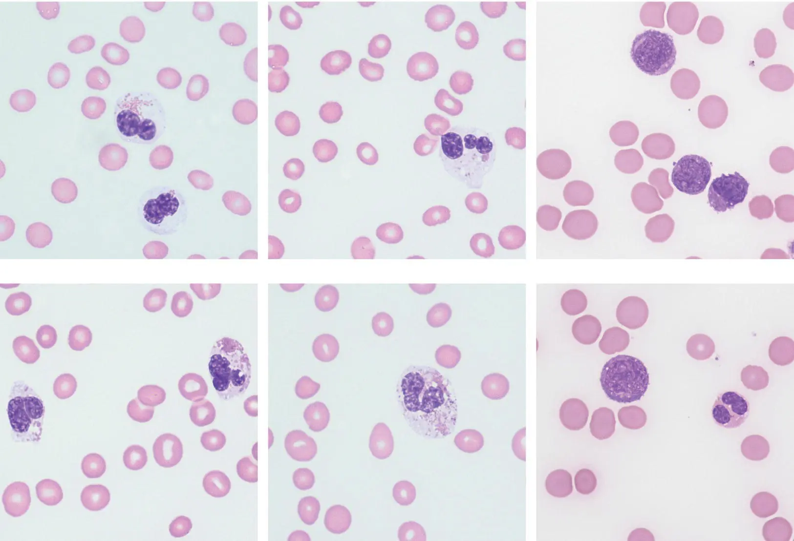
A 70‐year‐old man developed therapy‐related acute myeloid leukaemia (t‐AML) some years after starting chlorambucil therapy for chronic lymphocytic leukaemia. His FBC showed Hb 63 g/l, WBC 48.9 × 10 9/l, neutrophils 1.0 × 10 9/l, lymphocytes 5.5 × 10 9/l, eosinophils 19.6 × 10 9/l, monocytes 1.4 × 10 9/l, myelocytes 0.4 × 10 9/l, blast cells 21 × 10 9/l and platelet count 11 × 10 9/l. There were two nucleated red blood cells per 100 leucocytes. Many of the eosinophils were cytologically abnormal: 25% were hypogranular, 46% were vacuolated and 17% were both hypogranular and vacuolated (all images ×100 objective). Occasional eosinophils had fine basophilic granules (bottom centre) or non‐lobated nuclei. Neutrophils were cytologically normal. The myeloblasts were small and compact with little cytoplasm and prominent nucleoli (right images).
Clonal eosinophils in myeloid neoplasms tend to have more marked cytological abnormalities than those in reactive eosinophilia but there is considerable overlap. It is therefore essential for cytological abnormalities to be assessed together with other disease features. When there is an increase in blast cells, as in the current patient, or when there are clearly dysplastic features in other lineages, such as hypogranular neutrophils, the diagnosis is straightforward. However, if an increase in cytologically abnormal eosinophils occurs without other morphological clues, cytogenetic and molecular analysis may well be needed to confirm the diagnosis of a myeloid neoplasm.
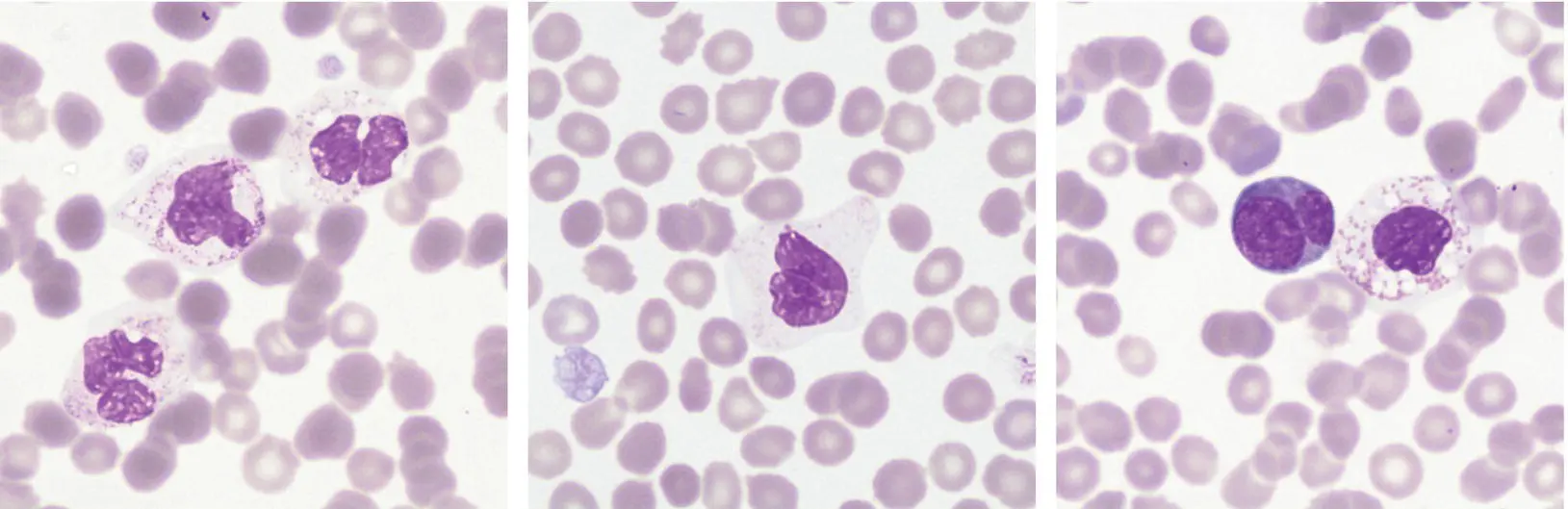
The images above (×100 objective) show hypogranular and vacuolated eosinophils; the cell in the centre image is virtually agranular. Note also the dysplastic platelet (centre) and the megakaryoblast (right); these latter two features together with the eosinophil morphology suggest a clonal haemopoietic disorder, in this case primary myelofibrosis.
Therapy‐related AML is an unfortunate consequence of bone marrow exposure to chemotherapy or radiotherapy used as treatment for neoplastic disease in haematology and oncology practice. As more of these conditions are cured with modern therapy, often using a combination of surgery, chemotherapy and radiotherapy, in breast cancer for example, more patients are surviving to develop therapy‐related myeloid neoplasms. It is not uncommon within haematology to see therapy‐related AML resulting from treatment of another haematological disease, as in the patient described here. We have seen such cases as a result of previous successful treatment for Hodgkin lymphoma, diffuse large B‐cell lymphoma, blastic plasmacytoid dendritic cell neoplasm and acute promyelocytic leukaemia. To be cured of a good‐prognosis leukaemia and develop a poor‐prognosis leukaemia as a consequence is particularly difficult to face.
MCQ
1 Therapy‐related acute leukaemia:Can be associated with 5q−, monosomy 5, 7q− and monosomy 7Can be lymphoblastic or myeloidHas an equally poor prognosis, regardless of the causative agent, the blast percentage or any cytogenetic abnormalities presentHas identical characteristics whether it follows alkylating agents or topoisomerase II inhibitorsIncreases in incidence with the age of exposure to the causative agentFor answers and discussion, see page 206.
Конец ознакомительного фрагмента.
Текст предоставлен ООО «ЛитРес».
Прочитайте эту книгу целиком, купив полную легальную версию на ЛитРес.
Безопасно оплатить книгу можно банковской картой Visa, MasterCard, Maestro, со счета мобильного телефона, с платежного терминала, в салоне МТС или Связной, через PayPal, WebMoney, Яндекс.Деньги, QIWI Кошелек, бонусными картами или другим удобным Вам способом.
Интервал:
Закладка:
Похожие книги на «Haematology»
Представляем Вашему вниманию похожие книги на «Haematology» списком для выбора. Мы отобрали схожую по названию и смыслу литературу в надежде предоставить читателям больше вариантов отыскать новые, интересные, ещё непрочитанные произведения.


Обсуждение, отзывы о книге «Haematology» и просто собственные мнения читателей. Оставьте ваши комментарии, напишите, что Вы думаете о произведении, его смысле или главных героях. Укажите что конкретно понравилось, а что нет, и почему Вы так считаете.