Practical Procedures in Implant Dentistry
Здесь есть возможность читать онлайн «Practical Procedures in Implant Dentistry» — ознакомительный отрывок электронной книги совершенно бесплатно, а после прочтения отрывка купить полную версию. В некоторых случаях можно слушать аудио, скачать через торрент в формате fb2 и присутствует краткое содержание. Жанр: unrecognised, на английском языке. Описание произведения, (предисловие) а так же отзывы посетителей доступны на портале библиотеки ЛибКат.
- Название:Practical Procedures in Implant Dentistry
- Автор:
- Жанр:
- Год:неизвестен
- ISBN:нет данных
- Рейтинг книги:3 / 5. Голосов: 1
-
Избранное:Добавить в избранное
- Отзывы:
-
Ваша оценка:
Practical Procedures in Implant Dentistry: краткое содержание, описание и аннотация
Предлагаем к чтению аннотацию, описание, краткое содержание или предисловие (зависит от того, что написал сам автор книги «Practical Procedures in Implant Dentistry»). Если вы не нашли необходимую информацию о книге — напишите в комментариях, мы постараемся отыскать её.
covers core topics such as:
Rationale and assessment for implant placement and restoration, including the diagnostic records and surgical considerations required for optimal planning and risk management Incision design considerations and flap management, with an essential knowledge of regional neuro-vascular structures Implant placement, encompassing the timing of the placement, bone requirements and understanding the importance of the peri-implant interface for soft tissue stability Impression techniques, loading protocols, digital workflows and the aesthetic considerations of implants Prosthetic rehabilitation of single tooth implants to fully edentulous workflows, including discussions of soft tissue support, biomechanics and occlusal verification Perfect for both general dental practitioners and specialists in implant dentistry,
is also a valuable reference to senior undergraduate and postgraduate dental students.
Practical Procedures in Implant Dentistry — читать онлайн ознакомительный отрывок
Ниже представлен текст книги, разбитый по страницам. Система сохранения места последней прочитанной страницы, позволяет с удобством читать онлайн бесплатно книгу «Practical Procedures in Implant Dentistry», без необходимости каждый раз заново искать на чём Вы остановились. Поставьте закладку, и сможете в любой момент перейти на страницу, на которой закончили чтение.
Интервал:
Закладка:
Although the digital workflow is constantly evolving, historically it began with this approach:
1 Intra‐oral scanning (or conventional impressions)
2 Construction of digital radiographic guides with radiopaque marker
3 CBCT (small FOV)
4 Conversion of radiographic guide to surgical guide based on the findings of the CBCT.
Modern digital workflows involve a streamlined approach which allows creation of the surgical guide being designed and milled from an initial intra‐oral scan and CBCT:
1 Intra‐oral scanning (or conventional impressions)
2 CBCT (small FOV)
3 Merging the intra‐oral scan with the CBCT, design and construction of a surgical guide.
3.2.2 Photography
The following sets of photographs are a minimum standard:
Full face (frontal): This image is shot at the same level as the patient and should cover their whole head. This vertical angle is important for majority of the images taken in dental photography. The interpupillary line and long axis of teeth is used to align the camera ( Figure 3.4).
Full smile – frontal, right, and left lateral view: This view shows the lips as well as the teeth visible for this angle. The upper lateral incisor is centred on the slide. The contralateral central incisor should be visible and possibly the lateral incisor and canine ( Figures 3.5– 3.7).
Retracted anterior view: This is an intra‐oral photograph using retractors held by the patient, with the teeth together or slightly apart ( Figures 3.8and 3.9).
Upper and lower right and left lateral retracted view: The image is centred on the lateral incisor so that it is in the centre of the picture. The retractor is pulled to side that the picture is being taken of, while the contralateral retractor is loosely held which allows the photograph to extend further posteriorly to capture the posterior teeth ( Figures 3.10and 3.11).
Upper and lower occlusal retracted view (use mirror): This is a reflected view from a high‐quality mirror, with as many teeth as possible included. Keep the mirror clear of fogging by warming it or using an air–water syringe. The mouth should be opened as wide as possible to allow the best mirror position. In the lower jaw is exactly the same as with the upper teeth but the patient needs to be asked to keep their tongue back so that is does not obscure the teeth ( Figures 3.12and 3.13).

Figure 3.4 Full face frontal. This image is shot at the same height as the patient, including their whole head, and can be taken with lips at rest and also in broad smile.
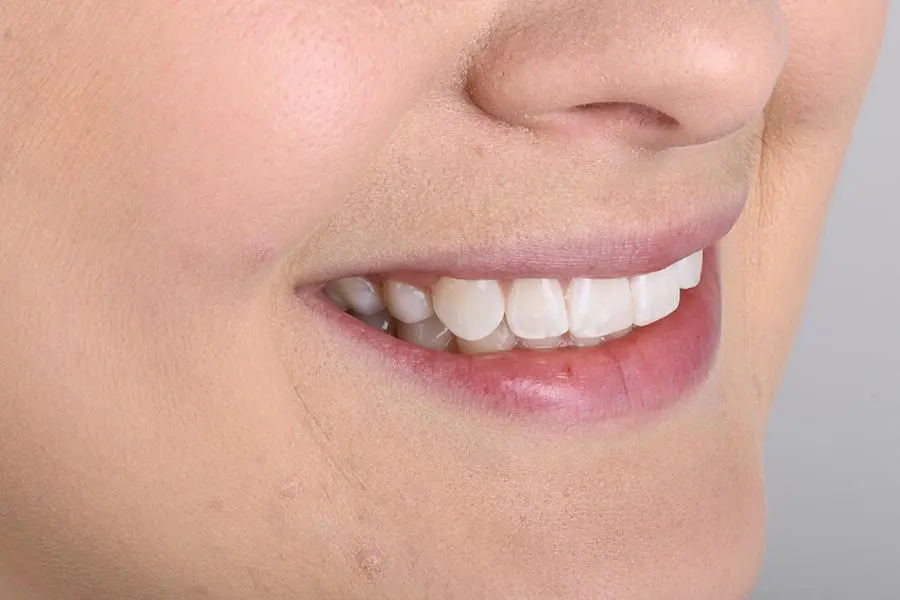
Figure 3.5 Right lateral smile.
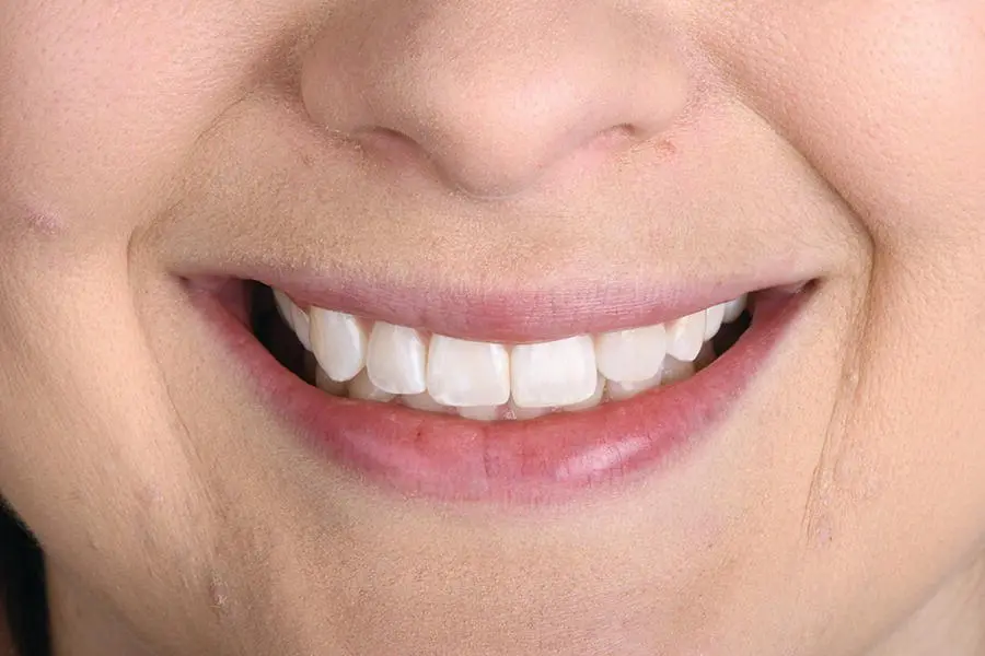
Figure 3.6Frontal smile.
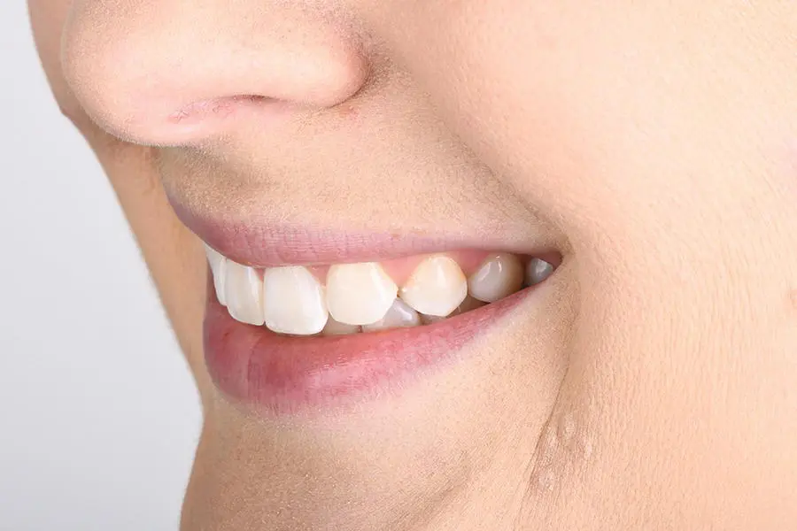
Figure 3.7 Left lateral smile.
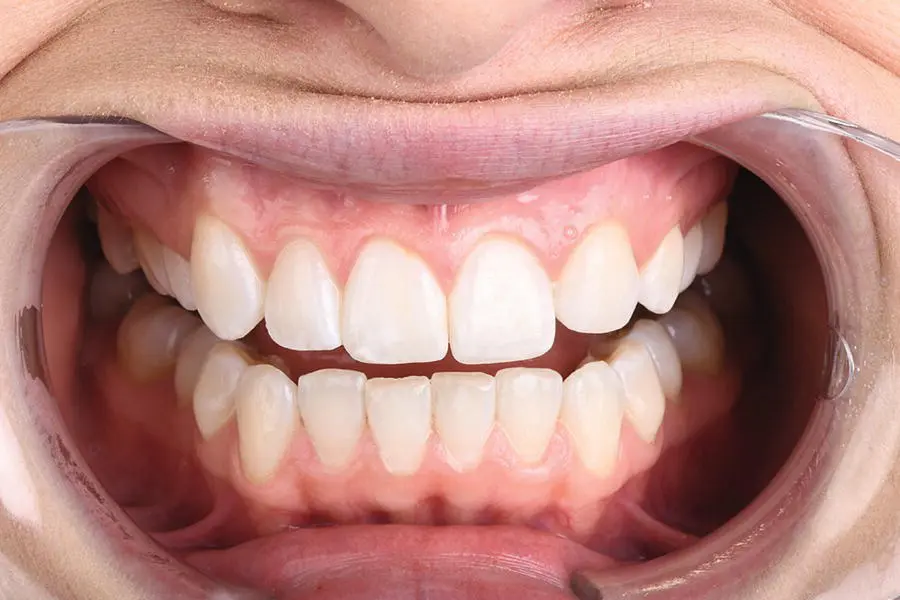
Figure 3.8 Retracted frontal shot with teeth apart.
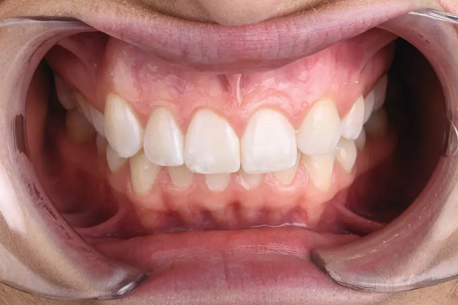
Figure 3.9 Retracted frontal shot with teeth in maximum intercuspation.
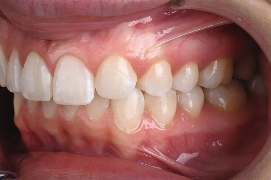
Figure 3.10 Retracted left photograph displaying left side of teeth. The left lateral incisor should be in the centre of the photograph.
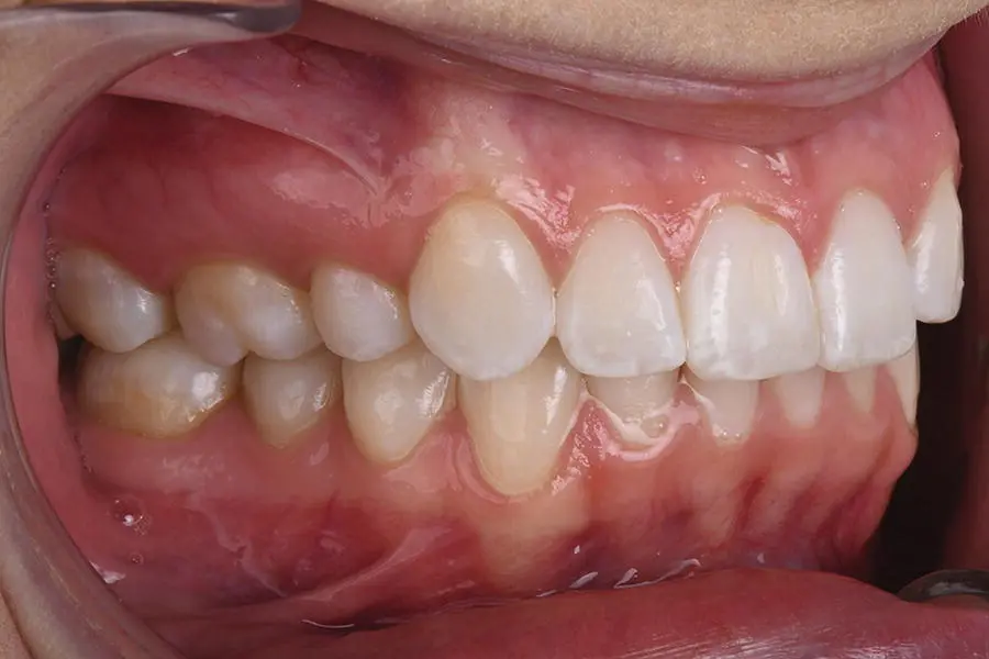
Figure 3.11 Retracted right photograph displaying right side of teeth. The right lateral incisor should be in the centre of the photograph.
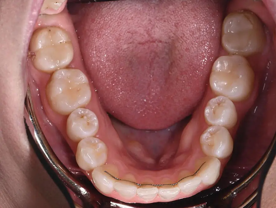
Figure 3.12 Occlusal photograph of mandibular teeth using a photographic mirror.
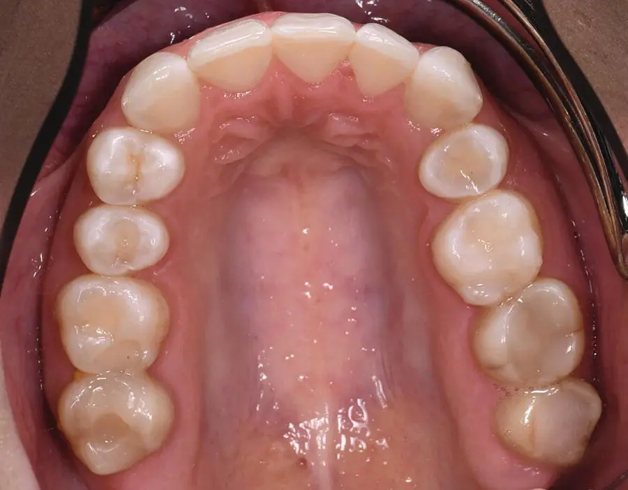
Figure 3.13 Occlusal photograph of maxillary teeth using a photographic mirror.
3.3 Tips
Conventional periapical and panoramic imaging is still very useful for treatment planning. Although CBCT is essential in planning, it can be difficult to appreciate the crown and root position of teeth adjacent to the planned site, because any given slice will vary. Clinicians who rely upon one slice during placement may encounter issues with proximity of the implant to the adjacent teeth.
When using surgical templates, ensure windows are cut out of the template adjacent to the implant sites to allow the clinician to visualise that the template fits accurately. These windows can be located in different parts of the template as well as in close proximity to the osteotomy site.
Practise the ALARA principle in radiographic imaging and, when possible, reduce the FOV to the ROI. Most modern CBCT machines are able to reduce FOV, thus lowering any dosage to patients.
References
1 1 Fortes, J., de Oliveira‐Santos, C., Matsumoto, W. et al. (2018). Influence of 2D vs 3D imaging and professional experience on dental implant treatment planning. Clin. Oral Investig. 23: 929–936.
2 2 Harris, D., Horner, K., Gröndahl, K. et al. (2012). E.A.O. guidelines for the use of diagnostic imaging in implant dentistry 2011. A consensus workshop organized by the European Association for Osseointegration at the Medical University of Warsaw. Clin. Oral Implants Res. 23: 1243–1253.
3 3 Tischler, M. (2010). Treatment planning implant dentistry: an overview for the general dentist. Gen. Dent. 58 (5): 368–374.
4 4 Tahmaseb, A., Wismeijer, D., Coucke, W., and Derksen, W. (2014). Computer technology applications in surgical implant dentistry: a systematic review. Int. J. Oral Maxillofac. Implants 29 (Suppl): 25–42.
4 Medico‐Legal Considerations and Risk Management
Christopher C.K. Ho
4.1 Principles
As dental professionals we possess a duty of care to exercise appropriate knowledge, skill, and care to our patients. There are ethical obligations attached to membership of the profession to provide an optimal level of care. The doctor/patient relationship is underpinned by two fundamental principles: ‘beneficence’, doing good and acting in the patient's best interests, and ‘non‐maleficence’, doing no harm [1]. The Latin phrase primum non nocere , or first do no harm, is one of the fundamental principles in healthcare practice. It is important to gain the necessary informed consent for patients so that they not only understand the advantages and disadvantages of treatment, but also any risks or inadvertent outcomes that may occur.
Читать дальшеИнтервал:
Закладка:
Похожие книги на «Practical Procedures in Implant Dentistry»
Представляем Вашему вниманию похожие книги на «Practical Procedures in Implant Dentistry» списком для выбора. Мы отобрали схожую по названию и смыслу литературу в надежде предоставить читателям больше вариантов отыскать новые, интересные, ещё непрочитанные произведения.


Обсуждение, отзывы о книге «Practical Procedures in Implant Dentistry» и просто собственные мнения читателей. Оставьте ваши комментарии, напишите, что Вы думаете о произведении, его смысле или главных героях. Укажите что конкретно понравилось, а что нет, и почему Вы так считаете.