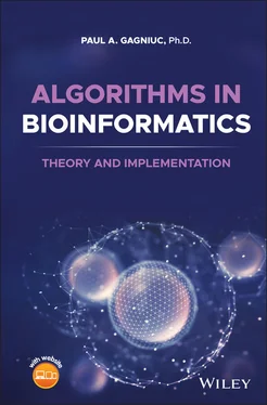
Paul A. Gagniuc - Algorithms in Bioinformatics
Здесь есть возможность читать онлайн «Paul A. Gagniuc - Algorithms in Bioinformatics» — ознакомительный отрывок электронной книги совершенно бесплатно, а после прочтения отрывка купить полную версию. В некоторых случаях можно слушать аудио, скачать через торрент в формате fb2 и присутствует краткое содержание. Жанр: unrecognised, на английском языке. Описание произведения, (предисловие) а так же отзывы посетителей доступны на портале библиотеки ЛибКат.
- Название:Algorithms in Bioinformatics
- Автор:
- Жанр:
- Год:неизвестен
- ISBN:нет данных
- Рейтинг книги:3 / 5. Голосов: 1
-
Избранное:Добавить в избранное
- Отзывы:
-
Ваша оценка:
- 60
- 1
- 2
- 3
- 4
- 5
Algorithms in Bioinformatics: краткое содержание, описание и аннотация
Предлагаем к чтению аннотацию, описание, краткое содержание или предисловие (зависит от того, что написал сам автор книги «Algorithms in Bioinformatics»). Если вы не нашли необходимую информацию о книге — напишите в комментариях, мы постараемся отыскать её.
Explore a comprehensive and insightful treatment of the practical application of bioinformatic algorithms in a variety of fields Algorithms in Bioinformatics: Theory and Implementation
global
local sequence alignment, forced alignment
Sequence logos, Markov chains
Self-Sequence alignment, Objective Digital Stains
Spectral Forecast
Discrete Probability Detector
Self-sequence
Objective Digital Stains
Spectral Forecast
Algorithms in Bioinformatics: Theory and Implementation
Algorithms in Bioinformatics — читать онлайн ознакомительный отрывок
Ниже представлен текст книги, разбитый по страницам. Система сохранения места последней прочитанной страницы, позволяет с удобством читать онлайн бесплатно книгу «Algorithms in Bioinformatics», без необходимости каждый раз заново искать на чём Вы остановились. Поставьте закладку, и сможете в любой момент перейти на страницу, на которой закончили чтение.
Интервал:
Закладка:
1.12.5 Control and Division of Organelles
Both genome and genome-less organelles divide. But how ? Regular bacterial fission (division) uses a dynamin-related protein (DRP) to constrict the membrane at its inner face [121]. However, DRPs are also essential for mitochondrial and peroxisomal fission [122]. Fission is required to provide a population of organelles for daughter cells during mitosis. In contrast to bacterial fission, mitochondria use dynamin and DPRs to constrict the outer membrane (a ring) from the cytosolic face [121]. For instance, the unicellular eukaryote Trichomonas vaginalis is a parasite that uses hydrogenosomes instead of regular mitochondria. The role of DRPs in the division of the hydrogenosome is similar to that described for peroxisomes and mitochondria [123]. Moreover, plastids use similar mechanisms and a plant-specific DPR [124, 125]. In eukaryotes, dynamin and DPRs have their genes stored in the nuclear genome. Thus, control over the division of membrane-bound organelles is held by the nuclear genome. Genes encoding DPRs were once present in the symbiotic α-proteobacterium ancestors of mitochondria or in the symbiotic cyanobacterium ancestors of the plastids. In other words, the alternative self-assembly of a complementary dynamin constriction mechanism on the outer membrane, allowed a transition of the organellar fission genes to the nuclear genome for synchronization of cell division. Moreover, this complementary mechanism allowed a reductive evolution up to the point of complete genome elimination in some organelles, such as in the case of hydrogenosomes. But how do organelle genes physically get into the nucleus to recombine with chromosomal DNA? Insertion of DNA from organelle genomes into the nucleus is DNA-mediated (RNA-mediated insertions may also occur) through a process called HGT [126]. As a side note, peroxisomes are single-membrane organelles that catalyze the breakdown of very-long-chain fatty acids through beta-oxidation [127]. Peroxisomes are a hybrid of mitochondrial and ER-derived (ER –endoplasmic reticulum) pre-peroxisomes [128].
1.12.6 The Horizontal Gene Transfer
The symbiosis between organisms is not possible without the HGT. The HGT is the weak “glue” that unites all species and it has important evolutionary implications [129]. In the future, these implications may very well undo the classification discussed above for the tree of life and how we understand the origin of life on Earth. The HGT refers to the transmission of DNA between different genomes, whereas the vertical gene transfer (VGT) is made between generations by sexual or asexual reproduction. The way in which the classification for the tree of life works is largely based on the VGT concept; thus, one can imagine the issue. HGT was first observed as a phenomenon in Streptococcus pneumoniae species by Frederick Griffith in 1928 [130]. The main observation made by Frederick Griffith was that virulence (pathogenicity) in this species of bacterium is transmitted by contact or proximity. This was an important revelation for the later field of genetics. Since then, increasing evidence shows that DNA fragments of different sizes may be exchanged between the kingdoms of life, to a greater or lesser extent [129]. Not long ago, the transfer of genetic information from the members of the Agrobacterium genus to eukaryote cells was seen as an extraordinary and rare process [131, 132]. Today, evidence indicates clearly that transfer of genetic information between species and inside different cell compartments is a common process, which takes place over the evolutionary time. For instance, bacteria have acquired genetic material from eukaryotic hosts and vice versa [133]. Viruses contain genes derived from their eukaryotic hosts and vice versa [134]. In plants, for instance, the HGT between genomes takes place through intracellular transfer of DNA among the nuclear, mitochondrial, and plastid genomes. The transfer of mitochondrial genes to the nucleus is known to be an ongoing evolutionary process. However, evidence also shows a HGT of mitochondrial DNA to the plastid genome [135]. Moreover, expression of a transferred nuclear gene in a mitochondrial genome was also observed [136]. For example, the orf164 gene in the mitochondrial genome of Arabidopsis is derived from the nuclear ARF17 gene that codes for an auxin-responsive protein [136]. Thus, the transfer of DNA segments from any location to any other location seems to be a rule across all life. However, HGT is most frequent between closely related species with similar genome features and less frequent otherwise [137]. In other words, HGT is a process that occurs at different frequencies between prokaryotes, between eukaryotes, between prokaryotes and eukaryotes and vice versa [138]. Perhaps, the importance of HGT goes as far as the emergence of new species (speciation) [139, 140].
1.12.7 On the Mechanisms of Horizontal Gene Transfer
Understanding of the mechanisms and vectors underpinning HGT across the kingdoms of life is still limited. Mobile genetic elements (MGEs) represent the main known vectors for HGT [137]. Well-known HGT events often include, but are not limited to, transposable elements (TE), plasmids or bacteriophage elements [141]. The behavior of a MGE has a certain degree of stochasticity and may incorporate a complete gene(s) or may include only sections of a gene, or with a high probability none of the two. Sections of genes transferred by MGEs decay in time and are recognized in bioinformatic analyzes as pseudogenes (nonfunctional genes) [126]. Among the MGEs, TE can best show the level of complexity that a DNA fragment can exhibit. The TE were first observed in Zea mays (corn) by Barbara McClintock in 1950 [142]. The main observation made by Barbara McClintock was that the genetic material can jump from one place to another within a genome. The insertion of TEs into the coding pigment-genes was responsible for unstable phenotypes on the kernels of a maize ear (kernels of different colors). Note that each kernel is an embryo produced from an individual fertilization and one ear of corn contains around 800 kernels positioned in 16 rows.
1.13 Origins of Eukaryotic Multicellularity
Above the evolutionary time, cells of multicellular organisms evolved a series of states (cell types). The mechanisms that lead to the formation of such states are unknown. Biology includes several competing hypotheses on the origin of eukaryotic multicellularity; all of them based on observations made on the behavior of current species. These hypotheses suggest multiple pathways that can lead to multicellular organisms; some pathways more successful than others. Moreover, these competing hypotheses may all be valid. Note that only a few general notions are mentioned here.
1.13.1 Colonies Inside an Early Unicellular Common Ancestor
One of the hypotheses for multicellularity suggests a repeated division of the nucleus within the same unicellular organism and a subsequent formation of membranes in between the nuclei. A reminiscent coenocytic behavior can be seen in multicellular eukaryotic organisms, for instance, in the eggs (0.51 ± 0.003 mm) laid by the well-known Drosophila melanogaster (vinegar fly). The initial stages of the vinegar fly eggs contain multiple nuclei in a common cytoplasmic space (the entire volume of the egg) [143]. Only a few stages of development later, the cell membranes around the floating nuclei start to appear almost simultaneously to constitute the initial cells of the larva [143].
1.13.2 Colonies of Early Unicellular Common Ancestors
A second hypothesis for the origin of multicellularity proposes that unicellular organisms may aggregate to form unitary colonies that can achieve multicellularity and cell specialization over time. According to this theory, multicellularity emerged from cooperation between unicellular organisms. Examples of cooperation among organisms have been observed in nature at different scales and in various forms. One of the simplest integrated multicellular organisms is Tetrabaena socialis in which four identical cells constitute the individual [144]. The nuclear genome of T. socialis dictates the number of cells in the colony [145]. Another example is the choanoflagellate (Greek and Latin –khoánē, “funnel”; flagellate, “flagellum”) Salpingoeca rosetta, which can exist as a unicellular organism or it can switch to form multicellular spherical colonies called rosettes (form bridges between cells by incomplete cytokinesis), showing a primitive level of cell differentiation and specialization. Formation of multicellular colonies is induced by different signal molecules. The source of such signal molecules can originate from individuals of the same species (i.e. slime molds) or from individuals of different species (i.e. bacterium species) [146]. In the case of S. rosetta , the signal molecules for colony formation originates from the food source, namely the Algoriphagus machipongonensis bacterium (phylum Bacteroidetes ) [147, 148]. Choanoflagellates, sponges and algae of the genus Volvox are more complex examples of first evolutionary stages that indicate the border between colonial organisms and multicellular organisms. Choanoflagellates are the closest relative of metazoans (all animals composed of cells differentiated into tissues and organs) [149, 150]. Some genes required for multicellularity in animals, such as genes for adhesion, genes for signaling, and genes for extracellular matrix formation, are also found in choanoflagellates [151]. This suggests that these genes may have evolved in a common ancestor before the transition to multicellularity in animals [152]. Sponges are one of the oldest primitive multicellular organisms in the fossil record. Choanoflagellates are small single-celled protists, partially similar in shape and function with some of the sponges cells (choanocytes) [153]. Many associations have been made in the past between choanocytes and choanoflagellates. However, the transcriptome of sponge choanocytes is the least similar to the transcriptomes of choanoflagellates and is significantly enriched in genes unique to either animals or sponges alone [154]. Slime molds are also interesting examples, which can indicate how some multicellular organisms formed. Slime molds are unrelated eukaryotic organisms that can live as single cells. In certain conditions (i.e. starvation), single cells of the same species can aggregate to form multicellular reproductive structures [155]. For instance, the multicellular aggregate (a slug-like mass of a few thousand cells called a grex ) of amoebae Dictyostelium discoideum can show cellular adhesion, cellular specialization, tissue organization, and coordination that allows for mechanical movement [156, 157]. Although the behavior of D. discoideum is not necessarily a close example of the process that led to multicellular organisms, it can certainly serve as a clue for detailed research on the emergence of multicellularity.
Читать дальшеИнтервал:
Закладка:
Похожие книги на «Algorithms in Bioinformatics»
Представляем Вашему вниманию похожие книги на «Algorithms in Bioinformatics» списком для выбора. Мы отобрали схожую по названию и смыслу литературу в надежде предоставить читателям больше вариантов отыскать новые, интересные, ещё непрочитанные произведения.
Обсуждение, отзывы о книге «Algorithms in Bioinformatics» и просто собственные мнения читателей. Оставьте ваши комментарии, напишите, что Вы думаете о произведении, его смысле или главных героях. Укажите что конкретно понравилось, а что нет, и почему Вы так считаете.
