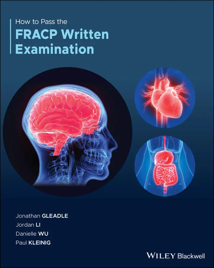
Jonathan Gleadle - How to Pass the FRACP Written Examination
Здесь есть возможность читать онлайн «Jonathan Gleadle - How to Pass the FRACP Written Examination» — ознакомительный отрывок электронной книги совершенно бесплатно, а после прочтения отрывка купить полную версию. В некоторых случаях можно слушать аудио, скачать через торрент в формате fb2 и присутствует краткое содержание. Жанр: unrecognised, на английском языке. Описание произведения, (предисловие) а так же отзывы посетителей доступны на портале библиотеки ЛибКат.
- Название:How to Pass the FRACP Written Examination
- Автор:
- Жанр:
- Год:неизвестен
- ISBN:нет данных
- Рейтинг книги:5 / 5. Голосов: 1
-
Избранное:Добавить в избранное
- Отзывы:
-
Ваша оценка:
How to Pass the FRACP Written Examination: краткое содержание, описание и аннотация
Предлагаем к чтению аннотацию, описание, краткое содержание или предисловие (зависит от того, что написал сам автор книги «How to Pass the FRACP Written Examination»). Если вы не нашли необходимую информацию о книге — напишите в комментариях, мы постараемся отыскать её.
Provides a thorough review of the latest FRACP basic training syllabus Features QR codes embedded in the text to enable quick access to all references Offers tips, hints, advice, and examination strategies from previous candidates Provides numerous questions grounded in clinically relevant cases Covers of areas of medicine that are new, contemporary, and evolving Covering both the ‘Basic Sciences’ and ‘Clinical Practice’ of the latest exam
is an essential companion for FRACP candidates as well as those looking to refresh, improve, or update their knowledge of the FRACP syllabus.
How to Pass the FRACP Written Examination — читать онлайн ознакомительный отрывок
Ниже представлен текст книги, разбитый по страницам. Система сохранения места последней прочитанной страницы, позволяет с удобством читать онлайн бесплатно книгу «How to Pass the FRACP Written Examination», без необходимости каждый раз заново искать на чём Вы остановились. Поставьте закладку, и сможете в любой момент перейти на страницу, на которой закончили чтение.
Интервал:
Закладка:
A dual‐chamber pacemaker does not provide symptomatic relief or protect patients from ventricular arrhythmias and sudden cardiac death. Recent clinical trials have demonstrated SGLT2 inhibitors reduces mortality and hospitalisation in patients with type 2 diabetes and established cardiovascular disease. However, it is currently contraindicated in patients with eGFR <30 ml/min/1.73 m 2. Adding a beta blocker in an elderly patient with severe airway disease and insulin dependent diabetes may need careful consideration and is not the most effective treatment for this patient.

Normand C, Linde C, Singh J, Dickstein K.Ref: Indications for Cardiac Resynchronization Therapy. A Comparison of the Major International Guidelines JACC: Heart Failure. 2018;6(Issue 4):308‐316., April 2018
https://www.sciencedirect.com/science/article/pii/S2213177918301203
16. Answer: C
SGLT2 inhibitor (SGLT2i) works by inhibiting SGLT2 in the proximal convoluted tubule, to prevent reabsorption of glucose and facilitate its excretion in urine. As glucose is excreted in urine, its plasma levels fall leading to an improvement in glycaemic parameters. This mechanism of action of SGLT2i is dependent on blood glucose levels and has minimal potential for hypoglycaemia. SGLT2i's mode of action depends upon normal renal glomerular–tubular function and their efficacy is reduced in persons with renal impairment. SGLT2i are not prescribed on their own but can be used in combination with other diabetes medications. SGLT2i may also cause modest weight loss and reduce systolic blood pressure, which are beneficial for patients with heart failure.
Thiazolidinediones may cause water retention, weight gain, or worsen heart failure and are contraindicated in patients with New York Heart Association (NYHA) class II–IV heart failure. They should be used cautiously in patients with NYHA class I heart failure.

Atherton J, Sindone A, De Pasquale C, et al. National Heart Foundation of Australia and Cardiac Society of Australia and New Zealand: Australian clinical guidelines for the management of heart failure 2018. Medical Journal of Australia. 2018;209(8):363–369.
https://onlinelibrary.wiley.com/doi/abs/10.5694/mja18.00647?sid=nlm%3Apubmed
17. Answer: B
This patient's clinical presentation, risk factors, and echocardiogram findings are consistent with heart failure with preserved ejection fraction (HFpEF). No therapy has been proven to reduce mortality in patients with HFpEF.
HFpEF predominantly affects elderly (>65 years) hypertensive women. Other risk factors include obesity, coronary artery disease, diabetes, atrial fibrillation, and hyperlipidaemia. It is established that the prevalence of HFpEF among patients with heart failure averages 47% and its prevalence in the community is estimated to be 1.1% to 5.5% of the general population. The prevalence of HFpEF has increased over the last two decades, due to ageing population, increasing prevalence of risk factors, such as hypertension, diabetes, and increased survival.
The diagnosis of HFpEF can be challenging because the symptoms and signs are non‐specific, hence it is a strictly clinical diagnosis. For patients presenting with heart failure and relatively normal LV ejection fraction (LVEF), valvular heart disease, infiltrative cardiomyopathies, pericardial disease, high‐output heart failure, chronic pulmonary disease, and pulmonary arterial hypertension should be excluded. An elevated BNP or NT‐proBNP on its own is insufficient for the diagnosis or exclusion of HFpEF.
Echocardiogram is the imaging modality of choice to establish the diagnosis of HFpEF by criteria; exclude valvular, right‐sided, or pericardial disease; and assess for other differential diagnoses. The following are the echocardiographic criteria recommended by the European Society of Cardiology for the diagnosis of HFpEF:
LVEF ≥50%
LV end‐diastolic volume index (LVEDI) <97 mL/m2
Raised LV filling pressure is indicated by a ratio of mitral early diastolic inflow velocity to mitral early annular lengthening velocity (E/e')>15.
Cardiac catheterisation is the gold standard for the diagnosis of HFpEF. Criteria for raised LV filling pressure include LV end‐diastolic pressure >16 mmHg or a mean pulmonary capillary wedge pressure >12 mmHg.
In contrast to heart failure with reduced ejection fraction, there is limited clinical trial evidence guiding the treatment of HFpEF. At present, no therapy including ACE inhibitors, angiotensin receptor blockers, β‐blockers and aldosterone antagonists has demonstrated mortality benefit in patients with HFpEF.
Managing comorbidities including hypertension, obesity, OSA, rate control for AF, and diabetes, are the mainstay of HFpEF treatment strategies. Diuretics can be used to reduce congestion and improve symptoms. Low‐dose spironolactone is recommended to reduce hospital admission on the basis of the TOPCAT trial results. Sildenafil is an inhibitor of phosphodiesterase‐5 that increases cGMP levels by blocking catabolism. Increased availability of cGMP could provide benefits for both vascular and myocardial remodelling, including attenuating hypertrophy, fibrosis, and impaired cardiac relaxation. In the RELAX trial, sildenafil did not improve 6 min walk distance or quality of life. Ivabradine is a selective sinus node If sodium channel inhibitor that reduces HR without affecting contractility. It can increase peak VO 2and reduced exercise mitral early diastolic velocity/mitral annular velocity (E/e') ratio.

Harper A, Patel H, Lyon A.AR. Heart failure with preserved ejection fraction. Clinical Medicine.Clin Med (Lond). 2018;18(Suppl 2): s24–s29. https://pubmed.ncbi.nlm.nih.gov/29700089/
18. Answer: D
Hypertrophic cardiomyopathy (HOCM) is a common monogenic cardiovascular disorder with a prevalence of 1 in 500. It is diagnosed with echocardiogram and MRI which shows a hypertrophic left ventricule without dilatation and it is not associated with another cardiac, systemic, metabolic, or syndromic disease. HOCM is diverse in clinical, phenotypic expression, and natural history. It is often underdiagnosed.
HOCM is inherited in an autosomal dominant pattern. It is associated with mutations (nucleotide sequence variants) in 11 or more genes encoding proteins of thick and thin myofilament contractile components of the cardiac sarcomere (Z‐disk), with beta‐myosin heavy chain and myosin‐binding protein C genes most commonly involved.
HOCM is predominantly an obstructive disease, with 70% of patients having mechanical impedance to left ventricular outflow (gradients ≥30 mm Hg) at rest or with exertion. Left ventricular outflow obstruction is usually produced by mitral‐valve systolic anterior motion and septal contact due to flow drag, causing complications such as mitral regurgitation, diastolic dysfunction, atrial fibrillation, congestive heart failure, and ventricular tachyarrhythmias.
HOCM is associated with increased sudden cardiac death associated with ventricular tachyarrhythmias, disorganised myocardial architecture, interstitial collagen deposition, and replacement scarring after myocyte death as a consequence of coronary microvascular mediated flow dysfunction and ischemia. Patients with a diagnosis of HOCM are often disqualified from participating in intensive competitive sports due to high risks of sudden cardiac death.
Читать дальшеИнтервал:
Закладка:
Похожие книги на «How to Pass the FRACP Written Examination»
Представляем Вашему вниманию похожие книги на «How to Pass the FRACP Written Examination» списком для выбора. Мы отобрали схожую по названию и смыслу литературу в надежде предоставить читателям больше вариантов отыскать новые, интересные, ещё непрочитанные произведения.


Обсуждение, отзывы о книге «How to Pass the FRACP Written Examination» и просто собственные мнения читателей. Оставьте ваши комментарии, напишите, что Вы думаете о произведении, его смысле или главных героях. Укажите что конкретно понравилось, а что нет, и почему Вы так считаете.