S. R. Prabhu - Handbook of Oral Pathology and Oral Medicine
Здесь есть возможность читать онлайн «S. R. Prabhu - Handbook of Oral Pathology and Oral Medicine» — ознакомительный отрывок электронной книги совершенно бесплатно, а после прочтения отрывка купить полную версию. В некоторых случаях можно слушать аудио, скачать через торрент в формате fb2 и присутствует краткое содержание. Жанр: unrecognised, на английском языке. Описание произведения, (предисловие) а так же отзывы посетителей доступны на портале библиотеки ЛибКат.
- Название:Handbook of Oral Pathology and Oral Medicine
- Автор:
- Жанр:
- Год:неизвестен
- ISBN:нет данных
- Рейтинг книги:5 / 5. Голосов: 1
-
Избранное:Добавить в избранное
- Отзывы:
-
Ваша оценка:
Handbook of Oral Pathology and Oral Medicine: краткое содержание, описание и аннотация
Предлагаем к чтению аннотацию, описание, краткое содержание или предисловие (зависит от того, что написал сам автор книги «Handbook of Oral Pathology and Oral Medicine»). Если вы не нашли необходимую информацию о книге — напишите в комментариях, мы постараемся отыскать её.
Discover a concise overview of the most common oral diseases in a reader-friendly book Handbook of Oral Pathology and Oral Medicine
Handbook of Oral Pathology and Oral Medicine
Handbook of Oral Pathology and Oral Medicine — читать онлайн ознакомительный отрывок
Ниже представлен текст книги, разбитый по страницам. Система сохранения места последней прочитанной страницы, позволяет с удобством читать онлайн бесплатно книгу «Handbook of Oral Pathology and Oral Medicine», без необходимости каждый раз заново искать на чём Вы остановились. Поставьте закладку, и сможете в любой момент перейти на страницу, на которой закончили чтение.
Интервал:
Закладка:
1 Chapter 2 Table 2.1 American Dental Association caries classification system: Site Defi...
List of Illustrations
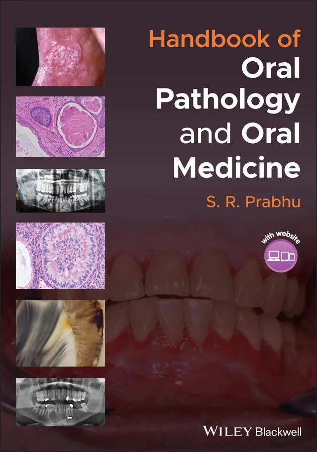
1 Chapter 1 Figure 1.1 Hypodontia: clinical photograph of missing maxillary lateral inci... Figure 1.2 Supernumerary premolars located lingual to the mandibular first a... Figure 1.3 Microdontia: maxillary left lateral incisor (‘peg lateral’) is co... Figure 1.4 (a) Gemination; mandibular right incisors show gemination. Note t... Figure 1.5 (a)Taurodontism of the mandibular first molar shows abnormally la... Figure 1.6 Amelogenesis imperfecta (hypocalcified type); the enamel is stain... Figure 1.7 Dentinogenesis imperfecta. (a) Note tooth wear and opalescent cro... Figure 1.8 Dentinal dysplasia radiograph showing absence of roots Figure 1.9 (a) Mesioangular impaction of the mandibular third molar. (b) Dis... Figure 1.10 (a) Dens invaginatus; radiograph showing dens invaginatus in a p... Figure 1.11 Yellow‐brown discoloration of maxillary incisors due to fluorosi... Figure 1.12 Tetracycline‐induced grey/brown discolouration of deciduous teet... Figure 1.13 Enamel pearl on the cementum Figure 1.14 Periapical radiograph of talon cusp on a partially erupted upper... Figure 1.15 (a) Hutchinson's incisors of congenital syphilis. Note screwdriv... Figure 1.16 An extracted mandibular molar with three roots.
2 Chapter 2 Figure 2.1 Rampant caries Figure 2.2 Early childhood caries Figure 2.3 Caries in a methamphetamine user Figure 2.4 Radiation caries Figure 2.5 (a) Early approximal enamel caries. Undecalcified section of a pr... Figure 2.6 Dentine caries. (a) Carious tooth with clinical crown lost to dec...
3 Chapter 3 Figure 3.1 Chronic hyperplastic pulpitis (pulp polyp) presenting as a fleshy... Figure 3.2 Periapical granuloma. (a) Radiolucent lesion of periapical granul... Figure 3.3 Apical abscess. (a) Presenting as a fluctuant gingival swelling; ... Figure 3.4 Condensing osteitis. Note the increased radiodensity around the r...
4 Chapter 4 Figure 4.1 (a) Generalized attrition of the incisal and occlusal surfaces. (... Figure 4.2 Resorption. (a) External: cropped orthopantomograph shows externa... Figure 4.3 Hypercementosis; extracted tooth with hypercementosis at the tip ... Figure 4.4 Cracked tooth syndrome; fractured premolar tooth (black arrows) v...
5 Chapter 5 Figure 5.1 Chronic gingivitis: clinical appearance of chronic marginal gingi... Figure 5.2 (a) Necrotizing ulcerative gingivitis shows papillary and gingiva...Figure 5.3 (a) Plasma cell gingivitis. Note swollen, oedematous red gingiva ...Figure 5.4 Desquamative gingivitis in a patient with mucous membrane pemphig...Figure 5.5 Generalized chronic periodontitis. (a) Disease in a 55‐year‐old w...Figure 5.6 (a) Generalized aggressive periodontitis (rapidly progressive per...Figure 5.7 Fibrous epulis. (a) Peripheral fibroma presenting as firm, pink, ...Figure 5.8 Peripheral ossifying/cementifying fibroma. (a) Pink nodular lesio...Figure 5.9 (a) Peripheral giant cell granuloma (giant cell epulis). This hig...Figure 5.10 Angiogranuloma. (a) a red lobulated mass is connected through th...Figure 5.11 Inflammatory gingival hyperplasia induced by plaque in an otherw...Figure 5.12 Generalized gingival enlargement with erythema and oedema in a p...Figure 5.13 Gingival enlargement caused by nifedipine, an antihypertensive d...Figure 5.14 Familial gingival hyperplasia. Firm fibrous gingival enlargement...Figure 5.15 (a) Gingival abscess: acute gingival abscess showing an erythema...Figure 5.16 Pericoronal abscess. Note a pus‐filled swelling partially coveri...Figure 5.17 Gingival enlargement in granulomatosis with polyangiitis (Wegene...Figure 5.18 Gingival enlargement in a patient with acute lymphocytic leukaem...Figure 5.19 Shiny, soft, bulbous, spongy gingival enlargement in a patient w...
6 Chapter 6Figure 6.1 Cropped orthopantomogram of acute suppurative osteomyelitis of th...Figure 6.2 Chronic suppurative osteomyelitis. Orthopantogram showing radiolu...Figure 6.3 (a) Focal sclerosing osteomyelitis: an intraoral radiograph showi...Figure 6.4 (a) Proliferative periostitis of the mandible: preoperative mandi...Figure 6.5 Cervicofacial cellulitis from odontogenic infection. Note diffuse...Figure 6.6 (a) Osteoradionecrosis of the mandible. (a) Note the exposed bone...Figure 6.7 Medication‐related osteonecrosis of the mandible (black arrow) in...
7 Chapter 7Figure 7.1 Radicular cyst. (a) Intraoral radiograph of a radicular cyst loca...Figure 7.2 Dentigerous cyst. (a) Cropped orthopantogram showing a dentigerou...Figure 7.3 Eruption cyst associated with tooth 11. Bluish surface of the swe...Figure 7.4 Odontogenic keratocyst. (a) Cropped Panoramic radiograph demonstr...Figure 7.5 (a) Lateral periodontal cyst. Cropped orthopantogram showing well...Figure 7.6 Calcifying odontogenic cyst. (a) Cropped orthopantogram showing a...Figure 7.7 Nasopalatine duct cyst. (a) Clinical photograph shows a palatal s...Figure 7.8 (a) Solitary bone cyst (simple bone cyst, black arrows). Note cha...
8 Chapter 8Figure 8.1 Ameloblastoma. (a) Pantomogram showing multilocular appearance of...Figure 8.2 Unicystic ameloblastoma. (a) Panoramic radiograph demonstrating r...Figure 8.3 Squamous odontogenic tumour. Bland irregularly shaped odontogenic...Figure 8.4 Calcifying epithelial odontogenic tumour. (a) Cropped panoramic r...Figure 8.5 Adenomatoid odontogenic tumour. (a) Radiograph showing an unerupt...Figure 8.6 Ameloblastic fibroma. (a) Orthopantomogram showing a well‐defined...Figure 8.7 Ameloblastic fibro‐odontome. (a). Cropped panoramic radiograph sh...Figure 8.8 (a) Odontome: Radiograph of a complex odontome. Note dense radiop...Figure 8.9 Dentinogenic ghost cell tumour. (a) An orthopantomogram showing a...Figure 8.10 Odontogenic myxoma. (a) orthopantomogram showing a poorly define...Figure 8.11 Odontogenic fibroma. Photomicrograph showing cellular fibrocolla...Figure 8.12 Cementoblastoma. (a) Round radiopacity with radiolucent rim at t...
9 Chapter 9Figure 9.1 Osteoma. (a). Clinical photograph of a 21 year old male with a sl...Figure 9.2 Gardner’s syndrome. (a) Extraoral osteomas: multiple diffuse bila...Figure 9.3 Central haemangioma. (a) Occlusal radiograph revealing a well‐def...Figure 9.4 Melanotic neuroectodermal tumour of infancy. (a) Clinical photogr...Figure 9.5 Osteosarcoma. (a) Radiographical findings in a case of chondrobla...Figure 9.6 Chondrosarcoma. (a) Coronal computed tomography revealing the pre...Figure 9.7 (a) Ewing's sarcoma. Cropped panoramic radiograph shows radioluce...Figure 9.8 Multiple myeloma. (a) Radiograph shows multiple punched‐out radio...Figure 9.9 Plasmacytoma. (a) Orthopantomogram revealing ill‐defined radioluc...Figure 9.10 Burkitt's lymphoma. (a) Tumour in a seven‐year‐old Nigerian boy ...
10 Chapter 10Figure 10.1 Ossifying fibroma. (a) Orthopantomograph showing a well‐defined ...Figure 10.2 (a) Periapical cemental dysplasia; radiograph showing partly min...Figure 10.3 (a) Periapical cemental dysplasia; photomicrograph showing woven...Figure 10.4 Central giant cell granuloma. (a) Cropped panoramic radiograph d...
11 Chapter 11Figure 11.1 Osteogenesis imperfecta. (a) Characteristic blue sclera in a pat...Figure 11.2 Cleidocranial dysplasia. (a) Extreme hyperadduction of shoulders...Figure 11.3 Cherubism. (a) An orthopantomogram showing multilocular appearan...Figure 11.4 Acromegaly. (a) Facial features: bulging forehead (frontal bossi...Figure 11.5 Brown tumour of hyperparathyroidism. (a) Orthopantomogram with l...Figure 11.6 Paget's Disease of bone. (a) Computed tomography, coronal view, ...Figure 11.7 Fibrous dysplasia of the mandible. (a) Panoramic radiograph show...Figure 11.8 (a) Torus mandibularis. Note the bilaterally symmetrical nodular...
12 Chapter 12Figure 12.1 Leukoedema of the left buccal mucosa showing grey‐white, diffuse...Figure 12.2 AnkyloglossiaFigure 12.3 Geographic tongue.Figure 12.4 Hairy tongueFigure 12.5 Fissured tongueFigure 12.6 Lingual thyroid. (a) The dome shaped red fleshy mass at the base...Figure 12.7 Macroglossia with crenations along the margins and loss of papil...Figure 12.8 Bifid tongueFigure 12.9 Bifid uvula.Figure 12.10 (a–c) Small, incomplete cleft lip (a), complete unilateral clef...Figure 12.11 Calibre persistent labial artery. (a). Note a linear elevated p...Figure 12.12 Epstein pearl in a five‐week‐old infantFigure 12.13 Oral dermoid cyst. (a) Presenting as a smooth‐surfaced, soft ti...Figure 12.14 Lingual varicosities on the ventral tongue.Figure 12.15 Lymphoid aggregate located at the posterolateral surface of the...Figure 12.16 Parotid papilla appearing as a nodule on the buccal mucosa.Figure 12.17 Prominent circumvallate papillae on the posterior tongue (yello...Figure 12.18 Generalized black‐brown physiological melanin hyperpigmentation...
Читать дальшеИнтервал:
Закладка:
Похожие книги на «Handbook of Oral Pathology and Oral Medicine»
Представляем Вашему вниманию похожие книги на «Handbook of Oral Pathology and Oral Medicine» списком для выбора. Мы отобрали схожую по названию и смыслу литературу в надежде предоставить читателям больше вариантов отыскать новые, интересные, ещё непрочитанные произведения.


Обсуждение, отзывы о книге «Handbook of Oral Pathology and Oral Medicine» и просто собственные мнения читателей. Оставьте ваши комментарии, напишите, что Вы думаете о произведении, его смысле или главных героях. Укажите что конкретно понравилось, а что нет, и почему Вы так считаете.