Atlas of Endoscopic Ultrasonography
Здесь есть возможность читать онлайн «Atlas of Endoscopic Ultrasonography» — ознакомительный отрывок электронной книги совершенно бесплатно, а после прочтения отрывка купить полную версию. В некоторых случаях можно слушать аудио, скачать через торрент в формате fb2 и присутствует краткое содержание. Жанр: unrecognised, на английском языке. Описание произведения, (предисловие) а так же отзывы посетителей доступны на портале библиотеки ЛибКат.
- Название:Atlas of Endoscopic Ultrasonography
- Автор:
- Жанр:
- Год:неизвестен
- ISBN:нет данных
- Рейтинг книги:4 / 5. Голосов: 1
-
Избранное:Добавить в избранное
- Отзывы:
-
Ваша оценка:
Atlas of Endoscopic Ultrasonography: краткое содержание, описание и аннотация
Предлагаем к чтению аннотацию, описание, краткое содержание или предисловие (зависит от того, что написал сам автор книги «Atlas of Endoscopic Ultrasonography»). Если вы не нашли необходимую информацию о книге — напишите в комментариях, мы постараемся отыскать её.
Atlas of Endoscopic Ultrasonography Atlas of Endoscopic Ultrasonography, Second Edition
Atlas of Endoscopic Ultrasonography, Second Edition
Atlas of Endoscopic Ultrasonography — читать онлайн ознакомительный отрывок
Ниже представлен текст книги, разбитый по страницам. Система сохранения места последней прочитанной страницы, позволяет с удобством читать онлайн бесплатно книгу «Atlas of Endoscopic Ultrasonography», без необходимости каждый раз заново искать на чём Вы остановились. Поставьте закладку, и сможете в любой момент перейти на страницу, на которой закончили чтение.
Интервал:
Закладка:
In this second edition of the Atlas, we are grateful that the renowned Brazilian endoscopist Everson Artifon has joined our team, along with our previous editor Brenna Casey who has continued with the Atlas. Our previous editor, John Deutsch, has retired and fortunately his timeless and superb chapters related to learning EUS anatomy are retained.
In this edition, we are excited to have expanded our international panel of world class endosonographers as contributing authors to provide a variety of styles and approaches to EUS. Our authors include some of the “first‐generation” pioneers of endoscopic ultrasound as well as the next generation of interventional EUS pioneers who are improving the imaging abilities of new and enhanced EUS technology and expanding the breadth of interventional techniques. We are especially pleased to offer many new sections on “How to do” aspects of interventional and therapeutic EUS procedures.
We hope this Atlas will appeal to a wide spectrum of endosonographers, from those who are beginning their training to those who are looking to expand their horizons with therapeutic techniques.
Finally, we want to thank our families, colleagues, editors, authors, and especially Jenny Seward from our publisher, Wiley, for all their support without whom this Atlas could not be possible.
Frank Gress MD
Thomas Savides MD
About the Companion Website
This book is accompanied by a companion website:
www.wiley.com/go/gress/atlas

Videos showing procedures described in the book. (All videos are referenced in the text at the end of each chapter.)
All figures from the book available for downloading
1 Normal Human Anatomy
John C. Deutsch
Essentia Health Care Systems, Duluth, MN, USA
Introduction
The Visible Human Project at the University of Colorado has generated large volumes of human anatomy data. The original information is captured by slowly abrading away frozen human cadavers in a transaxial manner and capturing the anatomy by digital imaging. The digital data is compiled and then over the years is manipulated by scientists at the University’s Center for Human Simulation to allow access to identified cross‐sections in any plane as well as to models which can be lifted from the data set. Details regarding the Visible Human Project and its applications to gastroenterology and endosonography have been previously described.
This atlas is fortunate to be able to use the interactive anatomy resources developed by Vic Spitzer, Karl Reinig, David Rubenstein, and others to create movies that help explain what takes place during endoscopic ultrasound (EUS) evaluations. Since EUS is a “real‐time” examination, it seems reasonable to present this section primarily as “real‐time” videos. The videos can be viewed over and over, allowing endosonographers to look not only at the highlighted structures, but also at structures they might visualize during EUS that are not specifically identified on the selected video.
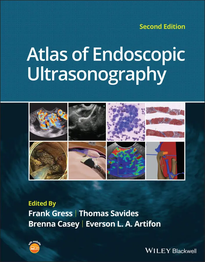
This chapter uses the terms “radial array orientation” to describe planar anatomy which would be found perpendicular to a line going through the digestive tract (as would be generated by a radial array echoendoscope, Figure 1.1) and “linear array orientation” for planar anatomy generated parallel to a line going through the digestive tract (as would be generated by a linear array echoendoscope, Figure 1.2).
Normal EUS anatomy from the esophagus
Radial array orientation ( Video 1.1)
Video 1.1starts with Visible Human Models of the left atrium (purple), trachea and bronchi (light blue), aorta and pulmonary arteries (red), vena cava (dark blue), and the esophagus (brown). A plane is shown passing through the esophagus. This plane contains the transaxial cross‐sectional anatomy images which then follow, starting in the oropharynx and going caudally. The upper esophageal sphincter (UES) is identified. As the images proceed distally, the trachea and esophagus can be followed to a point where the brachiocephalic left carotid and left subclavian arteries are evident just above the aortic arch. Below the aortic arch is the aortopulmonary window. The azygos arch can be seen exiting the superior vena cava (SVC). This occurs just above the tracheal bifurcation. The esophagus (labeled as “E”) is surrounded by the descending aorta, the vertebrae, and the trachea. The thoracic duct (not labeled) is visible between the aorta and vertebrae, inferior to the esophagus. Going distally, the pulmonary artery becomes prominent. The region between the right mainstem bronchus (RMB) and left mainstem bronchus (LMB) is the subcarinal space. The video progresses to a level where the left atrium surrounds the superior aspect of the esophagus and then the video ends as the esophagus passes the gastroesophageal junction.
An image plane cross‐section taken from a radial array orientation at the level of the subcarinal space is shown in Figure 1.3.
Linear array orientation ( Video 1.2)
Video 1.2starts with the same models as above (the left atrium [purple], trachea and bronchi [light blue], aorta and pulmonary arteries [red], vena cava [dark blue], and the esophagus [brown]). The plane shows potential ways that cross‐sectional anatomy can be generated. The video then shows a sagittal image with the descending aorta inferior to the esophagus, much as what is done during linear array EUS. In this orientation the pulmonary artery (PA) and left atrium are superior. The image plane is rotated to bring the left atrium and pulmonary artery to the inferior side of the esophagus. The models are then shown again, and the plane is moved in the caudal and cephalad directions, much as during EUS.
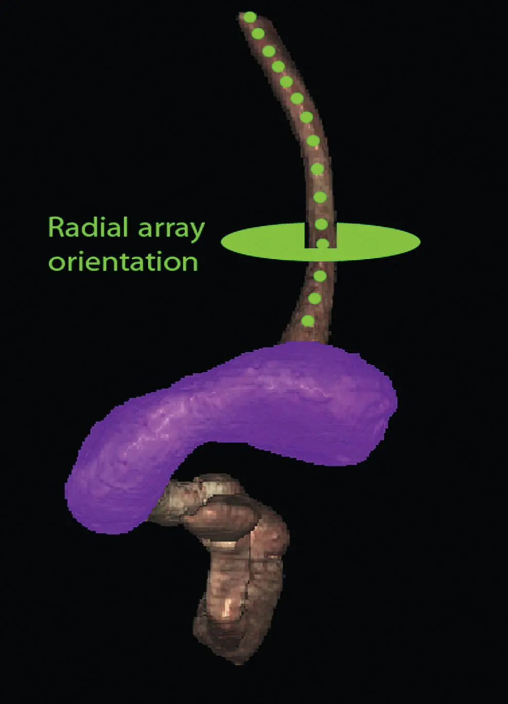
Figure 1.1 Visible Human Model of esophagus, stomach, and duodenum. The green circle shows a plane perpendicular to the axis and is similar to a plane developed during radial array endosonography.
Normal EUS anatomy from the stomach
Radial array orientation ( Video 1.3)
Endoscopic ultrasound of the stomach differs from EUS at other sites since the stomach does not constrain the endoscope tightly. It is important to follow anatomical structures (such as in a station approach) to avoid getting lost.
The video shows models of the stomach, esophagus, duodenum, gallbladder, pancreas (brown), the aorta, splenic artery, hepatic artery and left gastric artery (red), adrenal glands (pink), and splenic, superior mesenteric veins (dark blue) as viewed from behind. A plane is passed that is similar to the image plane generated during radial array EUS. The resultant cross‐sectional anatomy starts at the level of the gastroesophageal junction, with the aorta and inferior vena cava (IVC) labeled. The aorta (which is collapsed) is followed, which brings the pancreas and left adrenal gland into view. The first artery that comes off the aorta in the abdomen is the celiac artery. There is a trifurcation into the splenic, hepatic, and left gastric arteries (LGA), although the LGA is generally smaller and difficult to see. It is shown in the video at the “x” just before the bifurcation into the celiac and hepatic arteries as identified.
Figure 1.2 Visible Human Model of esophagus, stomach, and duodenum. The red circle shows a plane parallel to the axis and is similar to a plane developed during linear array endosonography.
Читать дальшеИнтервал:
Закладка:
Похожие книги на «Atlas of Endoscopic Ultrasonography»
Представляем Вашему вниманию похожие книги на «Atlas of Endoscopic Ultrasonography» списком для выбора. Мы отобрали схожую по названию и смыслу литературу в надежде предоставить читателям больше вариантов отыскать новые, интересные, ещё непрочитанные произведения.


Обсуждение, отзывы о книге «Atlas of Endoscopic Ultrasonography» и просто собственные мнения читателей. Оставьте ваши комментарии, напишите, что Вы думаете о произведении, его смысле или главных героях. Укажите что конкретно понравилось, а что нет, и почему Вы так считаете.