Istvan Urban - Vertical 2 - The Next Level of Hard and Soft Tissue Augmentation
Здесь есть возможность читать онлайн «Istvan Urban - Vertical 2 - The Next Level of Hard and Soft Tissue Augmentation» — ознакомительный отрывок электронной книги совершенно бесплатно, а после прочтения отрывка купить полную версию. В некоторых случаях можно слушать аудио, скачать через торрент в формате fb2 и присутствует краткое содержание. Жанр: unrecognised, на английском языке. Описание произведения, (предисловие) а так же отзывы посетителей доступны на портале библиотеки ЛибКат.
- Название:Vertical 2: The Next Level of Hard and Soft Tissue Augmentation
- Автор:
- Жанр:
- Год:неизвестен
- ISBN:нет данных
- Рейтинг книги:4 / 5. Голосов: 1
-
Избранное:Добавить в избранное
- Отзывы:
-
Ваша оценка:
Vertical 2: The Next Level of Hard and Soft Tissue Augmentation: краткое содержание, описание и аннотация
Предлагаем к чтению аннотацию, описание, краткое содержание или предисловие (зависит от того, что написал сам автор книги «Vertical 2: The Next Level of Hard and Soft Tissue Augmentation»). Если вы не нашли необходимую информацию о книге — напишите в комментариях, мы постараемся отыскать её.
A major part of this book comprises full-color, step-by-step images of patient cases. At times, reading it is like watching a surgical video, where the author 'stops the video' to discuss with you, the reader, what he is thinking and doing at that step, what his next step will be, and the reason for it.
Included again are the well-appreciated 'Lessons learned' sections, where the learning objectives are emphasized and further notes given, including ways to further improve the techniques. The section on the mandible is more detailed in this book, with the focus on larger defects and the different surgical steps in native, fibrotic, and scarred tissue types around the mental nerve during flap advancement.
In addition, light is shed on the detail in treating the anterior maxilla, which has not been published previously. It includes treatment options such as the fast track, the safe track, and the technical track of soft tissue reconstruction in conjunction with bone grafting as well as papilla reconstructions after bone regeneration. The section on the posterior maxilla hopes to resolve issues such as the management and complications of combined ridge and sinus grafting, including difficulties such as the lack of buccal, crestal or nasal bony walls of the posterior maxilla before bone grafting.
In this must-have new publication, the procedures are kept simple, repeatable, and biologically sound. The techniques presented are not overcomplicated; they are simple treatment strategies with lower complication rates and more predictability in the final outcome.
Vertical 2: The Next Level of Hard and Soft Tissue Augmentation — читать онлайн ознакомительный отрывок
Ниже представлен текст книги, разбитый по страницам. Система сохранения места последней прочитанной страницы, позволяет с удобством читать онлайн бесплатно книгу «Vertical 2: The Next Level of Hard and Soft Tissue Augmentation», без необходимости каждый раз заново искать на чём Вы остановились. Поставьте закладку, и сможете в любой момент перейти на страницу, на которой закончили чтение.
Интервал:
Закладка:
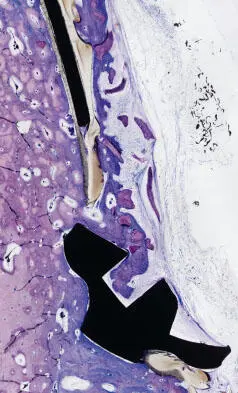
Fig 1-4 When compared with titanium, the PTFE-membrane demonstrated similar biocompatibility.
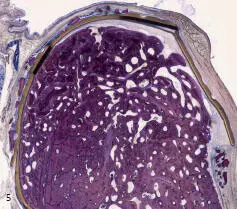
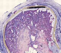
Figs 1-5 and 1-6 Histologic results of vertical augmentation. Note the excellent new bone formation and the well-incorporated xenograft particles.
Bone growth using a xenogenic bone graft
In a preclinical in vivo setting, a chronic vertical defect was treated using a xenograft. After 17 weeks of healing, the following was found: Emerging from the defect bed, the bone growth of a moderate to marked amount showed similar signs of remodeling, resulting in significant vertical ridge augmentation. The bone filler was markedly osteointegrated, showed slight signs of degradation, and demonstrated definite signs of osteoconduction (bone growth on the surface of the granules). The newly formed bone harbored numerous osteoblasts ( Figs 1-5
and 1-6
).
The epifluorescence analysis showed a marked grade of mineralization activity at different time points (OTC and XO), respectively. The signs of mineralization activity were visible at the newly formed and remodeled harversian systems (numerous concentric labeled rings). The outer circumferential bone lamellae were not fully formed, as is shown by their irregular shape. Two distinct and spaced lines of labeling (first OTC, then XO) indicated a marked vertical bone growth ( Figs 1-7
to 1-10
).
Figures 1-9
and 1-10
show that the xenograft particles are well incorporated in the newly formed trabecular bone. These images also demonstrate the phases of bone formation and maturation.
In the first phase, the newly formed ridge is present, but the cortical bone and the lacunae are not fully developed. This is referred to as ‘baby bone’ ( Figs 1-11
and 1-12
).
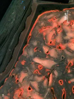
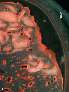
Figs 1-7 and 1-8 Epifluorescence analysis of a well-developed and mature bone after vertical augmentation.
In the next phase, the bone starts to further mature and corticalize, after which the outer layer becomes smooth and gains its final shape. Although the bone was good enough for the placement of implants, it would need about 3 more months to fully develop.
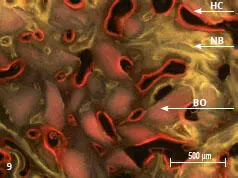
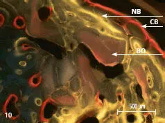
Figs 1-9 and 1-10 Epifluorescence analysis demonstrating the haversian canals and the cortical bone formation of the newly formed bone. The images demonstrate the incorporation of a biomaterial into the newly formed bone and the different time points of bone maturation. BO: anorganic bovine bone mineral; HC: haversian canal; NB: new bone; CB: cortical bone.
The healing time was 6 months ( Figs 1-13
and 1-14
). The implants were placed about a millimeter subcrestally. A tissue level implant placed into the bone with the polished collar 1 mm into the bone would be an excellent choice in the posterior region. The same patient had the other side grafted 10 months earlier. Due to scheduling issues, one side healed for longer than the other, but now the two phases of maturation can be compared. The ridge defects were similarly narrow ( Figs 1-15
to 1-21
).
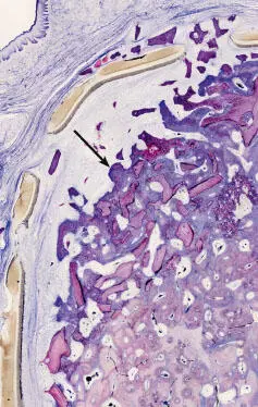
Fig 1-11 The outer surface of the ‘baby bone’ demonstrating irregularity and less maturity than the inner layer of the newly formed bone. This outer layer, referred to in this book as the ‘smear layer’ (see arrow), is about 1.5 mm in width. It will be remodeled and ‘shredded off’ during maturation.
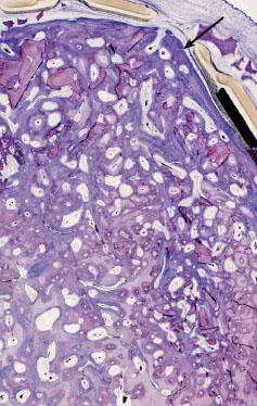
Fig 1-12 Image showing an area where corticalization has begun.
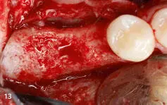
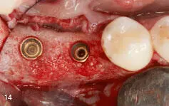
Figs 1-13 and 1-14 Clinical example of a posterior mandibular ridge augmentation using the Sausage technique. Note that some of the area is corticalized, whereas other parts are still in maturation.
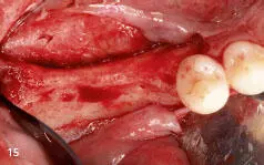
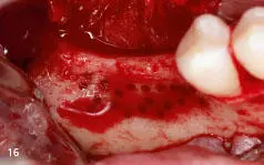
Figs 1-15 and 1-16 Occlusal and labial views of a narrow posterior mandibular ridge.
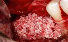
Fig 1-17 Labial view of the graft consisting of a 1:1 ratio of autogenous bone mixed with anorganic bovine bone mineral (ABBM).
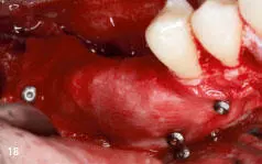
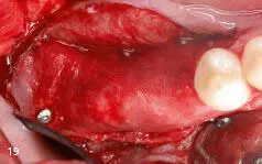
Figs 1-18 and 1-19 Labial and occlusal views of the fixated and stretched collagen membrane in place.
In this book, the smear layer will be highlighted, especially in the anterior maxilla chapters where it will be modified and preserved using the Mini Sausage technique as a secondary bone graft protecting the newly formed ridge.
The clinician should bear in mind that the smear layer will be either lost or modified. In most posterior cases, it is allowed to be shredded off, placing the implants deeper into the bone, whereas in the esthetic region, the Mini Sausage technique is used to prevent its resorption. These clinical procedures are exciting, and knowing the biology and dynamics of bone formation is essential to success.
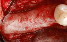
Fig 1-20 Occlusal view of the mature, corticalized, newly formed ridge.
Читать дальшеИнтервал:
Закладка:
Похожие книги на «Vertical 2: The Next Level of Hard and Soft Tissue Augmentation»
Представляем Вашему вниманию похожие книги на «Vertical 2: The Next Level of Hard and Soft Tissue Augmentation» списком для выбора. Мы отобрали схожую по названию и смыслу литературу в надежде предоставить читателям больше вариантов отыскать новые, интересные, ещё непрочитанные произведения.


Обсуждение, отзывы о книге «Vertical 2: The Next Level of Hard and Soft Tissue Augmentation» и просто собственные мнения читателей. Оставьте ваши комментарии, напишите, что Вы думаете о произведении, его смысле или главных героях. Укажите что конкретно понравилось, а что нет, и почему Вы так считаете.