Istvan Urban - Vertical 2 - The Next Level of Hard and Soft Tissue Augmentation
Здесь есть возможность читать онлайн «Istvan Urban - Vertical 2 - The Next Level of Hard and Soft Tissue Augmentation» — ознакомительный отрывок электронной книги совершенно бесплатно, а после прочтения отрывка купить полную версию. В некоторых случаях можно слушать аудио, скачать через торрент в формате fb2 и присутствует краткое содержание. Жанр: unrecognised, на английском языке. Описание произведения, (предисловие) а так же отзывы посетителей доступны на портале библиотеки ЛибКат.
- Название:Vertical 2: The Next Level of Hard and Soft Tissue Augmentation
- Автор:
- Жанр:
- Год:неизвестен
- ISBN:нет данных
- Рейтинг книги:4 / 5. Голосов: 1
-
Избранное:Добавить в избранное
- Отзывы:
-
Ваша оценка:
Vertical 2: The Next Level of Hard and Soft Tissue Augmentation: краткое содержание, описание и аннотация
Предлагаем к чтению аннотацию, описание, краткое содержание или предисловие (зависит от того, что написал сам автор книги «Vertical 2: The Next Level of Hard and Soft Tissue Augmentation»). Если вы не нашли необходимую информацию о книге — напишите в комментариях, мы постараемся отыскать её.
A major part of this book comprises full-color, step-by-step images of patient cases. At times, reading it is like watching a surgical video, where the author 'stops the video' to discuss with you, the reader, what he is thinking and doing at that step, what his next step will be, and the reason for it.
Included again are the well-appreciated 'Lessons learned' sections, where the learning objectives are emphasized and further notes given, including ways to further improve the techniques. The section on the mandible is more detailed in this book, with the focus on larger defects and the different surgical steps in native, fibrotic, and scarred tissue types around the mental nerve during flap advancement.
In addition, light is shed on the detail in treating the anterior maxilla, which has not been published previously. It includes treatment options such as the fast track, the safe track, and the technical track of soft tissue reconstruction in conjunction with bone grafting as well as papilla reconstructions after bone regeneration. The section on the posterior maxilla hopes to resolve issues such as the management and complications of combined ridge and sinus grafting, including difficulties such as the lack of buccal, crestal or nasal bony walls of the posterior maxilla before bone grafting.
In this must-have new publication, the procedures are kept simple, repeatable, and biologically sound. The techniques presented are not overcomplicated; they are simple treatment strategies with lower complication rates and more predictability in the final outcome.
Vertical 2: The Next Level of Hard and Soft Tissue Augmentation — читать онлайн ознакомительный отрывок
Ниже представлен текст книги, разбитый по страницам. Система сохранения места последней прочитанной страницы, позволяет с удобством читать онлайн бесплатно книгу «Vertical 2: The Next Level of Hard and Soft Tissue Augmentation», без необходимости каждый раз заново искать на чём Вы остановились. Поставьте закладку, и сможете в любой момент перейти на страницу, на которой закончили чтение.
Интервал:
Закладка:

Istvan Urban 2021
Contents
Introduction Introduction 1 The biology of vertically and horizontally augmented bone Vertical bone growth is very challenging, both biologically and technically. A recent meta-analysis by Urban et al 1 found that, regardless of the technique, about 4.5 mm of vertical bone gain was achieved in the included studies. However, complications occur least frequently when the guided bone regeneration (GBR) technique is used. The results of this study indicate the advantage of using GBR. The technical challenge of GBR is discussed extensively in this book, while the biologic challenge that may play a role in limiting vertical bone gain is discussed in this chapter. Based on the author’s experience, the amount of vertical bone gain is not limited by biology, but more by the clinician’s abilities. The biologic background of vertical bone gain was investigated by the author in preclinical settings.
Chapter 1 The biology of vertically and horizontally augmented bone
Chapter 2 Scientific evidence of vertical bone augmentation utilizing a titanium-reinforced polytetrafluoroethylene mesh 2 Scientific evidence of vertical bone augmentation utilizing a titanium-reinforced polytetrafluoroethylene mesh Introduction This chapter examines how a dense polytetrafluoroethylene (d-PTFE) mesh was investigated in the author’s clinical practice. The primary study objectives were: 1. To evaluate the amount of bone height gain in millimeters and as a percentage of the baseline deficiency as a result of vertical bone augmentation (VBA) using a titanium-reinforced PTFE mesh (RPM) with mixed autogenous and anorganic bovine bone mineral (ABBM) particulate grafts. 2. To examine the influence of defect location, baseline deficiency extent, and membrane exposure on augmentation height. 3. To report the incidence of surgical and postsurgical complications associated with this treatment.
The extreme vertical defect of the posterior mandible
Chapter 3 Reconstruction of the extreme posterior mandibular defect: surgical principles and anatomical considerations
Chapter 4 Reconstruction of an advanced posterior mandibular defect with scarred tissue
Chapter 5 Reconstruction of an advanced posterior mandibular defect with narrow basal bone
Chapter 6 Reconstruction of an advanced posterior mandibular defect with incomplete periodontal bone levels: the ‘pawn sacrifice’
Chapter 7 Reconstruction of an advanced posterior mandibular defect with the Lasagna technique using low-dose bone morphogenetic protein-
Anterior mandibular vertical augmentation
Chapter 8 Reconstruction of the advanced anterior mandibular defect: surgical principles and anatomical considerations
Chapter 9 Reconstruction of the advanced anterior mandibular defect: considerations for soft tissue reconstruction and preservation of the regenerated bone
Chapter 10 Reconstruction of the advanced anterior mandibular defect: importance of horizontal bone gain
Posterior maxilla
Chapter 11 Long-term results of implants placed in augmented sinuses with minimal and moderate remaining alveolar bone
Chapter 12 Difficulties and complications relating to sinus grafting: hemorrhage and sinus septa
Chapter 13 Difficulties in sinus augmentation and posterior maxillary reconstructions: missing labial sinus wall and ridge deficiencies
Chapter 14 Sinus graft infection and postoperative sinusitis
Chapter 15 The reconstruction of an extreme vertical defect in the posterior maxilla
Anterior maxillary vertical augmentation
Chapter 16 Introduction and clinical treatment guidelines
Chapter 17 Complex reconstruction of an anterior maxillary vertical defect
Anterior maxillary defect
Chapter 18 Extreme defect augmentation in the anterior maxilla
Soft tissue reconstruction in conjunction with bone grafting
Chapter 19 Reconstruction of a natural soft tissue architecture after bone regeneration
Chapter 20 The labial strip gingival graft
Chapter 21 The double strip graft
Chapter 22 Large open-healing connective tissue graft
Reconstruction of the interimplant papilla
Chapter 23 The double connective tissue graft
Chapter 24 The Ice-cube connective tissue graft
Chapter 25 The Iceberg connective tissue graft
Interproximal bone and soft tissue regeneration
Chapter 26 Vertical periodontal regeneration in combination with ridge augmentation
Ultimate esthetics
Chapter 27 Reconstructionof the bone and soft tissue in conjunction with preserving the mucogingival junction
Chapter 28 Complications
Introduction
1
The biology of vertically and horizontally augmented bone
Vertical bone growth is very challenging, both biologically and technically. A recent meta-analysis by Urban et al 1found that, regardless of the technique, about 4.5 mm of vertical bone gain was achieved in the included studies. However, complications occur least frequently when the guided bone regeneration (GBR) technique is used. The results of this study indicate the advantage of using GBR. The technical challenge of GBR is discussed extensively in this book, while the biologic challenge that may play a role in limiting vertical bone gain is discussed in this chapter. Based on the author’s experience, the amount of vertical bone gain is not limited by biology, but more by the clinician’s abilities.
The biologic background of vertical bone gain was investigated by the author in preclinical settings.
The histology of polytetrafluoroethylene (PTFE) membranes in an in vivo setting
The mucogingival tissue is primarily characterized by a moderate vascularized fibrotic reaction that surrounds the PTFE membrane. The membrane is usually internally and externally surrounded by connective tissue that is rich in fibers and poor in cells, with the fibers oriented parallel to the membrane. Membrane pores, if present, show the presence of a highly vascularized matrix of connective tissue and a dense network of collagen fibers that penetrates across the pores to fix on the internal side of the membrane or, in some cases, to the newly formed bone.
The inner layer, which is composed of expanded PTFE (e-PTFE), is always placed against the bone of the lingual and buccal sides. A slight number of macrophages admixed with a few lymphocytes, polymorphonuclear cells, giant cells/osteoclasts, and plasma cells were observed around the membrane. Deep at the lingual side of the ridge, the membrane was often in direct contact with the bone tissue, even showing slight signs of osteointegration ( Figs 1-1
to 1-4
).
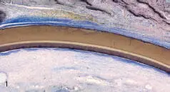

Figs 1-1 and 1-2 Parallel-oriented fibers around the membranes.
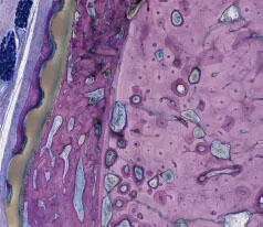
Fig 1-3 The membrane shows signs of osseointegration on both sides. Note the excellent biocompatibility of the polytetrafluoroethylene (PTFE) membrane.
Читать дальшеИнтервал:
Закладка:
Похожие книги на «Vertical 2: The Next Level of Hard and Soft Tissue Augmentation»
Представляем Вашему вниманию похожие книги на «Vertical 2: The Next Level of Hard and Soft Tissue Augmentation» списком для выбора. Мы отобрали схожую по названию и смыслу литературу в надежде предоставить читателям больше вариантов отыскать новые, интересные, ещё непрочитанные произведения.


Обсуждение, отзывы о книге «Vertical 2: The Next Level of Hard and Soft Tissue Augmentation» и просто собственные мнения читателей. Оставьте ваши комментарии, напишите, что Вы думаете о произведении, его смысле или главных героях. Укажите что конкретно понравилось, а что нет, и почему Вы так считаете.