Ghassem Ansari - Atlas of Cone Beam Computed Tomography
Здесь есть возможность читать онлайн «Ghassem Ansari - Atlas of Cone Beam Computed Tomography» — ознакомительный отрывок электронной книги совершенно бесплатно, а после прочтения отрывка купить полную версию. В некоторых случаях можно слушать аудио, скачать через торрент в формате fb2 и присутствует краткое содержание. Жанр: unrecognised, на английском языке. Описание произведения, (предисловие) а так же отзывы посетителей доступны на портале библиотеки ЛибКат.
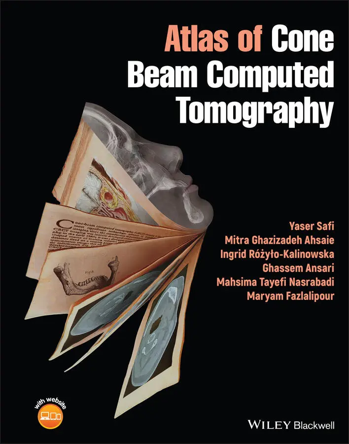
- Название:Atlas of Cone Beam Computed Tomography
- Автор:
- Жанр:
- Год:неизвестен
- ISBN:нет данных
- Рейтинг книги:5 / 5. Голосов: 1
-
Избранное:Добавить в избранное
- Отзывы:
-
Ваша оценка:
Atlas of Cone Beam Computed Tomography: краткое содержание, описание и аннотация
Предлагаем к чтению аннотацию, описание, краткое содержание или предисловие (зависит от того, что написал сам автор книги «Atlas of Cone Beam Computed Tomography»). Если вы не нашли необходимую информацию о книге — напишите в комментариях, мы постараемся отыскать её.
A thorough introduction to cone beam computed tomography, including in vivo and in vitro preparation and evaluation, indications in dentistry, and indications in medicine Comprehensive explorations of cone beam computed tomography artefacts and anatomic landmarks Practical discussions of cone beam computed tomography of dental structure, including normal anatomy, anomalies, and the difficulties of eruption In-depth examinations of cone beam computed tomography of pathological growth and development, including maxillofacial congenital and developmental anomalies Perfect for graduate dental students and postgraduate dental students in oral and maxillofacial radiology,
is also useful to general dentists, oral and maxillofacial radiologists, head and neck maxillofacial surgeons, head and neck radiologists, general radiologists, and ENT surgeons.
Atlas of Cone Beam Computed Tomography — читать онлайн ознакомительный отрывок
Ниже представлен текст книги, разбитый по страницам. Система сохранения места последней прочитанной страницы, позволяет с удобством читать онлайн бесплатно книгу «Atlas of Cone Beam Computed Tomography», без необходимости каждый раз заново искать на чём Вы остановились. Поставьте закладку, и сможете в любой момент перейти на страницу, на которой закончили чтение.
Интервал:
Закладка:
10 Chapter 10 Figure 10.1 Inflammatory pulpo‐periapical lesion. (a) Cross‐sectional view o...Figure 10.2 Radicular cyst. (a) Cross‐sectional view showing a well‐defined ...Figure 10.3 Radicular cyst. (a) Reformatted panoramic image showing a well‐d...Figure 10.4 Bilateral radicular cyst of primary molars. (a) Reformatted pano...Figure 10.5 Multiple cysts of jaw. Reformatted panoramic images from (a) max...Figure 10.6 Residual cyst. (a) Reformatted panoramic and (b) cross‐sectional...Figure 10.7 Healing process in residual odontogenic cyst. (a) Reformatted pa...Figure 10.8 Large infected radicular cyst. (a) Coronal, (b) sagittal, (c) ax...Figure 10.9 Periocoronitis. (a) Cropped panoramic reformat and (b) cross‐sec...Figure 10.10 Chronic osteomyelitis. (a) Axial, (b) cross‐sectional, (c) refo...Figure 10.11 Chronic osteomyelitis. (a) Axial image showing lytic destructio...Figure 10.12 Chronic osteomyelitis. (a) Reformatted panoramic image showing ...Figure 10.13 Chronic osteomyelitis caused by infected remained root. (a) Axi...Figure 10.14 Chronic osteomyelitis at the site of extracted right third mola...Figure 10.15 Hyperplastic follicle. (a) Reformatted panoramic image showing ...Figure 10.16 Dentigerous cyst. (a) Reformatted panoramic image with hash lin...Figure 10.17 Odontogenic kerato cyst (OKC). (a) Reformatted panoramic and (b...Figure 10.18 OKC. (a) Reformatted panoramic and (b) cross‐sectional images s...Figure 10.19 Small cyst‐like OKC. Axial view showing a well‐defined corticat...Figure 10.20 OKC. Note the scalloping feature between the teeth.Figure 10.21 Multiple OKCs in a patient with nevoid basal cell carcinoma syn...Figure 10.22 Healing OKC. (a) Reformatted panoramic and (b) cross‐sectional ...Figure 10.23 Lateral periodontal cyst (LPC). (a) Reformatted panoramic and (...Figure 10.24 Botryoid odontogenic cyst. (a) Axial, (b) cross‐sectional, and ...Figure 10.25 Bilateral buccal bifurcation cyst (BBC). (a) Reformatted panora...Figure 10.26 Glandular odontogenic cyst. (a) Axial, (b) cropped panoramic, a...Figure 10.27 Odontogenic myxoma in anterior mandible. (a) Reformatted panora...Figure 10.28 Ameloblastoma in posterior mandible. (a) Reformatted panoramic,...Figure 10.29 Unicystic ameloblastoma. The lesion has developed pericoronal t...Figure 10.30 Desmoplastic ameloblastoma. (a) Reformatted panoramic, (b) axia...Figure 10.31 Ameloblastic fibroma. (a) Reformatted panoramic, (b) cross‐sect...Figure 10.32 Ameloblastic fibro‐odontoma. (a) Reformatted panoramic and (b) ...Figure 10.33 Cystic odontoma. (a) Reformatted panoramic, (b) axial, and (c) ...Figure 10.34 Compound odontoma. (a) Reformatted panoramic, (b) maximum inten...Figure 10.35 Complex odontoma. (a) 3D surface rendering showing severe expan...Figure 10.36 Infection superimposed on complex odontoma. (a) Coronal, (b) sa...Figure 10.37 Hypercementosis. (a) Reformatted panoramic and (b) cross‐sectio...Figure 10.38 Cementoblastoma. (a) Reformatted panoramic image showing an amo...Figure 10.39 Periapical cemento osseous dysplasia (PCOD) at apex of left man...Figure 10.40 PCOD in early radiolucent stage. (a) Reformatted panoramic and ...Figure 10.41 Fibro‐osseous lesion inferior to IAN canal. (a) Reformatted pan...Figure 10.42 PCOD at site of previously extracted tooth. (a) Reformatted pan...Figure 10.43 PCOD at site of previously extracted teeth. (a) Reformatted pan...Figure 10.44 Focal cemento osseous dysplasia (FCOD). The lesion is radiopaqu...Figure 10.45 FCOD exposed at alveolar crest. The patient has a history of FC...Figure 10.46 Florid cemento ossoeus dysplasia (FLCOD). (a) Reformatted panor...Figure 10.47 Cemento ossifying fibroma (COF). (a) Reformatted panoramic, (b)...Figure 10.48 COF. (a) Serial axial and (b) serial coronal views showing an e... Figure 10.49 Bisphosphonate‐related osteonecrosis (BRONJ). (a) Reforma...Figure 10.50 Bone sclerosis and lamina dura thickening (arrows) due to consu...Figure 10.51 Osteoradionecrosis (ORN) in mandible. (a) Reformatted panoramic...Figure 10.52 ORN causing jaw fracture. (a) Reformatted panoramic and (b) 3D ...Figure 10.53 ORN in maxilla. (a) Reformatted panoramic image showing lytic o...Figure 10.54 Nasopalatine duct cyst. Sagittal views showing cystic formation...Figure 10.55 Simple bone cyst (SBC). (a) Reformatted panoramic, (b) sagittal...Figure 10.56 SBC developing within a focus of osseous dysplasia. (a) Reforma...Figure 10.57 Nasolabial cyst. (a) Axial and (b) 3D surface rendering images ...Figure 10.58 Stafne defect. (a) Axial and (b) 3D surface rendering images sh...Figure 10.59 Stafne defect in inferior border of mandible. (a) Reformatted p...Figure 10.60 Multiple dense bone islands (DBIs) in the mandible. (a) Reforma...Figure 10.61 Idiopathic osteosclerosis. (a) Reformatted panoramic image show...Figure 10.62 Sclerosing osteitis and root resorption. (a) Reformatted panora...Figure 10.63 Hyperostosis. (a) Axial and (b) 3D surface rendering images sho...Figure 10.64 Cortical hyperostosis on chin. A 15‐year‐old boy complained fro...Figure 10.65 Ossifying subperiosteal hematoma in a 14‐year‐old boy with a hi...Figure 10.66 Bilateral mandibular tori. (a) Axial and (b) 3D surface renderi...Figure 10.67 Frontal sinus osteoma. (a) Coronal and (b) sagittal views showi...Figure 10.68 Osteoma in the ethmoid sinus. (a) Coronal, (b) sagittal, (c) ax...Figure 10.69 Osteoma in maxillary sinus. Serial coronal CBCT images showing ...Figure 10.70 Osteoma. (a) Axial and (b) 3D reformatted panoramic images show...Figure 10.71 Multiple cortical osteoma. (a,b) 3D surface rendering showing m...Figure 10.72 Mixed cancellous and cortical osteoma. Axial view showing an os...Figure 10.73 Osteochondroma of condylar head. (a) Axial and (b) coronal view...Figure 10.74 Traumatic neuroma. Reformatted panoramic view of the mandible s...Figure 10.75 Neurilemoma. (a) Reformatted panoramic and (b) cross‐sectional ...Figure 10.76 Osteoid osteoma of temporal bone. (a) Coronal, (b) sagittal, (c...Figure 10.77 Osteoid osteoma in anterior mandible. (a) Axial, (b) cross‐sect...Figure 10.78 Osteoid osteoma in posterior mandible. (a) Reformatted panorami...Figure 10.79 Central giant cell granuloma (CGCG) in anterior mandible. (a) R...Figure 10.80 CGCG in anterior mandible. Same case as in Figure 10.80, 1 year...Figure 10.81 Aggressive CGCG in right maxilla. Coronal view showing destruct...Figure 10.82 Cherubism and CGCG in anterior mandible. (a) Conventional panor...Figure 10.83 Aneurysmal bone cyst (ABC). (a) Coronal, (b) sagittal, (c) axia...Figure 10.84 Desmoplastic fibroma of zygomatic bone. A 10‐year‐old patient p...Figure 10.85 Hemangioma of cheek soft tissue affecting maxilla. (a) Coronal ...Figure 10.86 Soft tissue arteriovenous malformation (AVM). (a) Axial and (b)...Figure 10.87 Soft tissue hemangioma. (a) Serial coronal and (b) 3D reformatt...Figure 10.88 Craniofacial fibrous dysplasia (FD). (a) Coronal, (b) sagittal,...Figure 10.89 Unifocal FD in left side of maxilla. (a,b) Reformatted panorami...Figure 10.90 FD in left side of mandible. (a) Reformatted panoramic image sh...Figure 10.91 Langerhans cell histiocytosis (LCH) in ramus. (a) Coronal, (b) ...Figure 10.92 LCH in alveolar process. (a) Axial, (b) cross‐sectional, (c) re...Figure 10.93 Patient with history of multiple myeloma and treatment with bis...Figure 10.94 Ewing sarcoma with history of chemotherapy. (a) Reformatted pan...Figure 10.95 Injection of steroids intralesional in CGCG as a treatment moda...Figure 10.96 Osteolytic osteosarcoma. (a) Reformatted panoramic image showin...Figure 10.97 Chondrosarcoma in anterior maxilla. (a) Reformatted panoramic a...Figure 10.98 Perineural extension malignancy to IAN canal. Reformatted panor...Figure 10.99 Pleomorphic adenoma (PA) in posterior palate. Patient presented...Figure 10.100 Central mucoepidermoid carcinoma (MEC). The patient complained...Figure 10.101 PA of right submandibular gland. (a) 3D surface rendering show...Figure 10.102 Acute Lymphatic Leukemia (ALL) in a 12‐year‐old boy with a his...Figure 10.103 Leukemia. (a,b) Reformatted panoramic images of the maxilla (a...Figure 10.104 Lymphoma. (a) Occlusal photography of maxilla showing expansio...Figure 10.105 Thalassemia. (a,b) Axial views of the mandible (a) and maxilla...Figure 10.106 Paget disease. (a) Reformatted panoramic, (b) cross‐sectional,...
Читать дальшеИнтервал:
Закладка:
Похожие книги на «Atlas of Cone Beam Computed Tomography»
Представляем Вашему вниманию похожие книги на «Atlas of Cone Beam Computed Tomography» списком для выбора. Мы отобрали схожую по названию и смыслу литературу в надежде предоставить читателям больше вариантов отыскать новые, интересные, ещё непрочитанные произведения.


Обсуждение, отзывы о книге «Atlas of Cone Beam Computed Tomography» и просто собственные мнения читателей. Оставьте ваши комментарии, напишите, что Вы думаете о произведении, его смысле или главных героях. Укажите что конкретно понравилось, а что нет, и почему Вы так считаете.