Ian Peate - Anatomy and Physiology for Nursing and Healthcare Students at a Glance
Здесь есть возможность читать онлайн «Ian Peate - Anatomy and Physiology for Nursing and Healthcare Students at a Glance» — ознакомительный отрывок электронной книги совершенно бесплатно, а после прочтения отрывка купить полную версию. В некоторых случаях можно слушать аудио, скачать через торрент в формате fb2 и присутствует краткое содержание. Жанр: unrecognised, на английском языке. Описание произведения, (предисловие) а так же отзывы посетителей доступны на портале библиотеки ЛибКат.
- Название:Anatomy and Physiology for Nursing and Healthcare Students at a Glance
- Автор:
- Жанр:
- Год:неизвестен
- ISBN:нет данных
- Рейтинг книги:5 / 5. Голосов: 1
-
Избранное:Добавить в избранное
- Отзывы:
-
Ваша оценка:
Anatomy and Physiology for Nursing and Healthcare Students at a Glance: краткое содержание, описание и аннотация
Предлагаем к чтению аннотацию, описание, краткое содержание или предисловие (зависит от того, что написал сам автор книги «Anatomy and Physiology for Nursing and Healthcare Students at a Glance»). Если вы не нашли необходимую информацию о книге — напишите в комментариях, мы постараемся отыскать её.
Everything you need to know about anatomy and physiology … An ideal introduction and revision guide for anatomy and physiology
www.wiley.com
www.wiley.com/email
All content reviewed by students for students
www.reviewnursingbooks.com
www.wiley.com/buy/9781119757207 at a Glance
at a Glance
At a Glance
Anatomy & Physiology for Nursing & Healthcare Students
Anatomy and Physiology for Nursing and Healthcare Students at a Glance — читать онлайн ознакомительный отрывок
Ниже представлен текст книги, разбитый по страницам. Система сохранения места последней прочитанной страницы, позволяет с удобством читать онлайн бесплатно книгу «Anatomy and Physiology for Nursing and Healthcare Students at a Glance», без необходимости каждый раз заново искать на чём Вы остановились. Поставьте закладку, и сможете в любой момент перейти на страницу, на которой закончили чтение.
Интервал:
Закладка:
Neurons are composed of an axon, dendrites and a cell body. Their function is to transmit nerve impulses. Nerve impulses only ever travel in one direction: from the receptive area – the dendrites – to the cell body and down the length of the axon.
Axon
Each neuron has only one axon, but the axon can branch to form an axon collateral (see Figure 10.3). The axon will also branch at its end into several axon terminals. The axon length may vary quite significantly, from very short to 100 cm long. The axon is much thicker and longer than the dendrites of a neuron. Larger neurons have a noticeably expanded region at the start of the axon, known as the axon hillock, which is the site of summation for incoming information. At any given moment, the combined influence of all axons that conduct impulses to a given neuron determines whether or not an action potential will be initiated at the axon hillock and disseminated along the axon.
Dendrites
Dendrites are the short branching processes that receive information. Generally, dendrites are very thin appendages, becoming narrower as they extend further away from the cell body (see Figure 10.3). Dendritic spines are short outgrowths that further increase the receptive surface area of a neuron. The surface of dendrite branches is covered with junctions designed for the reception of incoming information. Their branching processes provide a large surface area for this function. In sensory neurons, the dendrites frequently form part of the sensory receptors and in motor neurons they can be part of the synapse between one neuron and the next.
Cell body
The cell body (soma) is the central part of the neuron. It contains the nucleus of the cell and as such it is where most protein synthesis occurs. The nucleus ranges from 3 to 18 micrometers in diameter. Most of the neuron cell bodies (see Figure 10.3) are found inside the central nervous system, forming the grey matter. When there are clusters of cell bodies grouped together in the central nervous system, they are known as nuclei. Cell bodies found in the peripheral nervous system are called ganglia.
Myelin sheath
Oligodendrocytes and Schwann cells form the myelin sheaths that insulate axons in the central and peripheral nervous systems, respectively. Peripheral nerve axons and long or large axons are covered in a myelin sheath (see Figure 10.3). Myelin is a fatty material that protects the neuron as well as electrically insulating it, which speeds up impulse transmission. Schwann cells, wrapped in layers around the neuron within the peripheral nervous system, form the myelin sheath. The outermost part of the Schwann cell is its plasma membrane, called the neurilemma. There is a regular gap (about 1 μm) between adjacent Schwann cells called the nodes of Ranvier. Collateral axons can occur at the node. Some nerve fibres are unmyelinated and nerve impulse transmission in these fibres is significantly slower.
Cranial nerves
There are 12 pairs of cranial nerves emerging from the brain and supplying various structures, most of which are associated with the head and neck ( Figure 10.4). The cranial nerves vary in their functions; some are sensory nerves, containing only sensory fibres, some are motor nerves, containing only motor fibres, and some are mixed nerves, containing both sensory and motor fibres.
Clinical practice point
Multiple sclerosis is a disease that affects the central nervous system. The immune system attacks the myelin, the protective layer around nerve fibres, and causes inflammation and lesions. This makes it difficult for the brain to send signals to the rest of the body. The most common symptoms of multiple sclerosis include eye problems, numbness or tingling feelings, fatigue and pain. The symptoms can come and go and may change over time. They can be mild or more severe. The healthcare provider must ensure an individual assessment of needs is carried out and individual care plans are implemented and evaluated.
11 The structures of the brain
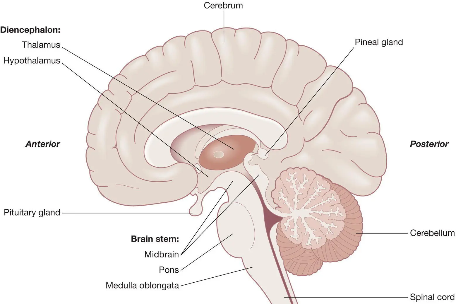
Figure 11.1 The cerebrum.
Source: Peate I, Wild K & Nair M (eds). Nursing Practice: Knowledge and Care (2014).
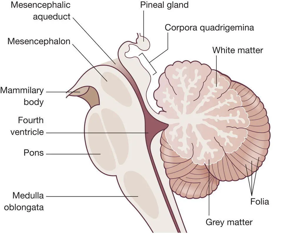
Figure 11.2 The cerebellum.
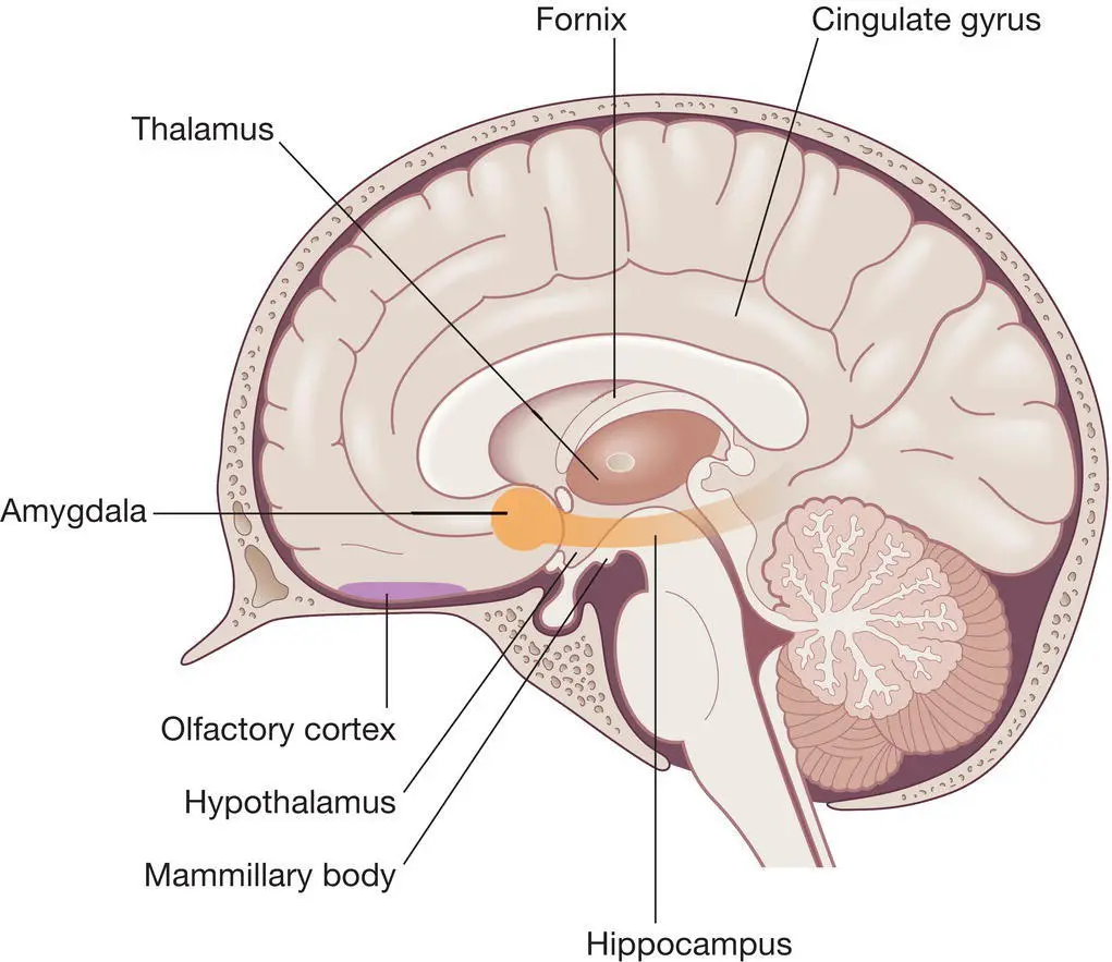
Figure 11.3 The limbic system.
The four major regions of the brain are:
the cerebrum
the diencephalon
the brainstem
the cerebellum.
Cerebrum
The cerebrum, also known as the telencephalon, is the largest and most highly developed part of the brain. It covers around two‐thirds of the brain mass and lies over and around most of the structures of the brain ( Figure 11.1). The cerebrum sits on top of the brainstem, with the cerebellum underneath the rear portion.
The cerebrum of an adult is divided into two large hemispheres (the left and right hemispheres). The surfaces of the cerebral hemispheres are highly folded and covered by a superficial layer of grey matter known as the cerebral cortex. The functions of the cerebrum include regulation of muscle contraction, memory storage and processing, production of speech, interpretation of taste, sound and memory for storage and processing.
Diencephalon
The diencephalon provides a functional link between the cerebral hemispheres and the rest of the central nervous system (CNS). It contains three paired structures: the thalamus, hypothalamus and epithalamus. Each component of the diencephalon has specialised functions that are integral to life.
Thalamus
The thalamus acts as a relay station for sensory impulses going to the cerebral cortex for integration and motor impulses entering and leaving the cerebral hemispheres. It also plays a role in memory.
Hypothalamus
The hypothalamus is closely associated with the pituitary gland and produces two hormones: antidiuretic hormone (ADH) and oxytocin. It is also the chief autonomic integration centre and is part of the limbic system, which is the emotional brain.
Epithalamus
The epithalamus is a small structure that is linked to the pineal gland, which secretes the hormone melatonin responsible for sleep/wake cycles.
Brainstem
The brainstem regulates vital cardiac and respiratory functions and acts as a vehicle for sensory information.
The structures that form the brainstem are involved in a range of activities that are essential for life. The brainstem is associated with the cranial nerves. The structures of the brainstem include the midbrain, pons and medulla oblongata (see Figure 11.1).
Midbrain
The midbrain contains nuclei that deal with auditory and visual information and reflexes. It also maintains consciousness and provides a conduction pathway connecting the cerebrum with the lower brain structures and spinal cord.
Pons
The pons connects and communicates with the cerebellum. It works with the medulla oblongata to control the depth and rate of respiration and contains nuclei that function in visceral and somatic motor control.
Читать дальшеИнтервал:
Закладка:
Похожие книги на «Anatomy and Physiology for Nursing and Healthcare Students at a Glance»
Представляем Вашему вниманию похожие книги на «Anatomy and Physiology for Nursing and Healthcare Students at a Glance» списком для выбора. Мы отобрали схожую по названию и смыслу литературу в надежде предоставить читателям больше вариантов отыскать новые, интересные, ещё непрочитанные произведения.


Обсуждение, отзывы о книге «Anatomy and Physiology for Nursing and Healthcare Students at a Glance» и просто собственные мнения читателей. Оставьте ваши комментарии, напишите, что Вы думаете о произведении, его смысле или главных героях. Укажите что конкретно понравилось, а что нет, и почему Вы так считаете.