Ian Peate - Anatomy and Physiology for Nursing and Healthcare Students at a Glance
Здесь есть возможность читать онлайн «Ian Peate - Anatomy and Physiology for Nursing and Healthcare Students at a Glance» — ознакомительный отрывок электронной книги совершенно бесплатно, а после прочтения отрывка купить полную версию. В некоторых случаях можно слушать аудио, скачать через торрент в формате fb2 и присутствует краткое содержание. Жанр: unrecognised, на английском языке. Описание произведения, (предисловие) а так же отзывы посетителей доступны на портале библиотеки ЛибКат.
- Название:Anatomy and Physiology for Nursing and Healthcare Students at a Glance
- Автор:
- Жанр:
- Год:неизвестен
- ISBN:нет данных
- Рейтинг книги:5 / 5. Голосов: 1
-
Избранное:Добавить в избранное
- Отзывы:
-
Ваша оценка:
Anatomy and Physiology for Nursing and Healthcare Students at a Glance: краткое содержание, описание и аннотация
Предлагаем к чтению аннотацию, описание, краткое содержание или предисловие (зависит от того, что написал сам автор книги «Anatomy and Physiology for Nursing and Healthcare Students at a Glance»). Если вы не нашли необходимую информацию о книге — напишите в комментариях, мы постараемся отыскать её.
Everything you need to know about anatomy and physiology … An ideal introduction and revision guide for anatomy and physiology
www.wiley.com
www.wiley.com/email
All content reviewed by students for students
www.reviewnursingbooks.com
www.wiley.com/buy/9781119757207 at a Glance
at a Glance
At a Glance
Anatomy & Physiology for Nursing & Healthcare Students
Anatomy and Physiology for Nursing and Healthcare Students at a Glance — читать онлайн ознакомительный отрывок
Ниже представлен текст книги, разбитый по страницам. Система сохранения места последней прочитанной страницы, позволяет с удобством читать онлайн бесплатно книгу «Anatomy and Physiology for Nursing and Healthcare Students at a Glance», без необходимости каждый раз заново искать на чём Вы остановились. Поставьте закладку, и сможете в любой момент перейти на страницу, на которой закончили чтение.
Интервал:
Закладка:
Nervous tissue
This is made up of neurons and glial cells. Its function is to receive and transmit neural impulses (reception and transmission of information). Two types of tissue are found in the nervous system: excitable cells (the neurons – initiate, receive, conduct and transmit information) and non‐excitable cells (the glial cells – supporting the neurons). A neuron is made up of two major parts: the cell body, containing the neuron’s nucleus, and cytoplasm and other organelles. Nerve processes are ‘finger‐like’ projections arising from the cell body and can conduct and transmit signals. There are two types: axons carrying signals away from the cell body and dendrites carrying signals toward the cell body. Neurons usually have one axon (this can be branched). Axons normally terminate at a synapse through which the signal is sent to the next cell, typically through a dendrite.
Connective tissue
There are many kinds of connective tissue and it is the most abundant type of tissue; connective tissue is typically used to fill empty spaces between other body tissues. The cells of connective tissue secrete substances that compose extracellular material, such as collagen and elastic fibres, creating a considerable spacing between these cells. Other important biological features include substance transportation, protection of the organism and insulation. Connective tissue (excluding blood) is found in organs supporting specialised tissues.
The matrix of areolar connective tissue is semi‐solid, containing adipocytes, mast cells and macrophages. Where there is a need to provide elasticity and tensile strength, areola tissue is present, for example under the skin, between muscles and in the alimentary canal. Adipose tissue supports the kidneys, brain and eyes and is related to energy intake and expenditure. Lymphoid tissue contains reticular cells and white blood cells, and is located in lymph tissue in the lymph nodes and lymphatic organs. Dense connective tissue (fibrous tissue composed of closely packed collagen fibres with little matrix) is found in ligaments, periosteum, muscle fascia and tendons. Blood is a fluid connective tissue. Cartilage is found as hyaline cartilage on the ends of the bones that form joints, the costal cartilage attaching the ribs to the sternum, and in the trachea, larynx and bronchi. Bone cells are surrounded by a matrix of collagen fibres with added strength provided by calcium and phosphate.
Muscle tissue
Muscle tissues are made of cells that permit contractions, generating movement. The function of muscle tissue is to pull bones (skeletal striated muscle), to contract and move viscera and vessels (smooth muscle), and make the heart beat (cardiac striated muscle). Muscle is involved each time we move, breathe, ingest food or urinate. The muscle cells comprise internal structures, called sarcomeres, in which myosin and actin molecules work to create contraction and movement.
There are three kinds of muscle in the body: skeletal, cardiac and smooth muscle. Skeletal muscle (striated muscle) is a voluntary muscle. Cells in skeletal muscle are long and thin with multiple nuclei. Cardiac muscle is only found in the heart, with the muscle fibres interlocking with each other to ensure that as one aspect of the muscle is stimulated, all other stimulated fibres contract sequentially. Cardiac muscle is not under voluntary control; the special cells of the sinoatrial node are responsible for sending out impulses resulting in cardiac contraction. Smooth muscle is involuntary, held together by connective tissue with bands of elastic protein wrapped around them. Smooth muscle is seen in the walls of hollow structures and vessels: blood vessels, ureters, urinary bladder, parts of the respiratory tract, ducts and glands of the alimentary tract.
Clinical practice point
A disruption of the structure of tissue is a sign of injury or disease. These changes can be detected through histology, which is the microscopic study of tissue appearance, organisation and function. When learning to care for people safely and effectively, this requires the nurse to appreciate the microscopic and macroscopic aspects of the human. Exploring how the body’s tissues function permits you to appreciate what can happen when cells, tissues and organ systems fail.
10 The brain and nerves
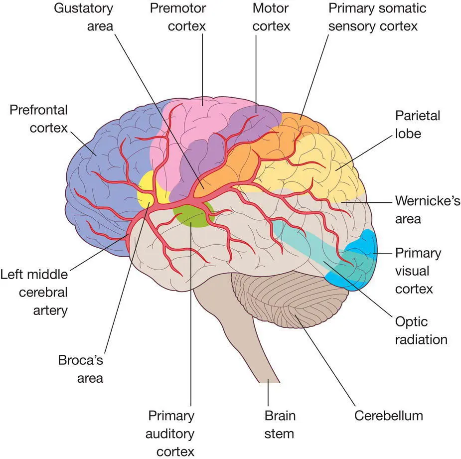
Figure 10.1 The brain.
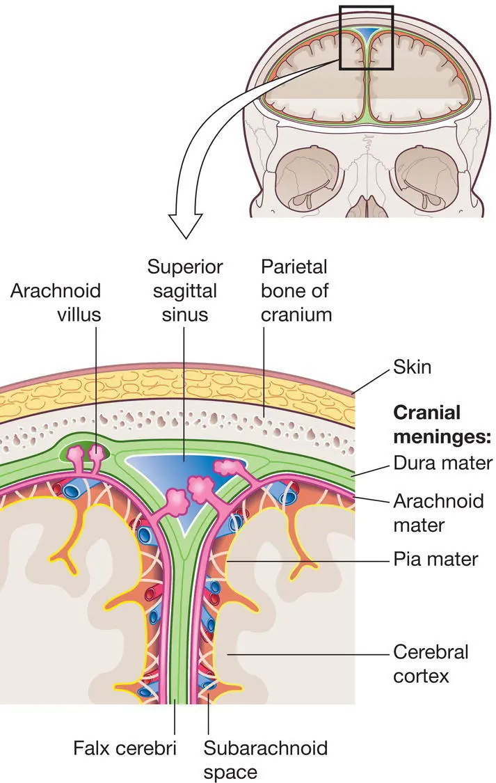
Figure 10.2 The meninges.
Source: Peate I, Wild K & Nair M (eds). Nursing Practice: Knowledge and Care (2014).
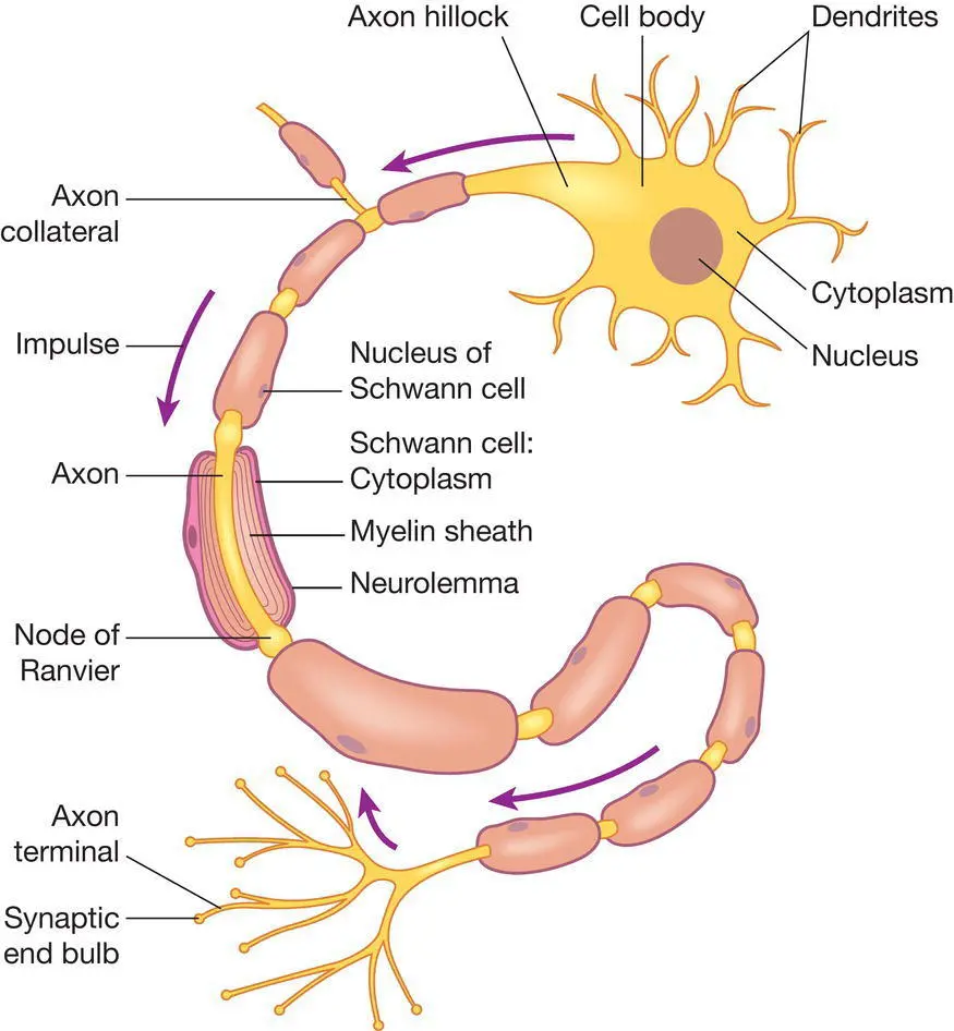
Figure 10.3 The neuron.
Source: Peate I, Wild K & Nair M (eds). Nursing Practice: Knowledge and Care (2014).
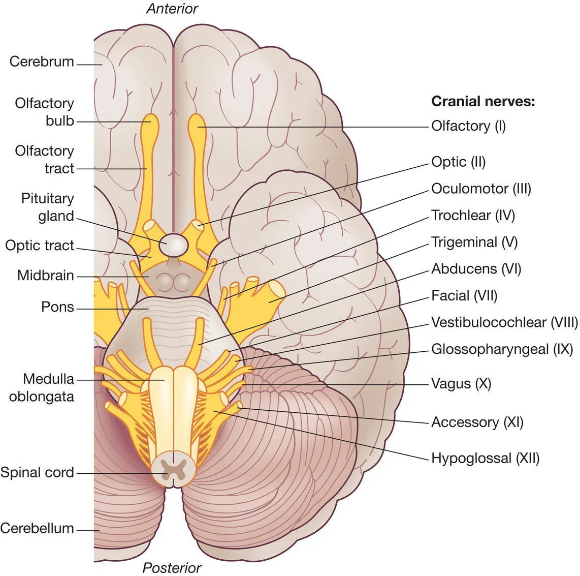
Figure 10.4 The cranial nerves.
Source: Peate I, Wild K & Nair M (eds). Nursing Practice: Knowledge and Care (2014).
The brain
The brain is one of the largest and most complex organs in the body. It is responsible for the integration of sensory information (for example, it interprets the senses); it also directs motor responses (it is the initiator of body movement and controller of behaviour) and is the centre of learning.
The brain weighs around 1400 grams. It is protected inside a bony shell (the cranium or skull) and washed by protective fluid (cerebrospinal fluid). It is the source of all those qualities that define us as humans ( Figure 10.1).
The brain receives 15% of the cardiac output. It has a system of autoregulation making sure that its blood supply is constant regardless of positional changes. Most of the expansion comes from the cerebral cortex, a convoluted layer of neural tissue covering the surface. The frontal lobes are particularly expanded and are involved in executive functions, such as self‐control, planning, reasoning and abstract thought.
The meninges
As nervous tissue can be easily damaged by pressure, it needs to be protected. The hair, skin and bone provide an outer layer of protection. Closest to the nervous tissue are the meninges which cover the delicate nervous tissue. They also protect the blood vessels that serve nervous tissue and they contain cerebrospinal fluid. The meninges are made up of three connective tissue layers: dura, arachnoid and pia maters ( Figure 10.2).
The cerebrospinal fluid
The cerebrospinal fluid (CSF) is produced by the choroid plexus located within the ventricles of the brain. Approximately 150 mL of CSF circulates around the brain, in the ventricles and around the spinal cord. Every 8 hours the CSF is replaced. The CSF provides a cushion to the brain to protect it from damage, it maintains a uniform pressure between the brain and spinal cord, and plays a small role in fluid and waste exchange between the brain and spinal cord.
The neuron
The functional unit of the brain is the neuron or nerve cell ( Figure 10.3). A neuron has a number of features in common with other cells, including a nucleus and mitochondria. However, because of its key role, it is well protected with some specialist modifications.
Читать дальшеИнтервал:
Закладка:
Похожие книги на «Anatomy and Physiology for Nursing and Healthcare Students at a Glance»
Представляем Вашему вниманию похожие книги на «Anatomy and Physiology for Nursing and Healthcare Students at a Glance» списком для выбора. Мы отобрали схожую по названию и смыслу литературу в надежде предоставить читателям больше вариантов отыскать новые, интересные, ещё непрочитанные произведения.


Обсуждение, отзывы о книге «Anatomy and Physiology for Nursing and Healthcare Students at a Glance» и просто собственные мнения читателей. Оставьте ваши комментарии, напишите, что Вы думаете о произведении, его смысле или главных героях. Укажите что конкретно понравилось, а что нет, и почему Вы так считаете.