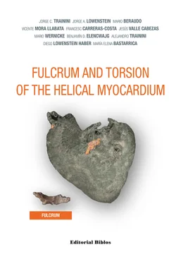
FULCRUM AND TORSION OF THE HELICAL MYOCARDIUM
In this new book, Trainini and his team attempt a step forward and make a series of proposals that come to complete cardiac anatomy, physiology and mechanics. Reading this new investigation is a pleasure that demands continuous attention, so that our “neuronal boxes” do not rebel against the effort it means to sometimes destroy what we have firmly installed in them. The text should be read slowly, as it was never easy to tread in swamp-limited grounds and, as in the ascent to the summit of a difficult mountain, stop now and then to take a breath and enjoy the view as we get near the peak, where we will see the final landscape of the new vision. Then we will be conscious that the effort was worthwhile.
In the first proposition of the research the authors study the anatomical and histological study of the myocardium, which defines a continuous, spiraling muscle, an integral piece that in order to fulfill its muscle function needs a supporting point, the cardiac fulcrum . The myocardium is inserted at its origin and end into this nucleus with tendinous, cartilaginous and even osseus structure, according to the specimen analyzed, located below and in front of the aorta. In this first proposition, splendidly documented, there is also an important contribution regarding the friction generated between the muscle layers in the mechanism of cardiac contraction
Afterward, the authors analyze the left ventricular endocardial and epicardial electrical activation sequence with three-dimensional electroanatomical mapping using a Carto navigation and mapping system, which allows a three-dimensional anatomical representation with electrical activation and propagation maps. Later on, the authors focus in the physiology and understanding of the suction mechanism that is explained about the persistent contraction of the ascending segment during the onset of the active protodiastolic phase. The authors describe a series of personal studies and investigations which define and clarify the three-stage cardiac mechanics with the concept of the scientifically-based suction phase, an active process that the authors explain as never before, integrating hydraulics and physics, disciplines not usually adequately incorporated to evaluate cardiac mechanics and the definition of mechanical suction pump.
Undoubtedly, the fast changes produced in cardiac imaging techniques will allow their use to understand the myocardial three-dimensional structure and its coupling with the new physiological concepts of cardiac mechanics.
We are facing a unique text, different, provocative. It is a creation that will demand a hundred percent of your attention, to sort out the new pathways that open before our eyes with original investigations and proposals in the limits of the unknown.
—Miguel Angel García Fernández
School of Medicine, Complutense University of Madrid, España
The true secret of cardiac mechanics is found deep inside the myocardium. At a glance, the cardiac muscle does not show its most hidden treasure. Only with an adequate dissection the mystery is unraveled and the cardiac fulcrum emerges between the muscle fibers. A chondroid anatomical structure that attaches the continuous myocardium at its ends. A support that allows torsion/detorsion motions. A supporting point working as the lever any muscle needs. This myocardium with helical structure explains a large part of cardiac pathophysiology. The beginning of this mystery led to explore along more complex pathwaysand evaluate a three-stage heart with an intermediate stage between systole and diastole, the suction phase.
Hidden mysteries, intrigues revealed after so many years of a classical cardiac anatomy, possible hypotheses and new concepts to ponder, are under the magnifying glass of this scientific study. Along these pages, the questions presented in this investigation were solved and explained in detail with the different studies performed. Other enigmas were jointly and interdisciplinary clarified from different perspectives. It was necessary to incorporate not strictly medical knowledge for the complex understanding of the involved mechanisms, which could not be revealed through a single discipline.
We never imagined that the beginning of this investigation would cause uncertainty and curiosity. Nothing more thrilling than submitting works to be evaluated and discussed collectively. New thoughts emerge from the critical and complementary view, from the possible to the real. Often something is built from simplicity, but the responsibility lies in sharing it to allow a common growth. This investigation has had a main objective, that of regarding a hypothesis with a new look that might contribute to the cardiac structure-function and solve pathologies. Fulcrum and Torsion of the Helical Myocardium invites to consider the cardiac organ as a true hydraulic pump.
JORGE C. TRAININI - JORGE A. LOWENSTEIN - MARIO BERAUDO - VICENTE MORA LLABATA - FRANCESC CARRERAS-COSTA - JESÚS VALLE CABEZAS - MARIO WERNICKE - BENJAMÍN D. ELENCWAJG - ALEJANDRO TRAININI - DIEGO LOWENSTEIN - MARÍA ELENA BASTARRICA
FULCRUM AND TORSION OF THE HELICAL MYOCARDIUM

3D-EAM: Three-dimensional electroanatomic mapping
APDSP: Active protodiastolic suction phase
CI: Cardiac index
CMR: Cardiac magnetic resonance
CO: Cardiac output
DCMP: Dynamic cardiomyoplasty
HDF: Hemodynamic forces
HR: Heart rate
LV: Left ventricle
LVEF: Left ventricular ejection fraction
LVSWI: Left ventricular systolic work index
MAP: Mean arterial pressure
NS: Not significant
PAP: Pulmonary artery pressure
PCP: Pulmonary capillary pressure
PVR: Pulmonary vascular resistance
RAP: Right atrial pressure
RV: Right ventricle
RVSWI: Right ventricular systolic work index
SI: Systolic index
SVR: Systemic vascular resistance
Miguel Angel García Fernández
Professor of Medicine-Cardiac Imaging. School of Medicine. Department of Medicine. Universidad Complutense de Madrid. Madrid, Spain.
Professor Tainini takes a step forward and again surprises us with his book Fulcrum and Torsion of the Helical Myocardium , which the reader has now in his hands and that could easily have the subtitle: “ Beyond the master Torrent ”….. and that is not a small thing.
As is well-known, Torrent Guasp with his mythical and original heart dissections, revolutionized the classical cardiac anatomy and mechanics found in our books for over 250 years. In a simple definition of his gigantic work, the ventricular myocardium was formed by a group of muscle fibers spatially arranged as a laterally flattened cord similar to a band that limited the two ventricular chambers and defined their functionality. All his studies converged in a radical contribution : diastolic suction as an active process, by the contraction off the myocardial band ascending segment .
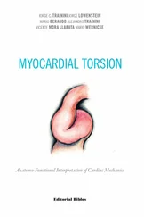
Undoubtedly, one of the most outstanding experts in Master Torrent Guasp’s theory is also Master Trainini, who had already delighted us with a splendid book written three years ago and leading a group of specialists: Myocardial torsion. Anatomo-functional investigation ”, achieving the most complete text created so far on cardiac anatomy and function under the “ lights of Torrent ”. This previous text came to respond and organize the answers of a long series of questions that emerged when physiology was approached with the master´s anatomy. The book, fitted as a magic puzzle, a large assortment of loose pieces that together provided solidity to Torrent’s theories.
Читать дальше
