Jorge C. Trainini - Fulcrum and Torsion of the Helical Myocardium
Здесь есть возможность читать онлайн «Jorge C. Trainini - Fulcrum and Torsion of the Helical Myocardium» — ознакомительный отрывок электронной книги совершенно бесплатно, а после прочтения отрывка купить полную версию. В некоторых случаях можно слушать аудио, скачать через торрент в формате fb2 и присутствует краткое содержание. Жанр: unrecognised, на английском языке. Описание произведения, (предисловие) а так же отзывы посетителей доступны на портале библиотеки ЛибКат.
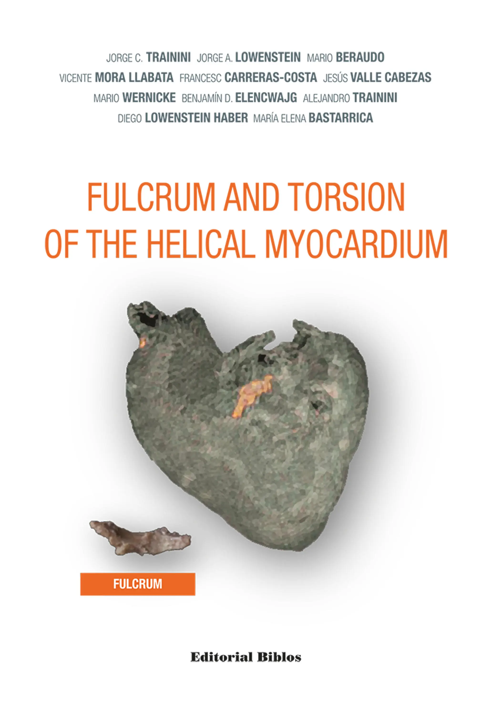
- Название:Fulcrum and Torsion of the Helical Myocardium
- Автор:
- Жанр:
- Год:неизвестен
- ISBN:нет данных
- Рейтинг книги:5 / 5. Голосов: 1
-
Избранное:Добавить в избранное
- Отзывы:
-
Ваша оценка:
Fulcrum and Torsion of the Helical Myocardium: краткое содержание, описание и аннотация
Предлагаем к чтению аннотацию, описание, краткое содержание или предисловие (зависит от того, что написал сам автор книги «Fulcrum and Torsion of the Helical Myocardium»). Если вы не нашли необходимую информацию о книге — напишите в комментариях, мы постараемся отыскать её.
Fulcrum and Torsion of the Helical Myocardium — читать онлайн ознакомительный отрывок
Ниже представлен текст книги, разбитый по страницам. Система сохранения места последней прочитанной страницы, позволяет с удобством читать онлайн бесплатно книгу «Fulcrum and Torsion of the Helical Myocardium», без необходимости каждый раз заново искать на чём Вы остановились. Поставьте закладку, и сможете в любой момент перейти на страницу, на которой закончили чтение.
Интервал:
Закладка:
3 Myocardial torsion constitutes the functional solution to eject the ventricular blood content with the necessary energy to supply the whole organism. In this way, the phylogenesis cardiac morphology is similar to ventricular mechanics, but lacks the comprehension of an electrical stimulus along its muscle pathways to correctly explain its motions. The studies undertaken on this topic aim to demonstrate the integrity of an essential cardiac structure-function. The analysis of left ventricular endo and epicardial electrical activation by means of three-dimensional electroanatomic mapping carried out in a series of patients, allowed us to address a transcendental question to analyze: How does myocardial torsion occur? Before answering this question, it is important to underline that throughout the whole book, when we speak of “myocardial torsion” it should be understood that it refers to an elastic and non-compressible material such as the myocardium. This means that this torsion, determined by the cardiac base and apex rotation in opposite directions implies a simultaneous longitudinal shortening. Although in the literature the terms twist, torsion and rotation have been used interchangeably, they do not mean the same thing, as will be seen later. “Torsion” is the term most frequently used to refer to cardiac motion, but we must not forget that the rotational motion described for the myocardium is accompanied by a simultaneous longitudinal shortening between base and apex.
4 The sliding motion between the myocardial segments during ventricular torsion-detorsion, implies that there must be an anti-friction mechanism to avoid the dissipation of the energy employed by the heart. Is there a specific histological explanation for this fact? Do Thebesian and Langer venous conduits play a role in this mechanism? Is there an organic lubricating source?
5 The development of the interventricular vortex studied by echocardiography is the consequence of torsion and of the necessary energy impulse the blood fluid requires to be ejected. Its flow dynamics can be understood through the physical theory of dissipative structures, which explains the organization of this intraventricular turbulence.
6 A passive cardiac full phase would be unfeasible due to the small pressure difference with the periphery. Ventricular filling was investigated as secondary to a generation of negative intraventricular pressure with energy expenditure during early diastole. The sudden lengthening of the left ventricular base-apex distance, after the ejective phase, produces a suction effect by an action similar to that of a “suction cup”: Could this mechanism be explained by the ascending segment persisting contraction during the first 100 ms of diastole? Does this mechanics allow us to consider suction during the early protodiastolic phase as an essential element in the physiology of the circulatory system, by being the continuity link between the pulmonary and systemic circulations?
7 According to the aforementioned bases, should a coupling phase be considered between systole and diastole where cardiac suction takes place?
8 In this three-stage heart (systole, suction, diastole): How does the energetic mechanism in the active suction phase act? Could the cardiac energy of suction and ejection be estimated? Should the left ventricular ejection fraction be considered a poorly reliable index? According to these considerations, would it not be more logical to speak of ejective cardiac energy as a parameter that summarizes the cardiac potential and to which all non-independent variables would concur?
9 Through the cardiac resynchronization procedure, Is it possible, to restore negative pressure to generate left ventricular suction with stimulation in the correct site, according to the path of the stimulus along the myocardial segments?
10 Could the understanding of this cardiac structure-function of the heart be of importance for surgical procedures of ventricular reduction and containment?
The methods used in this research to explain the hypothesis of the anatomo-functional integrity of the heart consisted of:
1 Cardiac dissection in bovine and human specimens.
2 Histological and histochemical analysis of the anatomical samples.
3 Left ventricular endocardial and epicardial electrical activation in humans by means of three-dimensional electroanatomic mapping.
4 The study of left ventricular suction physiology in dogs after removal of the right ventricle.
5 Measurement of left intraventricular pressure in ventricular resynchronization therapy.
6 Pathophysiological reinterpretation in experimental and clinical studies of heart failure (right ventricular bypass surgery, cardiomyoplasty, ventricular containment techniques, cardiac resynchronization, univentricular mechanical assistance, left ventricular repair).
7 Echocardiographic analysis to corroborate the research and usefulness of this knowledge in clinical practice since this technique has the ability to provide non-invasive knowledge in the complex mechanism of myocardial contraction.
8 Diffusion tensor sequences using cardiac magnetic resonance imaging to identify the orientation and deformation of myocardial fibers.
The findings and clinical data here presented are the result of experimental tests performed under the approval of all required regulatory authorities and under the informed consent of patients following the principles described in the current World Medical Association Declaration of Helsinki (2013). The experiments were conducted in accordance with the UK Animal Scientific Procedures Act 1996, the EU Directive 2010/63/EU for experimental animals and the “National Institutes of Health” guide for the care and use of laboratory animals (NIH Publication No. 8023, updated in 1978).
Конец ознакомительного фрагмента.
Текст предоставлен ООО «ЛитРес».
Прочитайте эту книгу целиком, купив полную легальную версию на ЛитРес.
Безопасно оплатить книгу можно банковской картой Visa, MasterCard, Maestro, со счета мобильного телефона, с платежного терминала, в салоне МТС или Связной, через PayPal, WebMoney, Яндекс.Деньги, QIWI Кошелек, бонусными картами или другим удобным Вам способом.
Интервал:
Закладка:
Похожие книги на «Fulcrum and Torsion of the Helical Myocardium»
Представляем Вашему вниманию похожие книги на «Fulcrum and Torsion of the Helical Myocardium» списком для выбора. Мы отобрали схожую по названию и смыслу литературу в надежде предоставить читателям больше вариантов отыскать новые, интересные, ещё непрочитанные произведения.


Обсуждение, отзывы о книге «Fulcrum and Torsion of the Helical Myocardium» и просто собственные мнения читателей. Оставьте ваши комментарии, напишите, что Вы думаете о произведении, его смысле или главных героях. Укажите что конкретно понравилось, а что нет, и почему Вы так считаете.