Predicting Heart Failure
Здесь есть возможность читать онлайн «Predicting Heart Failure» — ознакомительный отрывок электронной книги совершенно бесплатно, а после прочтения отрывка купить полную версию. В некоторых случаях можно слушать аудио, скачать через торрент в формате fb2 и присутствует краткое содержание. Жанр: unrecognised, на английском языке. Описание произведения, (предисловие) а так же отзывы посетителей доступны на портале библиотеки ЛибКат.

- Название:Predicting Heart Failure
- Автор:
- Жанр:
- Год:неизвестен
- ISBN:нет данных
- Рейтинг книги:3 / 5. Голосов: 1
-
Избранное:Добавить в избранное
- Отзывы:
-
Ваша оценка:
Predicting Heart Failure: краткое содержание, описание и аннотация
Предлагаем к чтению аннотацию, описание, краткое содержание или предисловие (зависит от того, что написал сам автор книги «Predicting Heart Failure»). Если вы не нашли необходимую информацию о книге — напишите в комментариях, мы постараемся отыскать её.
Predicting Heart Failure: Invasive, Non-Invasive, Machine Learning and Artificial Intelligence Based Methods
Predicting Heart Failure
Predicting Heart Failure: Invasive, Non-Invasive, Machine Learning and Artificial Intelligence Based Methods
Predicting Heart Failure — читать онлайн ознакомительный отрывок
Ниже представлен текст книги, разбитый по страницам. Система сохранения места последней прочитанной страницы, позволяет с удобством читать онлайн бесплатно книгу «Predicting Heart Failure», без необходимости каждый раз заново искать на чём Вы остановились. Поставьте закладку, и сможете в любой момент перейти на страницу, на которой закончили чтение.
Интервал:
Закладка:
2.3.11 Jugular Venous Pressure
The jugular venous pressure or jugular venous pulse estimation is a method for assessing central venous pressure. The central venous pressure alters with a change in the amount of blood flowing back to the heart as well as an increase or decrease in the capability of the heart to drive blood back to the circulatory system. The jugular venous pressure assessment procedure includes making the patient lie in a semi-recumbent position, turning the head of the patient to the left side (normally jugular venous pressure is assessed from the right side of the neck), and jugular venous pressure is measured by estimating the distance between the top point of the internal jugular vein and sternal angle. An elevated jugular venous pressure is linked with abnormal functioning of the right heart, heart failure conditions [13], and hypotension.
2.3.12 Carotid Pulse and Precordial Impulse
The carotid pulse is commonly a pressure signal sensed over the carotid artery when it passes near the neck. This pulse signifies the variations in the arterial blood pressure and volume with each heartbeat. It appears as a smooth rapid upstroke and a gradual downstroke with a brief interruption at the pulse peak. The abnormalities of the carotid pulse can be identified as an alteration in either pulse peak amplitude or a distortion of the upstroke or downstroke or both. These variations in carotid pulse contour reflect underlying cardiac abnormalities, such as aortic stenosis. However, it is generally confirmed only after detecting abnormal cardiac impulse or murmur.
Precordial impulses generally originate from the heart or great vessels visible or palpable on the anterior chest wall. All the precordial impulses including apex impulse, parasternal impulse, and pulmonary artery pulsation are observed and consideration is taken into account of the relevant location, size, and character, which including the duration, force, and contour.
2.3.13 Heart Sounds and Murmurs
Discrete bursts of auditory vibrations that differ in intensity (volume), frequency, quality, and duration are known as heart sounds, the sounds for a normal heartbeat being first heart sound (called lub) and second heart sound (called dub). The first heart sound consists of multiple high-frequency elements where only the first two among them can be heard normally. These two normally audible sound waves occur simultaneously when the mitral and tricuspid valves close. The first heart sound indicates the beginning of ventricular systole and the end of mechanical diastole. The second heart sound is described as a discrete burst of vibration that occurred due to the immediate closure of the aortic valve and pulmonic valve. The second heart sound consists of two components – the aortic component and the pulmonic component – with the aortic valve and pulmonic valve being the sources of the second heart sound. The second heart sound is produced at the end of ventricular contraction and it indicates the beginning of ventricular diastole and the end of mechanical systole.
Apart from the normal first and second heart sounds, unusual third and fourth heart sounds are found in healthy as well as unhealthy individuals. The third heart sound, or ventricular gallop, is an additional unusual sound produced after the first and second heart sound. It is generated when a large amount of blood strikes the left ventricle and happens when the mitral valves open after the second heart sound, allowing passive blood flow to the left ventricle. The fourth heart sound, or atrial gallop, is a low-pitched sound that occurs before the first heart sound as the ventricle fills late in its diastole due to atrial contraction. The fourth heart sound results from vibrations produced within the ventricle, and it is also known as the atrial sound because its development requires an effective atrial contraction.
Generally, the cardiologist examines the heart sound by cardiac auscultation technique using a stethoscope. The unusual pattern of heart sound can indicate a serious underlying heart condition. The first and second heart sound are common, while the third and fourth are unusual in healthy individuals. However, while the third heart sounds can be audible in healthy individuals below 40 years of age [14] and pregnant women [15], in people younger than 40 they are considered abnormal and a possible early indication of heart diseases including heart failure. The fourth heart sound is very rare in healthy individuals, but common in patients with heart diseases.
Heart murmurs are the type of abnormal sound generated in the heart as a result of the turbulent flow of blood. These murmurs are generated either by blood flow through an abnormal valve or by abnormalities in the internal chamber of the heart. In most cases, the murmurs are not serious and do not require quick medication or a hospital visit. However, in a few cases, the abnormal murmurs are due to heart valve problems present at birth, or caused by cardiac shunts or septal defects.
2.4 Evolution of the Devices for Conventional Clinical Cardiac Examination
Multiple devices have been used by physicians and paramedical experts to diagnose the existence of any heart defects or disease. Further, they continuously witness the evolution of these devices each year, in terms of size, accuracy, and specificity. This section discusses the very common devices used by the physicians and paramedical experts, and their brief history.
2.4.1 Stethoscope
The stethoscope is one of the common devices used by the physician for auscultation examination. Stethoscopes were first invented by a French physician, René Laënnec, in 1816. The prototype consisted of a rolled paper cone and a wooden tube, the purpose being to hear the heart’s sounds. In 1819, it was improved by replacing the wooden tube with a rubber tube. In the following years, many improvements were made to the design, with the introduction of the diaphragm, bell, and binaural version. In 1956, several further improvements were implemented, resulting in the current stethoscope now used by doctors [16]. The evolution of the rubber-based stethoscope is given in Figure 2.5.

Figure 2.5 Evolution of rubber-based stethoscopes.
The evolution of the stethoscope is not limited simply to design and appearance, but involves functionality, accuracy, and outcome. The first stethoscope only allowed doctors to hear the pumping of the heart through one ear, whereas the binaural stethoscope design, allowed its users to hear and compare sounds of each bell. Then came the stethoscope with its classic design of two sides and one bell, which helped physicians use it for the respiratory and cardiovascular systems. After that, the diaphragm was introduced, helping to distinguish low-pitched and high-pitched sounds for accurate diagnosis.
After several years, the development of digital stethoscopes began. A digital stethoscope (seee Figure 2.6) has the ability to convert acoustic sounds into electronic signals, which can be transmitted to a computer for further processing. The digital stethoscope has three different modules, namely data acquisition, preprocessing, and signal processing. The main benefit of these modules is to enhance the quality of the sound acquired [17]. Other recent improvements include embedded noise reduction and cancelation features that give better accuracy.
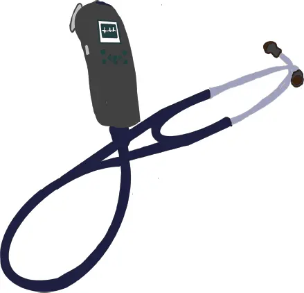
Figure 2.6 A digital stethoscope.
Читать дальшеИнтервал:
Закладка:
Похожие книги на «Predicting Heart Failure»
Представляем Вашему вниманию похожие книги на «Predicting Heart Failure» списком для выбора. Мы отобрали схожую по названию и смыслу литературу в надежде предоставить читателям больше вариантов отыскать новые, интересные, ещё непрочитанные произведения.


Обсуждение, отзывы о книге «Predicting Heart Failure» и просто собственные мнения читателей. Оставьте ваши комментарии, напишите, что Вы думаете о произведении, его смысле или главных героях. Укажите что конкретно понравилось, а что нет, и почему Вы так считаете.