Ridley's The Vulva
Здесь есть возможность читать онлайн «Ridley's The Vulva» — ознакомительный отрывок электронной книги совершенно бесплатно, а после прочтения отрывка купить полную версию. В некоторых случаях можно слушать аудио, скачать через торрент в формате fb2 и присутствует краткое содержание. Жанр: unrecognised, на английском языке. Описание произведения, (предисловие) а так же отзывы посетителей доступны на портале библиотеки ЛибКат.
- Название:Ridley's The Vulva
- Автор:
- Жанр:
- Год:неизвестен
- ISBN:нет данных
- Рейтинг книги:3 / 5. Голосов: 1
-
Избранное:Добавить в избранное
- Отзывы:
-
Ваша оценка:
Ridley's The Vulva: краткое содержание, описание и аннотация
Предлагаем к чтению аннотацию, описание, краткое содержание или предисловие (зависит от того, что написал сам автор книги «Ridley's The Vulva»). Если вы не нашли необходимую информацию о книге — напишите в комментариях, мы постараемся отыскать её.
Ridley’s The Vulva
Ridley’s The Vulva
Ridley's The Vulva — читать онлайн ознакомительный отрывок
Ниже представлен текст книги, разбитый по страницам. Система сохранения места последней прочитанной страницы, позволяет с удобством читать онлайн бесплатно книгу «Ridley's The Vulva», без необходимости каждый раз заново искать на чём Вы остановились. Поставьте закладку, и сможете в любой момент перейти на страницу, на которой закончили чтение.
Интервал:
Закладка:
Minor vestibular glands
The minor vestibular glands are small shallow glands usually less than 3 mm into the dermis and open directly to the surface. In postmortem studies, they vary in number from 1 to more than 100 [19].
Vestibular papillomatosis
Vestibular papillae are 1–5 mm thin projections that occur in the vestibule and inner labia minora, and are a normal variant ( Figure 2.7). It is suggested that they are the female equivalent of the tiny symmetrical projections found around the coronal sulcus known as penile pearly papules of the penis [20]. Originally, it was thought that the lesions were induced by the human papillomavirus (HPV), but there is now good evidence to the contrary [21, 22]. The normal glycogenation of the cells at the vestibule is often mistaken for koilocytosis, which is another reason for good communication with the pathologist.
Vestibular papillae can be distinguished from viral warts as they are soft and the same colour and texture as the surrounding mucosa. They are symmetrical in distribution and each papilla arises from a solitary base ( Figure 2.8), whereas viral warts often coalesce into a single base. Dermoscopy has also been used to distinguish the two entities [23] where the single base of each papilla is again confirmed. The application of 5% acetic acid does not produce acetowhitening in vestibular papillomatosis. They are usually asymptomatic, and no treatment is needed.
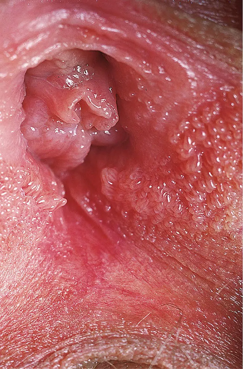
Figure 2.7 Vestibular papillae. Multiple filiform projections of the vestibular epithelium.
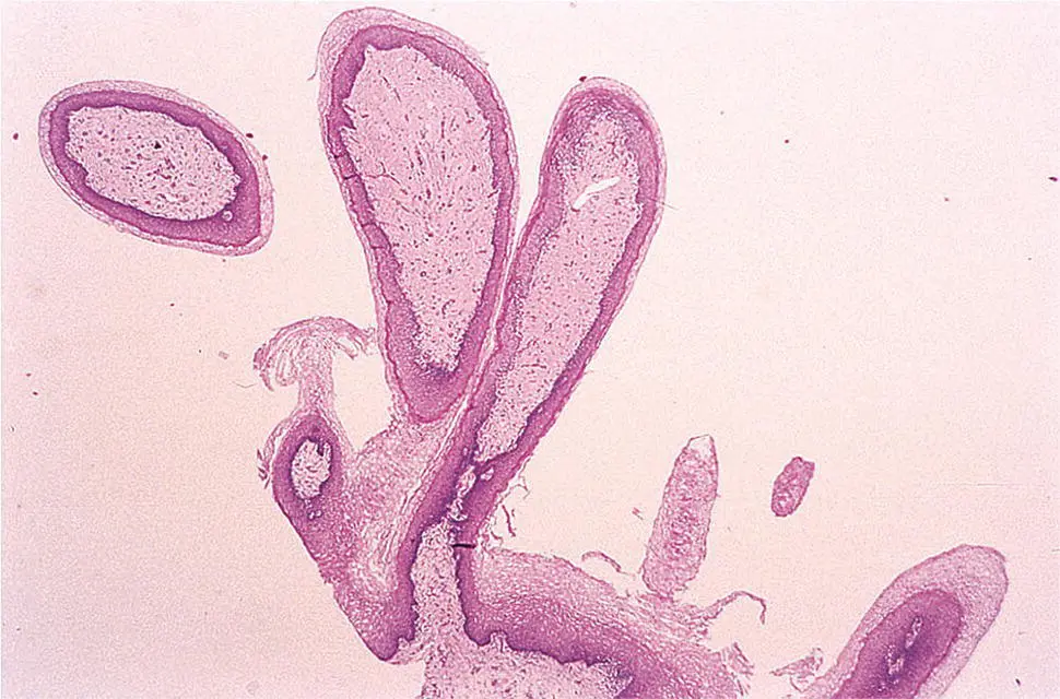
Figure 2.8 Histology of vestibular papillomatosis. Low power showing papillary projections with normal epithelium each arising from individual base.
Hymen
The hymen is a thin membrane of connective tissue surrounding the inner edge of the vestibule and the opening of the vagina. The appearance is again varied and can be a ring or semi‐circular fold. Once ruptured, an irregular ragged edge is left around the vaginal opening, and these small elevations are termed hymenal remnants ( Fig 2.9). Rupture can occur with exercise, tampon use, or sexual intercourse. It is clear that hymenal examination does not predict virginity status accurately or reliably and should never be performed for this purpose [24]. Developmental anomalies of the hymen are discussed in Chapter 1.
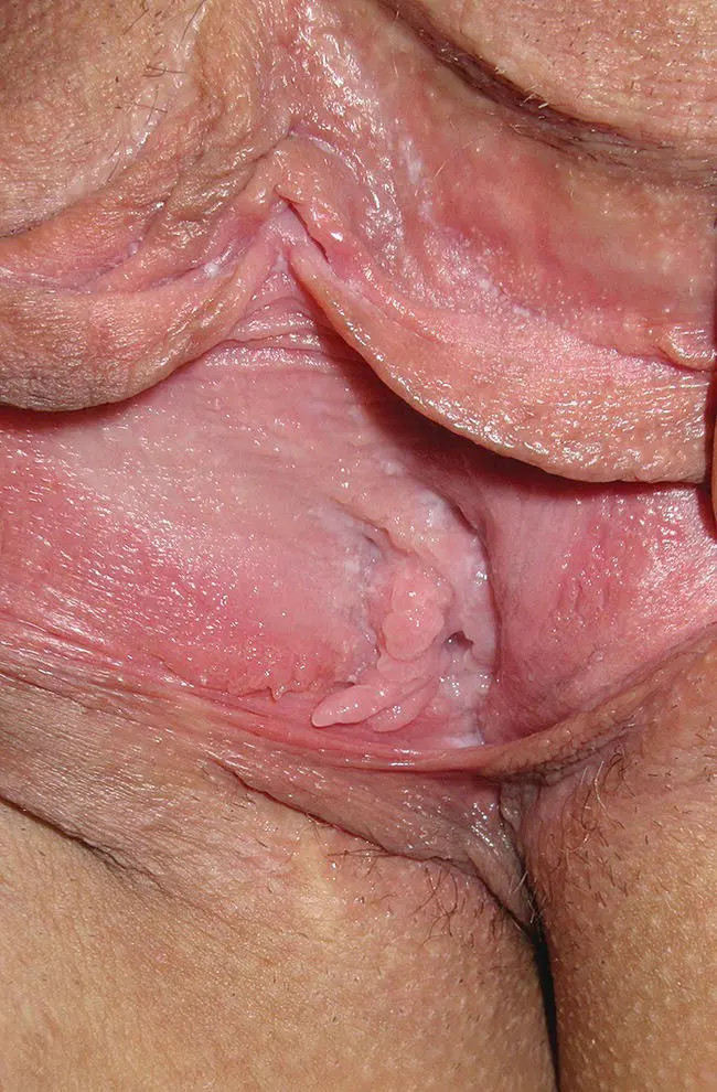
Figure 2.9 Hymenal remnants and tags.
The external urethral meatus and urethra
The female urethra is about 4 cm long and runs from the bladder downwards and forwards, embedded in the anterior wall of the vagina behind the symphysis pubis. The length can vary, and in a large study of 927 Caucasian women, the length was increased in the obese but reduced in those with a history of vaginal delivery [25]. After passing through the pelvic floor and perineal membrane, it ends at the external urethral orifice. The urethra is fixed at its origin by the pubovesical ligaments, throughout its length by the anterior wall of the vagina, and as it enters the perineum by the urogenital diaphragm. The external urethral orifice lies in the midline of the vestibule between the vagina and the clitoris. The orifice is easily seen, and on occasions there may be bright red projections of prolapsed urethral mucosa herniating out. These urethral caruncles are most commonly seen in post‐menopausal women and are thought to be due to reduced oestrogen ( Figure 2.10).
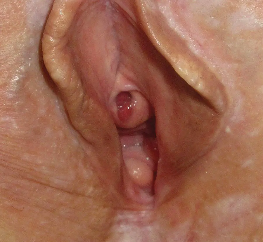
Figure 2.10 Urethral caruncle in a patient with lichen sclerosus.
Skene’s glands (paraurethral glands) are paired glands with their ducts opening on each side of the urethral orifice. Evidence suggests that they are analogous to the male prostate gland [26].
Associated structures
The vagina
The vagina is a fibromuscular tube about 7–10 cm long. It opens on the vulval vestibule and extends upwards and backwards, to be attached just above the lower margin of the uterine cervix. As the long axis of the vagina forms a right angle with the long axis of the normal anteverted uterus, the cervix projects downwards and backwards into the upper vagina. The vaginal fold around the periphery of the cervix is divided into anterior, posterior, and lateral fornices. The posterior wall of the vagina is about 2 cm longer than the anterior wall, but they are in contact with each other in the un‐distended vagina. This gives the vagina a crescentic or H‐shaped appearance in cross‐section. The outer wall of the vagina contains its vascular, lymphatic, and nerve supply.
The vagina is related anteriorly to the base of the bladder and to the urethra, which is embedded in its anterior wall. Posteriorly, the upper part of the vaginal wall is covered with peritoneum, and below the rectouterine pouch it is directly related to the ampulla of the rectum. In the perineum, it is separated from the anal canal by the perineal body ( Figure 2.11). The upper vagina gives attachment to the uterosacral ligaments posteriorly, the cardinal or transverse ligaments laterally, and the base of the bladder anteriorly, which itself is supported by the pubovesical ligaments. As the vagina passes through the pelvic floor, the most medial fibres of the pubococcygeus blend with its walls to form a supporting muscular sling. Below the pelvic floor the vagina is supported by the urogenital diaphragm, the perineal body, and the perineal musculature. Thus, the vagina has three compartments:
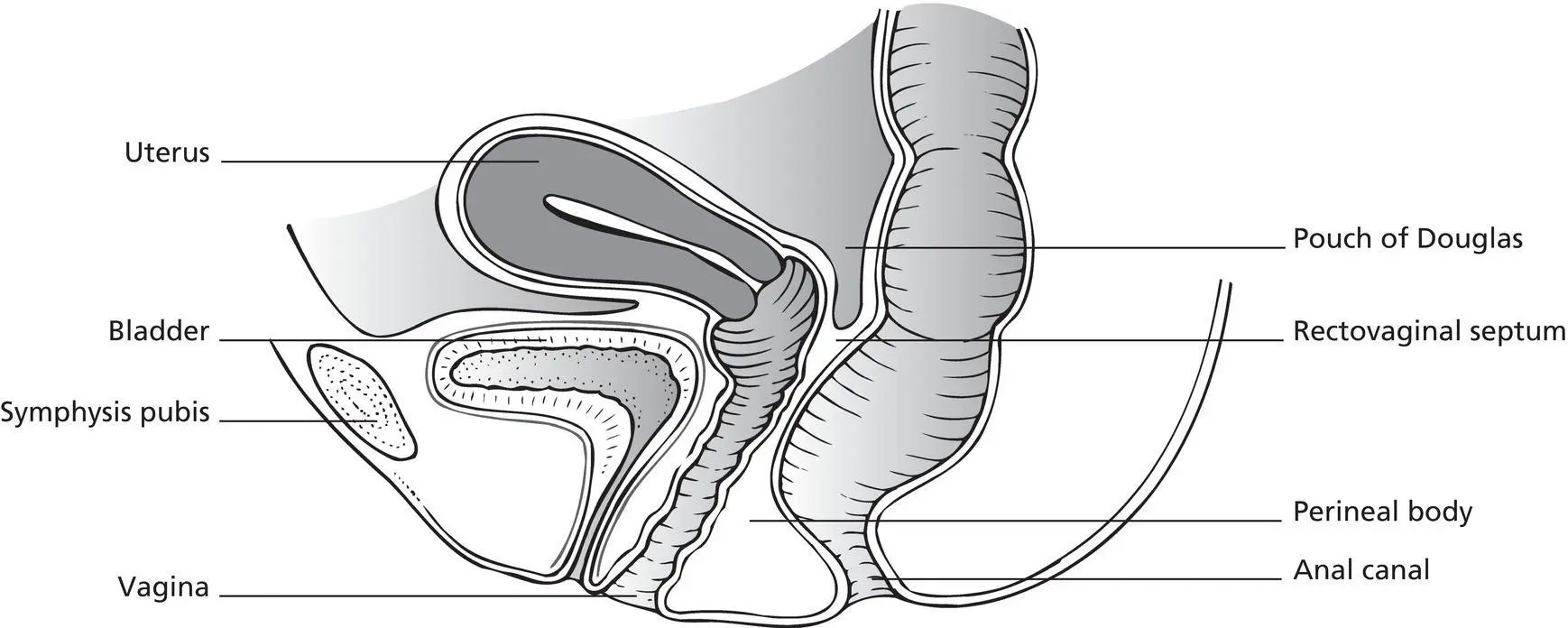
Figure 2.11 Midline section through pelvis and perineum.
upper, above the pelvic floor and related to the rectum
middle, which traverses the pelvic floor and urogenital diaphragm
lower in the perineum.
The perineum
The perineum is the outer diamond‐shaped area inferior to the sheet of muscle forming the pelvic floor, and is bounded by the symphysis pubis anteriorly, the ischial tuberosities laterally, and the coccyx posteriorly. It is an embryological junctional zone derived from the body wall ectoderm, hindgut endoderm, and the intervening mesoderm that surrounded the original cloacal membrane. It is further divided into an anterior urogenital triangle and posterior anal triangle. The vulva lies mainly within the anterior urogenital triangle but then extends anteriorly to the pubic symphysis. The anal canal and ischiorectal fossa occupy the posterior anal triangle. The perineal body is a fibromuscular mass lying between the upper half of the anterior anal wall and the entire posterior portion of the vagina. It is the central point where muscles attach to the ischial tuberosities.
The urogenital triangle
The urogenital triangle is contained within the subpubic arch and is divided into superficial and deep perineal pouches by the urogenital diaphragm.
Читать дальшеИнтервал:
Закладка:
Похожие книги на «Ridley's The Vulva»
Представляем Вашему вниманию похожие книги на «Ridley's The Vulva» списком для выбора. Мы отобрали схожую по названию и смыслу литературу в надежде предоставить читателям больше вариантов отыскать новые, интересные, ещё непрочитанные произведения.


Обсуждение, отзывы о книге «Ridley's The Vulva» и просто собственные мнения читателей. Оставьте ваши комментарии, напишите, что Вы думаете о произведении, его смысле или главных героях. Укажите что конкретно понравилось, а что нет, и почему Вы так считаете.