Ridley's The Vulva
Здесь есть возможность читать онлайн «Ridley's The Vulva» — ознакомительный отрывок электронной книги совершенно бесплатно, а после прочтения отрывка купить полную версию. В некоторых случаях можно слушать аудио, скачать через торрент в формате fb2 и присутствует краткое содержание. Жанр: unrecognised, на английском языке. Описание произведения, (предисловие) а так же отзывы посетителей доступны на портале библиотеки ЛибКат.
- Название:Ridley's The Vulva
- Автор:
- Жанр:
- Год:неизвестен
- ISBN:нет данных
- Рейтинг книги:3 / 5. Голосов: 1
-
Избранное:Добавить в избранное
- Отзывы:
-
Ваша оценка:
Ridley's The Vulva: краткое содержание, описание и аннотация
Предлагаем к чтению аннотацию, описание, краткое содержание или предисловие (зависит от того, что написал сам автор книги «Ridley's The Vulva»). Если вы не нашли необходимую информацию о книге — напишите в комментариях, мы постараемся отыскать её.
Ridley’s The Vulva
Ridley’s The Vulva
Ridley's The Vulva — читать онлайн ознакомительный отрывок
Ниже представлен текст книги, разбитый по страницам. Система сохранения места последней прочитанной страницы, позволяет с удобством читать онлайн бесплатно книгу «Ridley's The Vulva», без необходимости каждый раз заново искать на чём Вы остановились. Поставьте закладку, и сможете в любой момент перейти на страницу, на которой закончили чтение.
Интервал:
Закладка:
15 29 Schoemaker, M.J., Swerdlow, A.J., Higgins, C.D. et al. Cancer incidence in women with Turner syndrome in Great Britain: A national cohort study. Lancet Oncol. 2008; 9: 239–246.
16 30 Matsumoto, F., Matsuyama, S., Matsui, F. et al. Variation of Gonadal dysgenesis and Tumor risk in patients with 45,X/46, XY mosaicism. Urology. 2020; 137: 157–160.
17 33 Falhammar, H., Frisén, L., Hirschberg, A.L. et al. Increased cardiovascular and metabolic morbidity in patients with 21‐hydroxylase deficiency: A Swedish population‐Based National Cohort Study. J Clin Endocrinol Metab. 2015; 100(9): 3520–3528.
18 36 Conlon, J.L. Diethylstilbestrol: Potential health risks for women exposed in utero and their offspring. JAAPA. 2017; 30(2): 49–52.
19 37 Brunskill, P.J. The effects of fetal exposure to danazol. Br J Obstet Gynaecol. 1992; 99(3): 212–215.
20 39 Xue, M., Wang, X., Li, C. et al. Novel pathogenic mutations in disorders of sex development associated genes cause 46,XY complete gonadal dysgenesis. Gene. 2019; 718: 144072.
21 42 Oppelt, P.G., Lermann, J., Strick, R. et al. Malformations in a cohort of 284 women with Mayer‐Rokitansky‐Küster‐Hauser (MRKH) syndrome. Reprod Biol Endocrinol. 2012; 10: 57.
22 43 Rall, K., Eisenbeis, S., Henninger, V. et al. Typical and atypical associated findings in a group of 346 patients with Mayer‐Rokitansky‐Küster‐Hauser syndrome. J Pediatr Adolesc Gynecol. 2015; 28(5): 362–368.
23 44 Fontana, L., Gentilin, B., Fedele, L. et al. Genetics of Mayer‐Rokitansky‐Küster‐Hauser (MRKH) syndrome. Clin Genet. 2017; 91(2): 233‐246.
24 45 Backhouse, B., Hanna, C., Roberska, G. et al. Identification of candidate genes for Mayer‐Rokitansky‐Küster‐Hauser syndrome using genomic approaches. Sex Dev. 2019; 13(1): 26–34.
25 46 ACOG Committee Opinion No 728. Müllerian agenesis: Diagnosis, management, and treatment. Obstet Gynecol. 2018; 131(1): e35–42.
26 48 Edmonds, D.K. Congenital malformations of the genital tract and their management. Best Pract Res Clin Obstet Gynaecol. 2003; 17: 19–40.
27 55 Slavotinek, A.M. and Biesecker, L.G. Phenotypic overlap of McKusick‐Kaufman syndrome with Bardet‐Biedl syndrome: A literature review. Am J Med Genet. 2000; 95: 208–215.
28 72 Woo, L.L., Thomas, J.C., and Brock, J.W. Cloacal exstrophy: A comprehensive review of an uncommon problem. J Pediatr Urol. 2010; 6(2): 102–111.
29 73 Kubota, M., Osuga, Y., Kato, K. et al. Treatment guidelines for persistent cloaca, cloacal exstrophy and Mayer‐Rokitansky‐Küster‐Hauser syndrome for the appropriate transitional care of the patient. Surg Today. 2019; 49(12): 985–1002.
2 The Normal Vulva
Fiona M. Lewis
CHAPTER MENU
Normal vulval anatomy Mons pubis Labia majora Labia minora Sebaceous glands (Fordyce spots) The clitoris The vestibule Hart’s line Bartholin’s glands Minor vestibular glands Vestibular papillae Hymen External urethral meatus and urethra
Associated structuresVagina Perineum The urogenital diaphragm Anal triangle Pelvic floor Ischiococcygeus muscle Iliococcygeus muscle Pubococcygeus muscle The Inguinofemoral region
Blood supply of the vulva Internal pudendal artery Femoral artery Venous drainage Microscopic anatomy
Lymphatic drainage of the vulva The external iliac lymph nodes Medial group Anterior group Lateral group Microscopic anatomy
Nerve supply of the vulva Somatic innervation Autonomic (visceral) innervation Pudendal nerve Microscopic anatomy
Normal microscopic anatomy and histological featuresEpithelia of the vulva Mons pubis Labia majora Labia minora Clitoris Vestibule Vagina Other epithelial cell types Melanocytes Langerhans cells Merkel cells The Basement membrane zone The dermis
References
Vulval lesions are not well described in the early medical literature, but first appear in the writings of Severinus Pineus in the sixteenth century and van den Spieghel in the seventeenth century. The terms used to describe the anatomical structures are often related to their function. The word vulva is derived from the Latin word for ‘wrapping’. The vagina (sheath) and mons veneris (hill of Venus) are obvious descriptions. The clitoris is usually thought to come from the Greek kleitoris , meaning ‘key’ or ‘gatekeeper’. Hymen is derived from the Greek hymen , meaning membrane. The labia are probably so called because they surround the vaginal opening like lips (Latin labium – lip).
There is a very wide variation in the appearance of the normal vulva, and this chapter looks at the normal anatomy, anatomical variants, and normal histological features at different sites. It is vital to understand the normal before the abnormal is diagnosed, thereby avoiding unnecessary treatment and worry for the patient.
Normal vulval anatomy
The vulva consists of seven main parts: the mons pubis, the labia majora and minora, the vestibule of the vagina, the hymen, the clitoris, and the external urethral orifice ( Figure 2.1). All of these structures may vary in size and symmetry, but this is a subject that has been rather neglected. In a study of 59 textbooks, very little was included about this topic [1]. Patients often worry about normal variants and the quest for the ‘perfect’ vulva. Indeed, in a study of 33 women seeking labial reduction, all had labia minora within normal limits [2]. This incorrect perception was confirmed in another study of younger adolescent girls presenting for consideration of genital surgery. Again, none had documented abnormality [3]. Patients will also perceive an abnormality if the labia minora are visible, even though they are of normal size [4]. There has been more interest recently in the normal variation in appearance of the vulva in both popular culture and the scientific literature. The artist Jamie McCartney took plaster casts of 400 vulvas to create a sculpture which illustrates the wide range of appearance ( www.greatwallofvagina.co.uk), and this has been used as both an artistic and educational resource.
Several studies have addressed the normal vulval appearance in adults [ 5,6,7], prepubertal girls [8,9], and adolescents [ 10]. These studies include women of different ethnicities but the measurements are similar, and average measurements of the vulval structures in adults are shown in Table 2.1. It has been shown that genital dimensions have no effect on sexual function [11].
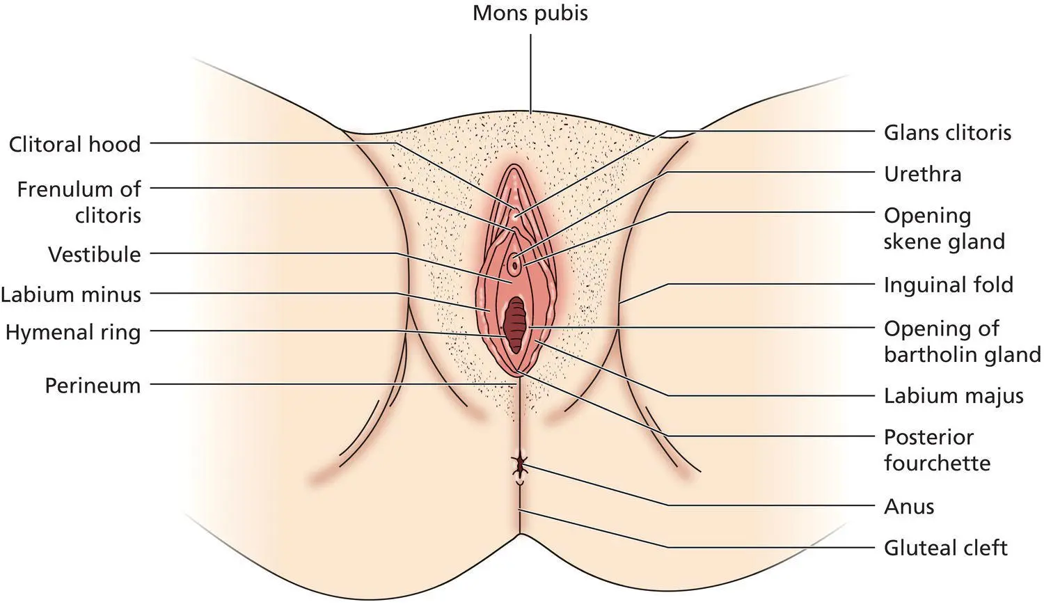
Figure 2.1 Anatomy of the normal vulva.
Mons pubis
The mons pubis (mons) in the adult female is a prominent pad of hair‐bearing skin and subcutaneous fat overlying the pubic symphysis. It forms an inverted triangle with the base being the anterior horizontal line of pubic hair growth. The average length of the base is 16 cm, and the height about 13 cm [12].
The character of pubic hair varies with ethnic background, as it is generally thicker in type 5 and 6 skin types. The normal hair density is 6–31 hairs/cm 2, but this, together with the rate of hair growth, reduces with age. About a third of women over the age of 60 have progressive loss of pubic hair [13]. There is no change in the thickness of the hair with age. In contrast to hair growth in the axillae and on the scalp, pubic hair growth is not altered during pregnancy [14].
Labia majora
The labia majora are two cutaneous folds that form the lateral boundaries of the pudendal cleft. They originate from the mons pubis anteriorly and merge with the perineal body posteriorly (the posterior labial commissure). The subcutaneous fat is mainly deposited in the medial aspects, and so they tend to flatten out as they reach the perineal body. The lateral surfaces of the labia majora are adjacent to the medial surfaces of the thighs and are separated from them by a deep groove, the genitocrural or inguinal fold. The medial surfaces may be in contact with each other, but may be separated by the labia minora if they are large. The size of the labia majora varies considerably. The length of the labia majora and introitus has been shown to be positively correlated with body mass index but inversely correlated with age [ 5].
Читать дальшеИнтервал:
Закладка:
Похожие книги на «Ridley's The Vulva»
Представляем Вашему вниманию похожие книги на «Ridley's The Vulva» списком для выбора. Мы отобрали схожую по названию и смыслу литературу в надежде предоставить читателям больше вариантов отыскать новые, интересные, ещё непрочитанные произведения.


Обсуждение, отзывы о книге «Ridley's The Vulva» и просто собственные мнения читателей. Оставьте ваши комментарии, напишите, что Вы думаете о произведении, его смысле или главных героях. Укажите что конкретно понравилось, а что нет, и почему Вы так считаете.