Successful Training in Gastrointestinal Endoscopy
Здесь есть возможность читать онлайн «Successful Training in Gastrointestinal Endoscopy» — ознакомительный отрывок электронной книги совершенно бесплатно, а после прочтения отрывка купить полную версию. В некоторых случаях можно слушать аудио, скачать через торрент в формате fb2 и присутствует краткое содержание. Жанр: unrecognised, на английском языке. Описание произведения, (предисловие) а так же отзывы посетителей доступны на портале библиотеки ЛибКат.
- Название:Successful Training in Gastrointestinal Endoscopy
- Автор:
- Жанр:
- Год:неизвестен
- ISBN:нет данных
- Рейтинг книги:3 / 5. Голосов: 1
-
Избранное:Добавить в избранное
- Отзывы:
-
Ваша оценка:
Successful Training in Gastrointestinal Endoscopy: краткое содержание, описание и аннотация
Предлагаем к чтению аннотацию, описание, краткое содержание или предисловие (зависит от того, что написал сам автор книги «Successful Training in Gastrointestinal Endoscopy»). Если вы не нашли необходимую информацию о книге — напишите в комментариях, мы постараемся отыскать её.
Teaches trainee gastroenterologists the endoscopic skills needed to meet the medical training requirements to practice gastroenterology and helps clinical specialists refresh their skills to pass their recertification Successful Training in Gastrointestinal Endoscopy, Second Edition
Successful Training in Gastrointestinal Endoscopy, Second Edition
Successful Training in Gastrointestinal Endoscopy — читать онлайн ознакомительный отрывок
Ниже представлен текст книги, разбитый по страницам. Система сохранения места последней прочитанной страницы, позволяет с удобством читать онлайн бесплатно книгу «Successful Training in Gastrointestinal Endoscopy», без необходимости каждый раз заново искать на чём Вы остановились. Поставьте закладку, и сможете в любой момент перейти на страницу, на которой закончили чтение.
Интервал:
Закладка:
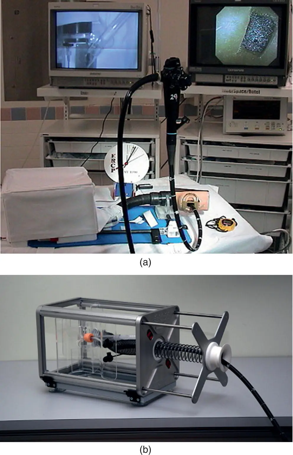
Figure 1.9 (a) Novel ERCP endotrainer introduced by Joseph Leung, MD that allows for simulated sphincterotomy and fluorosocpy equivalent. (b) Rome ERCP trainer designed by Dr. Guido Costamagna and colleagues allows for cannulation and endotherapy of bile and pancreatic ducts and ability to interchange papillae of different orientation and cannulation difficulty
(Photo courtesy of Cook Medical, Winston‐Salem, NC).
 In contrast, Dr. Christopher Thompson teamed up with MIT engineers to develop a static box model to allow for repetitive practice on five specific tasks designed to mimic component skill sets integral to performing colonoscopy [23]. In a departure from the paradigm to design sophisticated simulators to allow a trainee to practice complete procedures, the Thompson Endoscopic Skills Trainer (TEST) (Endosim, LLC. Boston, MA) is an example of using simulation to deconstruct complex procedures into core maneuvers (Video 1.2).
Ex vivo artificial tissue models: the “Phantom” Tübingen models
A further advance in endoscopic simulation was developed by Grund et al. at the University of Tübingen in Germany [24]. In this “Interphant” or “Phantom” model, artificial electrically conductive tissue called Artitex is used to fashion abnormalities such as polyps and strictures and incorporate this into static models. These “pathologies” are in place of the painted‐on abnormalities used in some of the pure static models mentioned above. Grund’s “Artitex” abnormalities are sewn directly into a three‐dimensional latex anatomical model ( Figure 1.10a,b). While these models generally lack a realistic representation of bowel wall compliance and motility, the integrated pathology appears realistic and allows practice in electrosurgical techniques.
In order to simulate the resistance to endoscope passage in an actual procedure, this colon model uses a semiflexible series of coils. In addition, to allow for a still wider possibility of simulated techniques, Grund’s model can incorporate real animal tissue into the existing framework. For example, using a chicken heart, they can fashion an ampulla of Vater replete with separate pancreatic and biliary orifices and insert this into their upper endoscopy simulator ( Figure 1.10b). The advantage of using this type of system is that several “polyp‐laden” colons and “chicken‐heart papillae” can be prepared in advance and quickly inserted into the chassis of the model during a training session, after the initially prepared material has been depleted.
The Tübingen simulators made possible the teaching of polypectomy and provided an excellent means of teaching therapeutic procedures such as argon plasma coagulation and simple therapeutic ERCP. In particular, the orientation of the man‐made papilla more closely resembled that of humans than the porcine papilla found in the Erlangen models described below. Pancreatic cannulation and endotherapy was possible, in contrast to the porcine tissue models in which the pancreatic orifice was not readily accessible. However, procedures that required submucosal injection were still not feasible.
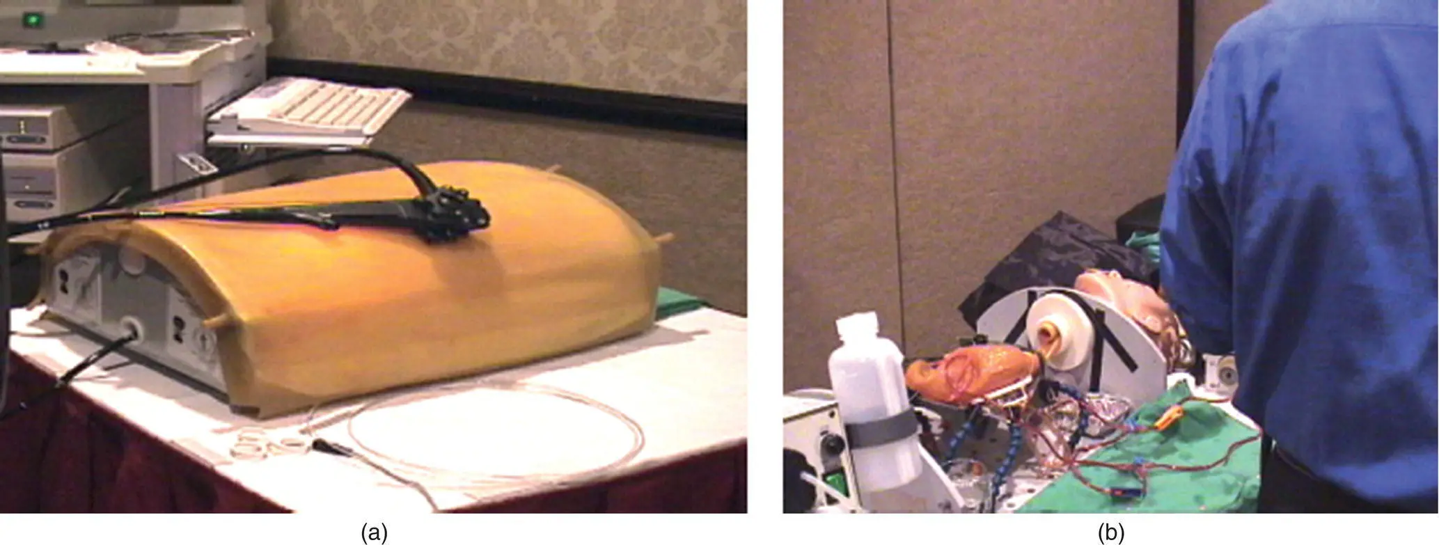
Figure 1.10 (a) Artificial tissue colonoscopy “Phantom” simulator, U. of Tübingen. (b) Combined artificial tissue “Phantom” upper GI simulator with integrated chicken heart tissue papilla for ERCP simulation.
While this model represented a technological advance over prior static models and added many new capabilities, there remained several limitations that hindered its more widespread use in training. The main drawbacks were that the pathology remained hand‐prepared and that the models were not mass‐produced. Therefore, the “Phantoms” have not been readily available and have required the presence of the Tübingen team if the device was to be used at a training course. The trade‐off for increased realism and the ability to start practicing therapeutic manipulations were significant increases in the logistical and cost obstacles to widespread use. Furthermore, models combining the real tissue abnormalities of the Tübingen model with the more accessible ex vivo animal tissue simulators described below now exist.
Ex vivo animal tissue simulators: Erlanger and EASIE models
In 1996, Hochberger and Neuman created an innovative simulator using pig organs obtained from a slaughterhouse and fastened to a plastic platform [25, 26]. In order to create a model that would allow training and practice in therapeutic techniques, Hochberger then created a highly realistic simulation of pulsatile arterial bleeding ( Figure 1.11a,b). This was accomplished by inserting tubes through the stomach, and sewing real arteries attached to a roller pump capable of pulsatile perfusion with a cherry‐colored saline solution. Following this, Hochberger developed representations of other pathologies for this model, including polyps, varices, and strictures [27, 28].
Currently, there are two basic model types based on these principles. The original Erlanger model features pig organs inserted into a dummy mannequin. This model has been used in the simulation of various laparoscopic surgical procedures [29]. Hochberger then created the compact‐EASIE model, a smaller portable, lightweight version using a tabletop platform. There are now several commercially available versions of this type of simulator in which only the organs needed for endoscopy simulation are secured to the platform plastic tray ( Figure 1.11a,b). For example, only the esophagus to the duodenal bulb may be needed for a specific training session, but the model has the flexibility to allow the liver and hepatobiliary tree for an ERCP simulation involving fluoroscopy. Multiple therapeutic procedures may be demonstrated, taught, and evaluated on both of these animal tissue‐based simulators. With the advent of portable, compact tabletop ex vivo models that can easily be shipped to a location along with pre‐prepared frozen organ packages, some of the obstacles to simulator availability have diminished.
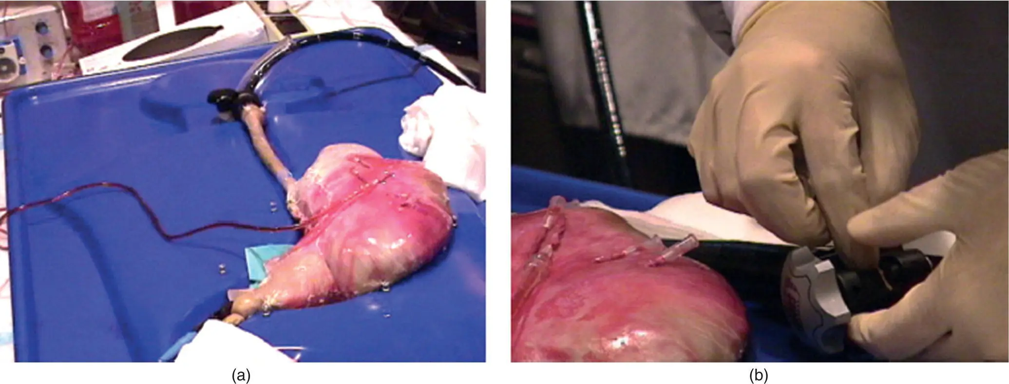
Figure 1.11 (a) Compact‐EASIE porcine model hemostasis simulator. (b) Close‐up view of porcine stomach with arteries sutured in attached to catheters for hook‐up to tubing connecting vessels to pump. The trainee puts together a band ligation device for varices treatment simulation.
While the most common application of the Hochberger model has been for hemostasis training, this group has conducted a number of training courses using the EASIE model in other areas, including EMR, stricture management, vital staining, polypectomy, and ERCP [28, 30–32]. A wide range of techniques can be demonstrated, including basic biliary cannulation, plastic stent insertion, choledochoscopy, laser lithotripsy, and placement of bilateral hilar metal stents (Videos 1.3 and 1.4). EASIE training has been shown to significantly improve technical skill in endoscopic hemostasis in gastroenterology trainees as compared to clinical endoscopy training alone [33]. As detailed in multiple chapters in this book, further adaptations of the ex vivo simulator have extended the use of this modality for training in colonoscopy, balloon‐assisted small bowel endoscopy, improved simulation of bile duct and pancreatic duct manipulation, EUS with FNA, and even NOTES ®[34–37].
Интервал:
Закладка:
Похожие книги на «Successful Training in Gastrointestinal Endoscopy»
Представляем Вашему вниманию похожие книги на «Successful Training in Gastrointestinal Endoscopy» списком для выбора. Мы отобрали схожую по названию и смыслу литературу в надежде предоставить читателям больше вариантов отыскать новые, интересные, ещё непрочитанные произведения.


Обсуждение, отзывы о книге «Successful Training in Gastrointestinal Endoscopy» и просто собственные мнения читателей. Оставьте ваши комментарии, напишите, что Вы думаете о произведении, его смысле или главных героях. Укажите что конкретно понравилось, а что нет, и почему Вы так считаете.