Paul M. Speight - Shear's Cysts of the Oral and Maxillofacial Regions
Здесь есть возможность читать онлайн «Paul M. Speight - Shear's Cysts of the Oral and Maxillofacial Regions» — ознакомительный отрывок электронной книги совершенно бесплатно, а после прочтения отрывка купить полную версию. В некоторых случаях можно слушать аудио, скачать через торрент в формате fb2 и присутствует краткое содержание. Жанр: unrecognised, на английском языке. Описание произведения, (предисловие) а так же отзывы посетителей доступны на портале библиотеки ЛибКат.
- Название:Shear's Cysts of the Oral and Maxillofacial Regions
- Автор:
- Жанр:
- Год:неизвестен
- ISBN:нет данных
- Рейтинг книги:5 / 5. Голосов: 1
-
Избранное:Добавить в избранное
- Отзывы:
-
Ваша оценка:
- 100
- 1
- 2
- 3
- 4
- 5
Shear's Cysts of the Oral and Maxillofacial Regions: краткое содержание, описание и аннотация
Предлагаем к чтению аннотацию, описание, краткое содержание или предисловие (зависит от того, что написал сам автор книги «Shear's Cysts of the Oral and Maxillofacial Regions»). Если вы не нашли необходимую информацию о книге — напишите в комментариях, мы постараемся отыскать её.
Shear’s Cysts of the Oral and Maxillofacial Regions
Shear’s Cysts of the Oral and Maxillofacial Regions Fifth Edition
Shear's Cysts of the Oral and Maxillofacial Regions — читать онлайн ознакомительный отрывок
Ниже представлен текст книги, разбитый по страницам. Система сохранения места последней прочитанной страницы, позволяет с удобством читать онлайн бесплатно книгу «Shear's Cysts of the Oral and Maxillofacial Regions», без необходимости каждый раз заново искать на чём Вы остановились. Поставьте закладку, и сможете в любой момент перейти на страницу, на которой закончили чтение.
Интервал:
Закладка:
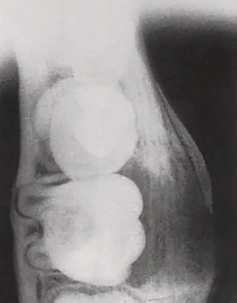
Buccal expansion is often apparent and with involvement of the periosteum new bone may be laid down, either as a single linear band or in a laminated pattern ( Figure 4.4). This pattern of laminated new bone is seen as a result of periostitis and is also seen in chronic osteomyelitis. Wolf and Hietanen (1990 ) noted this feature and suggested that clinically and radiographically, the mandibular buccal bifurcation cyst may be similar to osteomyelitis, and that in some cases the cyst may be a cause of chronic osteomyelitis, (sometimes referred to as Garré's osteomyelitis or periostitis ossificans).
CBCT is especially useful in the diagnosis of mandibular buccal bifurcation cysts because it allows the buccal expansion and extent of the lesion to be fully visualised (Ramos et al. 2012 ; Bautista et al. 2019 ; Dave et al. 2020 ). Magnetic resonance imaging (MRI) can also be useful, but although it has the advantage of lower radiation dosage, it has very limited use in routine examination of jaw lesions in dentistry. CBCT shows a hypodense, well‐demarcated, and often spherical cystic lesion with a corticated outline, which is always located on the buccal aspect of the tooth. CBCT and occlusal radiographs show that the affected tooth is tilted buccally and transverse views show the apices abutting and occasionally eroding the lingual cortical plate ( Figure 4.4; Bautista et al. 2019 ).
Inflammatory collateral cysts at other sites show similar radiological features. The four cases associated with premolars, reported by Morimoto et al. (2004 ), showed well‐demarcated radiolucencies overlying the buccal aspect of erupting and incompletely formed premolars. There was buccal expansion and in all cases the teeth were displaced lingually. Cysts in the globulomaxillary region present as a well‐demarcated radiolucency between the lateral incisor and the canine tooth. The cyst has an ‘inverted pear’ shape and the tooth roots are displaced and diverge. All eight cases reported by Vedtofte and Holmstrup (1989 ) showed these features and in all cases both the canine and incisor tooth were fully erupted.
Radiological Differential Diagnosis
The histological features of inflammatory collateral cysts are not specific, but taken together the clinical and radiological features should allow an accurate diagnosis ( Box 4.2). In virtually all cases the cysts arise on the buccal or disto‐buccal aspect of a partially erupted or recently erupted tooth with characteristic radiological features. The associated tooth is vital. Nevertheless, as discussed previously, there is great variation in the relative frequency of these cysts in different studies and it is apparent that some workers do not recognise it as an entity. For the most part this is probably due to lack of clear diagnostic criteria and uncertainty about the pathogenesis. The key features of these lesions and diagnostic criteria are suggested in Box 4.2.

Figure 4.4 Occlusal view of a mandibular buccal bifurcation cyst on a mandibular first molar tooth. There is resorption of bone on the buccal aspect of the tooth and buccal expansion. Subperiosteal new bone has been deposited buccally, giving a laminated appearance. Note also the displacement and tilting of the tooth, so that its apices abut onto, and appear to erode, the lingual cortical plate.
Source: Courtesy of Dr Douglas W Stoneman (Previously published: Dental Radiography and Photography (1983 ) 56, 1–14. Courtesy of Eastman Kodak Co.)
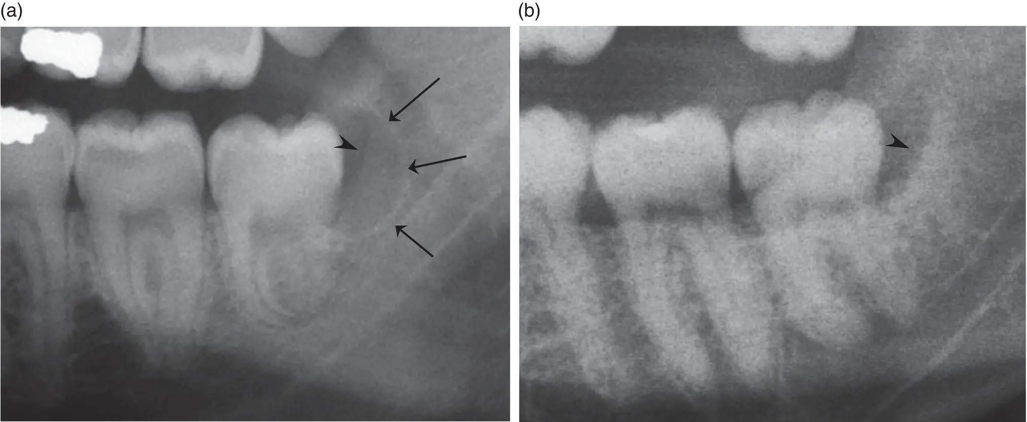
With regard to the paradental cyst, the presence of a pericoronal radiolucency associated with a partially erupted third molar is not specific, since a similar radiolucency may result from an inflamed or hyperplastic dental follicle or pericoronitis ( Figure 4.5). In such a case, the pathologist may be presented with fragments of inflamed tissue and a clinical diagnosis of ‘pericoronitis’, and therefore a diagnosis of paradental cyst may not be considered. In addition, some workers consider that a pericoronal radiolucency distal to a partially erupted third molar may arise as a result of lateral displacement of a dentigerous cyst during tooth eruption, or that a cyst that arises in a pericoronal relationship is a mere variant of dentigerous cyst. In this case, the final diagnosis may be of an ‘inflamed dentigerous cyst’.

Figure 4.5 Differential diagnosis of the paradental cyst. (a) The cyst is well demarcated and corticated (arrows) and is distinct from the distal follicular space (arrowhead). (b) Chronic pericoronitis on a vertically impacted and partially erupted third molar. The distal follicular space is dilated (arrowhead) and there is evidence of alveolar bone loss, but there is no cyst.
However, careful examination of radiographs will show that the paradental cyst is distinct from the pericoronal follicular tissues and the criteria for diagnosis must include the finding of a well‐demarcated cyst that is distinct from the distal aspect of the dental follicle ( Figures 4.2and 4.5a). If the follicular space is merely expanded, then the lesion may be a hyperplastic dental follicle, or bone loss associated with chronic pericoronitis. Figure 4.5b shows an expanded pericoronal space associated with a partially erupted but vertical third molar. There is also evidence of pocketing with alveolar bone loss. This appearance is consistent with chronic long‐standing pericoronitis, but without cyst formation. The relationship to the dentigerous cyst is discussed further below.
Both the paradental cyst and mandibular buccal bifurcation cyst overly the tooth roots and may resemble a periapical radicular cyst, or a radicular cyst lying lateral to the root associated with a lateral root canal. However, in the case of an inflammatory collateral cyst, the periodontal space and the lamina dura remain intact ( Figures 4.2 and 4.4), a feature that excludes an inflammatory cyst of endodontic origin.
A number of other cysts may on occasion arise on the lateral aspect of a tooth, including odontogenic keratocyst and glandular odontogenic cyst. However, these do not embrace the crown of partially erupted teeth, are not associated with pericoronitis, and have characteristic features that clearly distinguish them from inflammatory collateral cysts (see Chapters 7and 10). Developmental lateral periodontal cysts may also be considered, but the clinicopathological features are quite different (see Chapter 8). Although lateral periodontal cysts are most common in the mandible, they are found in the canine/premolar region and they lie lateral to the roots of fully erupted teeth in middle‐aged adults. They are not associated with inflammation.
A number of workers have drawn attention to the fact that the clinical and radiological features of mandibular buccal bifurcation cyst may be similar to the chronic form of osteomyelitis often referred to as Garré's osteomyelitis or periostitis ossificans (Stoneman and Worth 1983 ; Wolf and Hietanen 1990 ). This is typified by buccal expansion with laminated depositions of new subperiosteal bone – a feature that is also seen in cases of mandibular buccal bifurcation cyst ( Figure 4.4). Periostitis ossificans of the mandible is most often seen in association with first molars in young people and is usually secondary to periapical infection. The mandibular buccal bifurcation cyst is of inflammatory origin and it is probable that in some cases, persistent chronic inflammation may give rise to a low‐grade osteomyelitis with periostitis. However, a diagnosis of collateral cyst can be made on the basis of a vital tooth, a well‐defined buccal radiolucency, and evidence of continuity between the buccal periodontal pocket and the cyst lumen.
Читать дальшеИнтервал:
Закладка:
Похожие книги на «Shear's Cysts of the Oral and Maxillofacial Regions»
Представляем Вашему вниманию похожие книги на «Shear's Cysts of the Oral and Maxillofacial Regions» списком для выбора. Мы отобрали схожую по названию и смыслу литературу в надежде предоставить читателям больше вариантов отыскать новые, интересные, ещё непрочитанные произведения.
Обсуждение, отзывы о книге «Shear's Cysts of the Oral and Maxillofacial Regions» и просто собственные мнения читателей. Оставьте ваши комментарии, напишите, что Вы думаете о произведении, его смысле или главных героях. Укажите что конкретно понравилось, а что нет, и почему Вы так считаете.


