Paul M. Speight - Shear's Cysts of the Oral and Maxillofacial Regions
Здесь есть возможность читать онлайн «Paul M. Speight - Shear's Cysts of the Oral and Maxillofacial Regions» — ознакомительный отрывок электронной книги совершенно бесплатно, а после прочтения отрывка купить полную версию. В некоторых случаях можно слушать аудио, скачать через торрент в формате fb2 и присутствует краткое содержание. Жанр: unrecognised, на английском языке. Описание произведения, (предисловие) а так же отзывы посетителей доступны на портале библиотеки ЛибКат.
- Название:Shear's Cysts of the Oral and Maxillofacial Regions
- Автор:
- Жанр:
- Год:неизвестен
- ISBN:нет данных
- Рейтинг книги:5 / 5. Голосов: 1
-
Избранное:Добавить в избранное
- Отзывы:
-
Ваша оценка:
Shear's Cysts of the Oral and Maxillofacial Regions: краткое содержание, описание и аннотация
Предлагаем к чтению аннотацию, описание, краткое содержание или предисловие (зависит от того, что написал сам автор книги «Shear's Cysts of the Oral and Maxillofacial Regions»). Если вы не нашли необходимую информацию о книге — напишите в комментариях, мы постараемся отыскать её.
Shear’s Cysts of the Oral and Maxillofacial Regions
Shear’s Cysts of the Oral and Maxillofacial Regions Fifth Edition
Shear's Cysts of the Oral and Maxillofacial Regions — читать онлайн ознакомительный отрывок
Ниже представлен текст книги, разбитый по страницам. Система сохранения места последней прочитанной страницы, позволяет с удобством читать онлайн бесплатно книгу «Shear's Cysts of the Oral and Maxillofacial Regions», без необходимости каждый раз заново искать на чём Вы остановились. Поставьте закладку, и сможете в любой момент перейти на страницу, на которой закончили чтение.
Интервал:
Закладка:
The mandibular buccal bifurcation cyst is always situated on the buccal aspect of the root of the affected tooth and the tooth is usually tilted buccally so that the apices are adjacent to the lingual cortical plate, a feature that is best seen in occlusal radiographs ( Figure 4.4) or cone‐beam computed tomography (CBCT). The tooth is always vital, which allows a lateral radicular cyst to be excluded.
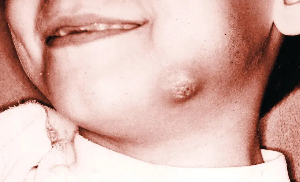
Figure 4.1 Young boy with mandibular buccal bifurcation cyst involving a recently erupted mandibular first permanent molar. Infection has extended through the bone, resulting in a facial abscess.
Source: Courtesy of Dr D.W. Stoneman.
With regard to inflammatory collateral cysts presenting at other sites, the clinical features are very similar to the mandibular buccal bifurcation cyst. Morimoto et al. (2004 ) found that all four of their cases on lower premolars presented with swelling and three were also painful. Inflammatory collateral cysts in the globulomaxillary region probably arise in association with the erupting canine and present as an inverted pear‐shaped radiolucency between the incisor and canine teeth, which are vital and show divergent roots. Of the eight cysts in the globulomaxillary region reported by Vedtofte and Holmstrup (1989 ), five were asymptomatic chance findings and three presented with signs of acute infection.
Radiological Features
The clinical features of inflammatory collateral cysts are not specific and an accurate diagnosis can only be made after consideration of the radiological features, which are characteristic ( Box 4.2).
The paradental cyst shows a number of quite specific but subtle features, which were first described by Craig (1976 ). On conventional radiographs the cyst presents as a well‐demarcated radiolucency, usually with a corticated margin. This is well illustrated in Figure 4.2, which shows radiographs from the paper by Vedtofte and Praetorius (1989 ). Of note is that the case illustrated in Figure 4.2a is of bilateral paradental cysts associated with second molars, in a 13‐year‐old girl where the third molars are absent. The cyst is usually displaced distally, but in all cases the radiolucency is superimposed over the buccal aspect of the tooth and overlies the roots and bifurcation area. Most paradental cysts are 10–15 mm in diameter (Philipsen et al. 2004 ) and rarely exceed 20 mm. Larger lesions may appear to be periapical, but it is important to note that the periodontal ligament space is not widened and the lamina dura is intact around the roots ( Figures 4.2and 4.3). Most cysts extend distally, but the distal element is well defined and is distinct from the distal follicular space ( Figure 4.2). This feature was first noted by Craig (1976 ) and was confirmed by Colgan et al. (2002 ), who identified it in 9 of their 15 cases and considered it to be an important and helpful diagnostic criterion, since it indicates that the dental follicle is not involved in the development of the cyst.
Box 4.2Inflammatory Collateral Cysts: Key Features and Diagnostic Criteria
Paradental cyst
Arises on the last standing mandibular molar – almost always a third molar
There is a history or presence of pericoronitis
May be swelling and discomfort, but often symptomless
The associated tooth is vital
Well‐demarcated and corticated radiolucency
Usually 10–15 mm in diameter
Lies on the buccal aspect of the tooth root and bifurcation, but often orientated distally
An important diagnostic feature is that the distal follicular space is intact and distinct from the cyst
The periodontal space and lamina dura are intact
Mandibular buccal bifurcation cyst
Arises on mandibular first or second molars
Often has symptoms – swelling, pain, and there may be suppuration
The associated tooth is vital
Well‐demarcated and corticated radiolucency
10–20 mm in diameter
Lies on the buccal aspect of the tooth root and bifurcation
The periodontal space and lamina dura are intact
Buccal expansion is common
Subperiosteal new bone (visible especially on occlusal radiographs) may be deposited in a laminated pattern
The tooth is tilted buccally and the root apices may abut onto the lingual cortical plate
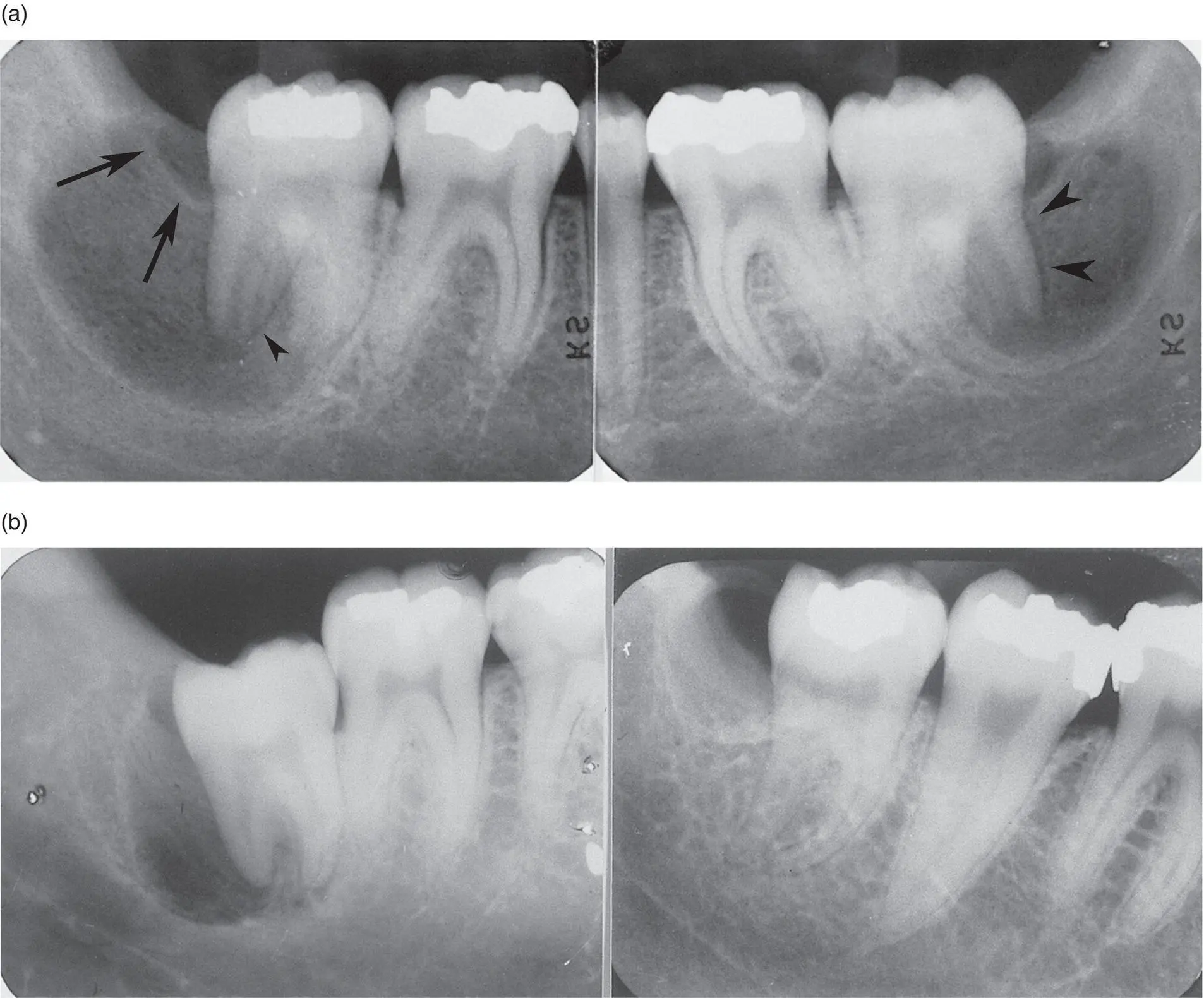
Figure 4.2 Paradental cysts. (a) Bilateral cysts in a 13‐year‐old associated with recently erupted second molars. The third molars were absent. (b) Two cysts in different patients associated with partially erupted (left) and recently erupted (right) third molars. In all cases, the cysts are distal and buccal to the involved teeth. The distal part of the cyst is separate from the distinct distal follicular space (arrows in a) and the periodontal ligament space and lamina dura are intact (arrowheads).
Source: Vedtofte P 1989, p. 182–188 / with permission of Elsevier. Courtesy of CV Mosby Co.
Although most lesions are located in a buccal or disto‐buccal location ( Figure 4.2), Colgan et al. (2002 ) suggested that the precise site of the cyst may depend on the angle of impaction of the associated tooth. A distoangular impaction was shown in 10 of their 15 teeth and all had cysts located towards the distal aspect. Two of their cysts were located towards the mesial aspect of the teeth, but both showed mesioangular impactions. Two teeth that were vertically impacted had cysts located purely on the buccal aspect. The vast majority of reports of paradental cysts record that cysts are located towards the distal aspect of the teeth and that a mesial orientation is rare (Craig 1976 ; Ackermann et al. 1987 ; Vedtofte and Praetorius 1989 ; Philipsen et al. 2004 ). Since most impacted third molars are mesioangular, these data suggest that a distally impacted tooth is more likely to give rise to a paradental cyst than a mesially angled tooth.
Radiological examination of the mandibular buccal bifurcation cyst should include occlusal and panoramic radiographs or CBCT. The cyst presents as a well‐demarcated and usually corticated radiolucency overlying the buccal aspects of the roots of an erupting or recently erupted first or second molar tooth ( Figure 4.3). The inferior margin of the cyst is concave and rarely the cyst may extend to the inferior border of the mandible, but does not lead to distortion of the lower border or to an external deformity ( Figure 4.3a). There may be involvement of the bone in the furcation and inter‐radicular bone may be lost. In the inter‐radicular region, therefore, the lamina dura may be lost, but the periodontal ligament space and the lamina dura are intact on the mesial and distal aspects of the roots ( Figure 4.3), a feature that enables distinction from a radicular cyst.
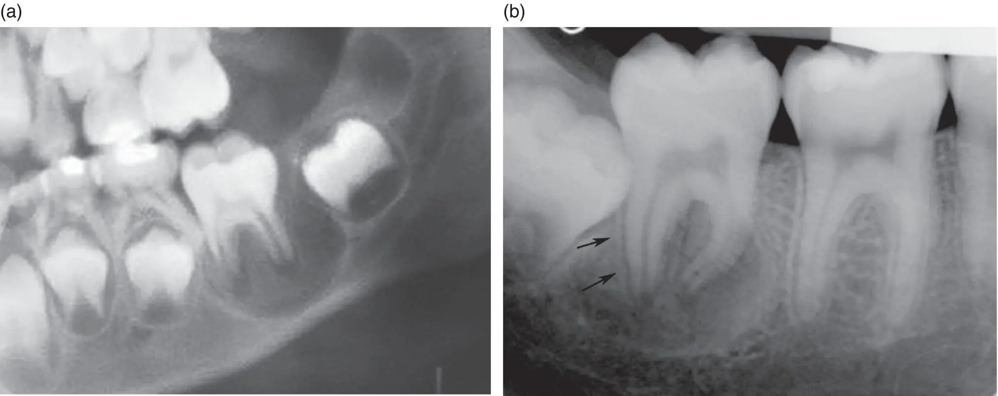
Figure 4.3 Mandibular buccal bifurcation cysts involving (a) an erupting first permanent molar and (b) a second permanent molar. The cysts are well demarcated and corticated and overly the buccal and periapical aspects of the tooth roots. The periodontal ligament and lamina dura are intact (arrows).
Sources: (a) Courtesy of Dr Douglas W Stoneman. (b) Courtesy of Prof Paul Speight (Previously published: El‐Naggar AK 2017, Courtesy of IARC).
Читать дальшеИнтервал:
Закладка:
Похожие книги на «Shear's Cysts of the Oral and Maxillofacial Regions»
Представляем Вашему вниманию похожие книги на «Shear's Cysts of the Oral and Maxillofacial Regions» списком для выбора. Мы отобрали схожую по названию и смыслу литературу в надежде предоставить читателям больше вариантов отыскать новые, интересные, ещё непрочитанные произведения.


Обсуждение, отзывы о книге «Shear's Cysts of the Oral and Maxillofacial Regions» и просто собственные мнения читателей. Оставьте ваши комментарии, напишите, что Вы думаете о произведении, его смысле или главных героях. Укажите что конкретно понравилось, а что нет, и почему Вы так считаете.