Paul M. Speight - Shear's Cysts of the Oral and Maxillofacial Regions
Здесь есть возможность читать онлайн «Paul M. Speight - Shear's Cysts of the Oral and Maxillofacial Regions» — ознакомительный отрывок электронной книги совершенно бесплатно, а после прочтения отрывка купить полную версию. В некоторых случаях можно слушать аудио, скачать через торрент в формате fb2 и присутствует краткое содержание. Жанр: unrecognised, на английском языке. Описание произведения, (предисловие) а так же отзывы посетителей доступны на портале библиотеки ЛибКат.
- Название:Shear's Cysts of the Oral and Maxillofacial Regions
- Автор:
- Жанр:
- Год:неизвестен
- ISBN:нет данных
- Рейтинг книги:5 / 5. Голосов: 1
-
Избранное:Добавить в избранное
- Отзывы:
-
Ваша оценка:
Shear's Cysts of the Oral and Maxillofacial Regions: краткое содержание, описание и аннотация
Предлагаем к чтению аннотацию, описание, краткое содержание или предисловие (зависит от того, что написал сам автор книги «Shear's Cysts of the Oral and Maxillofacial Regions»). Если вы не нашли необходимую информацию о книге — напишите в комментариях, мы постараемся отыскать её.
Shear’s Cysts of the Oral and Maxillofacial Regions
Shear’s Cysts of the Oral and Maxillofacial Regions Fifth Edition
Shear's Cysts of the Oral and Maxillofacial Regions — читать онлайн ознакомительный отрывок
Ниже представлен текст книги, разбитый по страницам. Система сохранения места последней прочитанной страницы, позволяет с удобством читать онлайн бесплатно книгу «Shear's Cysts of the Oral and Maxillofacial Regions», без необходимости каждый раз заново искать на чём Вы остановились. Поставьте закладку, и сможете в любой момент перейти на страницу, на которой закончили чтение.
Интервал:
Закладка:
Regardless of the finer details of the biology of these lesions, from the clinical point of view it is evident that a radiolucent lesion may persist and may produce symptoms, for a substantial period of time after extraction of the offending tooth. On biopsy, many show the typical features of a cyst and residual cyst remains an appropriate term for these lesions.
Radiological Features
The classic description of the radiological appearance of radicular cysts is that they are round or ovoid radiolucencies surrounded by a narrow, radiopaque or corticated margin that extends from the lamina dura of the involved tooth ( Figure 3.4; see also Figure 2.2). In infected or rapidly enlarging cysts, the corticated margin may not be present. A residual cyst is usually round to oval with a well‐demarcated and often corticated margin. They are found within an edentulous area of the jaws, at a site of a previous tooth extraction ( Figure 3.5). With a residual cyst, the differential diagnosis of keratocyst must be considered. A radicular cyst on the lateral margin of a root in association with an accessory root canal must be differentiated from a lateral periodontal cyst. Root resorption is not often seen on routine radiographs, but it may occur.
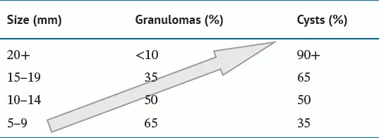
Despite these well‐described features, many studies have shown that it is not possible to reliably differentiate radiologically between a radicular cyst and a periapical granuloma. In one of the largest studies, which has not been repeated, Mortensen et al. (1970 ) examined histological material of 396 periapical lesions with a diameter of 5 mm or more, that had been classified pre‐operatively as cysts or granulomas on radiological evidence. A correct preliminary diagnosis had been made in 81% of 232 granulomas, but in only 48% of 164 cysts. They also showed that the relative number of granulomas decreased with increasing size of the lesion, whereas the relative number of cysts increased. Table 3.1summarises their data and shows that if a lesion measured between 10 and 14 mm in radiographic diameter, there was an equal chance of it being a granuloma or a cyst. About one‐third of lesions measuring 5–9 mm were cysts and one‐third of lesions measuring 15 mm or more were granulomas. However, they found that very large lesions, over 20 mm, were almost always cysts, a finding confirmed by others (Natkin et al. 1984 ).
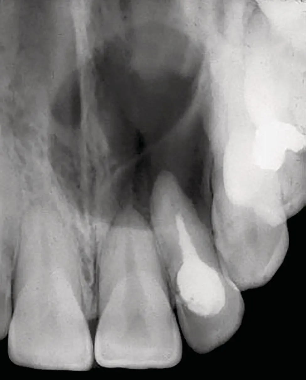
Figure 3.4 Radiograph of a radicular cyst. The lesion is a well‐defined radiolucency associated with the apex of a non‐vital root‐filled tooth.
Mortensen et al. (1970 ) also noted that many cysts had a diffuse radiographic margin and therefore lacked the cortication often described as typical for radicular cysts. This suggested that a corticated margin, although common, was not a specific diagnostic criterion for a cyst, a finding supported by others. Ricucci et al. (2006a ) examined 57 periapical radiolucent lesions and found that of 10 lesions with a corticated outline, only 3 were cysts. Conversely, 40 of the 47 lesions without cortication were granulomas.
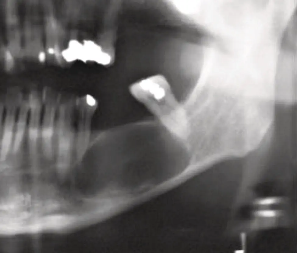
Figure 3.5 Radiograph of a residual cyst. The lesion is at the site of a previously extracted tooth. The lesion must be differentiated from an odontogenic keratocyst.
Table 3.1 The relationship between the size of a lesion on radiological examination and the histological diagnosis of a cyst or granuloma. The arrow illustrates the increasing chance of a lesion being a cyst.
Source: Based on Mortenson et al. (1970 ).
Shrout et al. (1993 ) used radiometric methods to analyse the grey levels on digitised images of periapical lesions. In a pilot study of only 10 mandibular lesions, they showed that analysis of grey levels could correctly identify 4 of 6 granulomas and all 4 cysts. They concluded that it may be feasible to differentiate between radicular cysts and periapical granulomas on the basis of radiographic density. With the advent of digital radiography and powerful software to routinely analyse images, it would be interesting to see if these findings could be confirmed. Early studies examining grey levels on images from cone beam computed tomography (CBCT) suggest that this may provide an accurate diagnosis (Simon et al. 2006 ). Measurement of conventional parameters on CBCT, however, have proved to be no more accurate than conventional X‐rays (Guo et al. 2013 ).
Although teeth may be resorbed by radicular cysts, there is a poor correlation between radiological evidence of resorption and actual tooth resorption on histology. Laux et al. (2000 ) compared the radiological and histological findings in 114 periapical lesions. Ninety three (81%) showed histological evidence of tooth resorption, but only 21 (19%) showed evidence of resorption on the radiographs. It should be noted, however, that only 30 of the 93 lesions with histological resorption showed dentine involvement. In the majority (63 cases) only cementum was involved and it was acknowledged that this would not normally be visible on a plain radiograph.
The data from these studies suggest that there are no specific radiological features that can be used to distinguish between a radicular cyst and a periapical granuloma. Clinicians know that it is useful in treatment planning to have an indication, a priori , whether a periapical lesion is a cyst or a granuloma. There are a number of key features that together can identify that a lesion is odontogenic and can be highly suggestive of a cyst ( Box 3.2). Both granulomas and cysts are associated with a heavily restored or carious tooth and lie within the periodontal ligament. Cysts are more often over 15 mm in diameter, more often have a corticated margin, and show a greater degree of radiopacity.
Box 3.2Clinical and Radiological Features: Key Facts
Always associated with a non‐vital tooth
Most commonly found on upper anterior teeth
Often symptomless and found on radiological examination
Firm or hard swelling, but large lesions may show ‘egg‐shell’ crackling
Residual cysts are found at sites of a previous tooth extraction
Radiology shows well‐demarcated, corticated lesion
Rarely greater than 30 mm in diameter
Pathogenesis
For any type of cyst to develop, three elements are needed: a source of epithelium, a stimulus for epithelial proliferation, and a mechanism of growth and bone resorption. In the case of a radicular cyst, the process is driven by an inflammatory response at the apex of a tooth ( Box 3.3). Although trauma, instrumentation, and irritation from filling materials may cause inflammation, in reality this is usually short lived and the chronic inflammation needed to initiate cyst formation almost always follows microbial infection in a dead pulp following caries. The presence of bacteria and their products in the root canal then leads to periapical inflammation and the formation of a periapical granuloma, which may then lead to cyst formation.
It must be noted, however, that although cysts are a direct sequela of periapical inflammation, cyst formation is not inevitable and radicular cysts are in fact quite rare relative to the prevalence of caries and periapical lesions. In an analysis of 256 periapical lesions, Nair et al. (1996 ) showed that only 15% were actually cysts, although a further 37% were granulomas with proliferating epithelium. Their criteria for diagnosis of a cyst was unusually stringent and depended on the ability to examine, in multiple serial sections, the entire specimen and to be able to visualise a distinct epithelial‐lined cavity. Few studies have undertaken such meticulous examination of lesions using serial sections, but those that have have confirmed the findings of Nair et al. (1996 ) that cysts form the minority of periapical lesions. The actual frequencies reported have been 17.1% (Simon 1980 ), 15.2% (Nair et al. 1996 ), 32.0% (Ricucci et al. 2006b ), and 24.2% (Ricucci et al. 2020 ).
Читать дальшеИнтервал:
Закладка:
Похожие книги на «Shear's Cysts of the Oral and Maxillofacial Regions»
Представляем Вашему вниманию похожие книги на «Shear's Cysts of the Oral and Maxillofacial Regions» списком для выбора. Мы отобрали схожую по названию и смыслу литературу в надежде предоставить читателям больше вариантов отыскать новые, интересные, ещё непрочитанные произведения.


Обсуждение, отзывы о книге «Shear's Cysts of the Oral and Maxillofacial Regions» и просто собственные мнения читателей. Оставьте ваши комментарии, напишите, что Вы думаете о произведении, его смысле или главных героях. Укажите что конкретно понравилось, а что нет, и почему Вы так считаете.