Alan Gunn - Parasitology
Здесь есть возможность читать онлайн «Alan Gunn - Parasitology» — ознакомительный отрывок электронной книги совершенно бесплатно, а после прочтения отрывка купить полную версию. В некоторых случаях можно слушать аудио, скачать через торрент в формате fb2 и присутствует краткое содержание. Жанр: unrecognised, на английском языке. Описание произведения, (предисловие) а так же отзывы посетителей доступны на портале библиотеки ЛибКат.
- Название:Parasitology
- Автор:
- Жанр:
- Год:неизвестен
- ISBN:нет данных
- Рейтинг книги:3 / 5. Голосов: 1
-
Избранное:Добавить в избранное
- Отзывы:
-
Ваша оценка:
Parasitology: краткое содержание, описание и аннотация
Предлагаем к чтению аннотацию, описание, краткое содержание или предисловие (зависит от того, что написал сам автор книги «Parasitology»). Если вы не нашли необходимую информацию о книге — напишите в комментариях, мы постараемся отыскать её.
Highly detailed textbook on parasites and parasite relationships Parasitology: An Integrated Approach
Parasitology: An Integrated Approach, 2nd edition
Parasitology — читать онлайн ознакомительный отрывок
Ниже представлен текст книги, разбитый по страницам. Система сохранения места последней прочитанной страницы, позволяет с удобством читать онлайн бесплатно книгу «Parasitology», без необходимости каждый раз заново искать на чём Вы остановились. Поставьте закладку, и сможете в любой момент перейти на страницу, на которой закончили чтение.
Интервал:
Закладка:
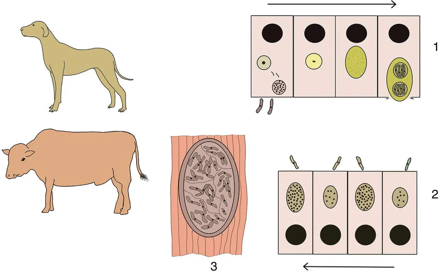
Figure 3.14 Life cycle of Sarcocystis bovicanis . 1: Digestion of a sarcocyst within the dog’s small intestine, releases bradyzoites that invade the gut epithelial cells and then make their way to the lamina propria region where they transform into either male or female gametes. After gamete fusion, the parasites undergo sporogony to form oocysts that contain two sporocysts. The oocysts are shed into the lumen of the dog’s gut and pass with the faeces. 2: A cow consumes the sporocysts/oocysts, and these release the sporozoites that invade its gut epithelial cells and make their way to the blood vessels. The parasites invade the endothelial cells of the blood vessels, transform into merozoites, and undergo four cycles of merogony. After each cycle, the newly formed merozoites infect new endothelial cells. 3: After the last cycle, the merozoites invade skeletal and cardiac muscle cells and transform into metrocytes, each of which divides asexually to form a sarcocyst. Eventually, the metrocytes cease producing new metrocytes and form bradyzoites. Completion of the life cycle requires a dog to consume flesh containing the bradyzoites. Drawings not to scale.
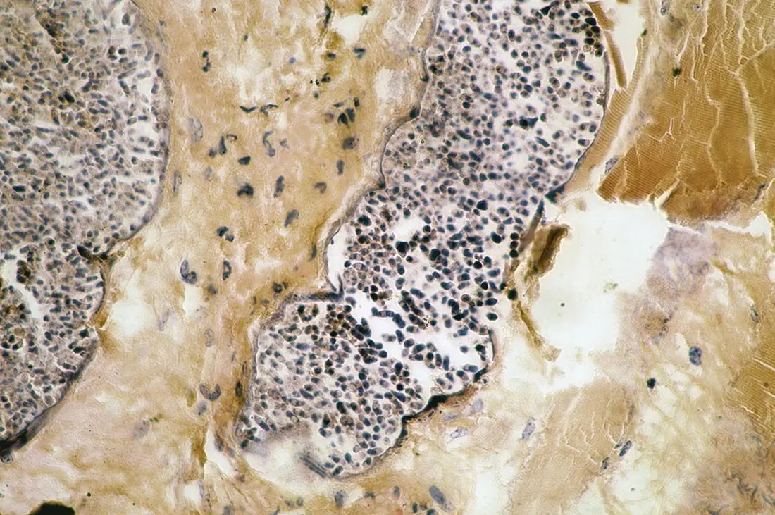
Figure 3.15 Transverse section through a sarcocysts of Sarcocystis muris in the trachea of a mouse.
Human Sarcocystis Infections
Humans are the definitive hosts for S. bovihominis , S. suihominis and several other species of Sarcocystis that we usually acquire from eating raw or poorly cooked meat. As definitive hosts to these parasites, we suffer from intestinal infections. The symptoms are non‐specific and typically include nausea and diarrhoea. The infections are usually self‐limiting and seldom serious.
We can also act as intermediate hosts for some Sarcocystis species. For example, we can act as an intermediate host for Sarcocystis nesbitti although in most cases the species responsible is uncertain. The definitive hosts for S. nesbitti are probably snakes or other reptiles. Therefore, in common with the other species for which we are intermediate hosts, we are ‘dead end’ hosts because few animals have the opportunity to eat us. Presumably, we suffer accidental infections with the sporocysts/oocysts through contamination of food or water and the normal intermediate hosts are other species of primates. The symptoms of infection depend upon the site at which the sarcocysts grow and their abundance. Typically, they induce inflammatory responses that result in pain, fever, and swelling at the infected site. There are reports of regular outbreaks of human sarcocystosis amongst tourists visiting parts of Malaysia (Fayer et al. 2015). Whether these link to one or more species of Sarcocystis is uncertain.
In intermediate hosts, the consequences of infection vary between species, the level of challenge, and the species of Sarcocystis parasitizing them. However, most pathology is usually associated with damage caused to the vascular epithelium during the second stage of merogony. Heavy infections of S. bovicanis in cattle can result in widespread haemorrhages afflicting virtually every organ in the body. This results in anaemia, emaciation, and the animal may become anorexic; abortion can occur in breeding cattle. The immune response results in lymphadenopathy and submandibular oedema whilst the hair at the end of the tail is often lost. Most infections in domestic livestock, however, are subclinical and not discovered until the sarcocysts are detected during meat hygiene inspections after the animal is slaughtered.
3.5.6 Genus Toxoplasma , Toxoplasma gondii
This genus contains only one species, Toxoplasma gondii . However, it has a remarkable host range and can probably infect all mammals and birds.
This intracellular parasite was initially described from a desert rodent, the North African gondi ( Ctenodactylus gondi ) [sometimes spelled ‘ gundi ’ – vowels are not used in written Arabic, so some words appear in various spellings when translated into English] in Tunisia 1908, but the life cycle was not established until 1969–1970. The name Toxoplasma has nothing to do with toxins but derives from the curved shape of the tachyzoite stage of the life cycle ( Figure 3.16). ‘ Toxon ’ ( Tόξoν ) is the Greek word for a ‘bow or something that is crescent shaped and ‘ plasma ’ ( πλάσμα ) is Greek for ‘creature’. Since these humble beginnings, it has become apparent that not only does T. gondii have a cosmopolitan distribution but also it has perhaps the widest host range of any parasite.
The life cycle of T. gondii has two parts and three infectious stages ( Figure 3.17). The two parts of the life cycle are the sexual cycle that occurs within cats and other felines that are the definitive hosts and the asexual cycle that occurs in the intermediate hosts – which are virtually any warm‐blooded animal, including us. The three infectious stages are the sporulated oocysts that contain the sporozoites, the tachyzoites, and the tissue cysts that contain bradyzoites: all three stages are infectious to both the feline definitive hosts and the intermediate hosts. Cats acquire their infection by consuming either sporulated oocysts passed in another cat’s faeces (e.g., through contamination) or an intermediate host containing the tachyzoites and bradyzoites. Following ingestion, the parasites invade the epithelial cells lining the cat’s small intestine and undergo a complex series of asexual divisions. The first step is multiplication by endodyogeny – this involves two daughter cells developing within a mother cell and consuming her before separation takes place. After endodyogeny comes reproduction by endopolygeny in which the mother cell produces several daughter cells simultaneously. Next, comes gametogenesis and the formation of microgametes and macrogametes. Fusion of the gametes results in the formation of a zygote, and this develops into an oocyst that is shed with the faeces whilst it is still unsporulated. Sporogony happens outside the host and takes about 2–3 days under ideal conditions. Fresh faeces are therefore not infectious because the oocysts first need to undergo sporogony to form sporocysts. The infectious sporulated oocysts are oval (10–13 × 9–11 μm) and contain two sporocysts each of which contains four sporozoites. The sporocysts can remain infectious in the environment for months and possibly even 1–2 years. The shedding of oocysts starts 3–10 days after initial infection and continues for only 1–2 weeks although some estimates suggest that a cat may shed up to 100 million oocysts in its faeces during that time. After an initial infection, a cat is usually resistant to re‐infection. The prolonged survival of the oocysts in the environment therefore compensates for the short period over which an infected cat sheds them in its faeces. In some cats, developmental stages may persist in a dormant state and give rise to another batch of oocysts following their subsequent reactivation. This could happen because of changes in the cat’s immune status. Immunity can also wane naturally with time and a cat may suffer another T. gondii infection several years after the first challenge.
Читать дальшеИнтервал:
Закладка:
Похожие книги на «Parasitology»
Представляем Вашему вниманию похожие книги на «Parasitology» списком для выбора. Мы отобрали схожую по названию и смыслу литературу в надежде предоставить читателям больше вариантов отыскать новые, интересные, ещё непрочитанные произведения.


Обсуждение, отзывы о книге «Parasitology» и просто собственные мнения читателей. Оставьте ваши комментарии, напишите, что Вы думаете о произведении, его смысле или главных героях. Укажите что конкретно понравилось, а что нет, и почему Вы так считаете.