Alan Gunn - Parasitology
Здесь есть возможность читать онлайн «Alan Gunn - Parasitology» — ознакомительный отрывок электронной книги совершенно бесплатно, а после прочтения отрывка купить полную версию. В некоторых случаях можно слушать аудио, скачать через торрент в формате fb2 и присутствует краткое содержание. Жанр: unrecognised, на английском языке. Описание произведения, (предисловие) а так же отзывы посетителей доступны на портале библиотеки ЛибКат.
- Название:Parasitology
- Автор:
- Жанр:
- Год:неизвестен
- ISBN:нет данных
- Рейтинг книги:3 / 5. Голосов: 1
-
Избранное:Добавить в избранное
- Отзывы:
-
Ваша оценка:
Parasitology: краткое содержание, описание и аннотация
Предлагаем к чтению аннотацию, описание, краткое содержание или предисловие (зависит от того, что написал сам автор книги «Parasitology»). Если вы не нашли необходимую информацию о книге — напишите в комментариях, мы постараемся отыскать её.
Highly detailed textbook on parasites and parasite relationships Parasitology: An Integrated Approach
Parasitology: An Integrated Approach, 2nd edition
Parasitology — читать онлайн ознакомительный отрывок
Ниже представлен текст книги, разбитый по страницам. Система сохранения места последней прочитанной страницы, позволяет с удобством читать онлайн бесплатно книгу «Parasitology», без необходимости каждый раз заново искать на чём Вы остановились. Поставьте закладку, и сможете в любой момент перейти на страницу, на которой закончили чтение.
Интервал:
Закладка:
3.5.3 Genus Cystoisospora
Members of this genus formerly belonged to the genus Isospora . The presence of a tissue cyst stage within their life cycle distinguishes them from parasites of the genus Isospora. They are monoxenous although some species (e.g., those infecting cats) exploit paratenic hosts. The genus includes several species that infect mammals and us. For example, Cystoisospora canis causes diarrhoea in puppies, Cystoisospora suis causes neonatal piglet diarrhoea, and Cystoisospora belli is an important human pathogen.
3.5.3.1 Cystoisospora (Isospora) belli
This species occurs throughout the world but is particularly common in tropical and subtropical countries. The first cases were recognised in people returning from the battlefields during the First World War, hence the species name (Latin bellum = war). It only infects humans and there do not appear to be any paratenic hosts (Dubey and Almeria 2019). It causes diarrhoea and is frequently associated with persons suffering from AIDS or immunosuppressive illnesses.
The life cycle begins when we ingest oocysts (17–37 × 8–21 μm) containing infective sporozoites ( Figure 3.13). In common with other members of the genus, each oocyst contains two sporocysts and each sporocyst contains four sporozoites. The sporozoites emerge in the small intestine and invade the epithelial cells where they transform into merozoites that divide to produce more merozoites that in turn divide in a cycle that repeats many times. This destroys the host cell and releases the parasites so that they can invade new host cells. In serious infections, this results in the loss of large areas of the gut lining and allows secondary invasion by gut microbes. Presumably, the parasites gain access to the circulation because tissue cysts develop in variety of organs distant from the gastrointestinal tract. For example, within the spleen, liver, and lymph nodes. Each cyst contains a single parasite (zoite) (i.e., they are monozoic) that resembles a typical coccidian sporozoite. The formation of tissue cysts usually occurs in immunocompromised patients but their role in pathology and relapses is uncertain. There is a suggestion that in pigs, the formation of tissue cysts by C. suis contributes to the maintenance of immunity to the parasite (Shrestha et al. 2015). Nowadays, few animals get the opportunity to eat humans, so the tissue cysts probably play no role in transmission of C. belli . The importance of tissue cysts in the transmission of other mammalian Cystoisospora species is uncertain.
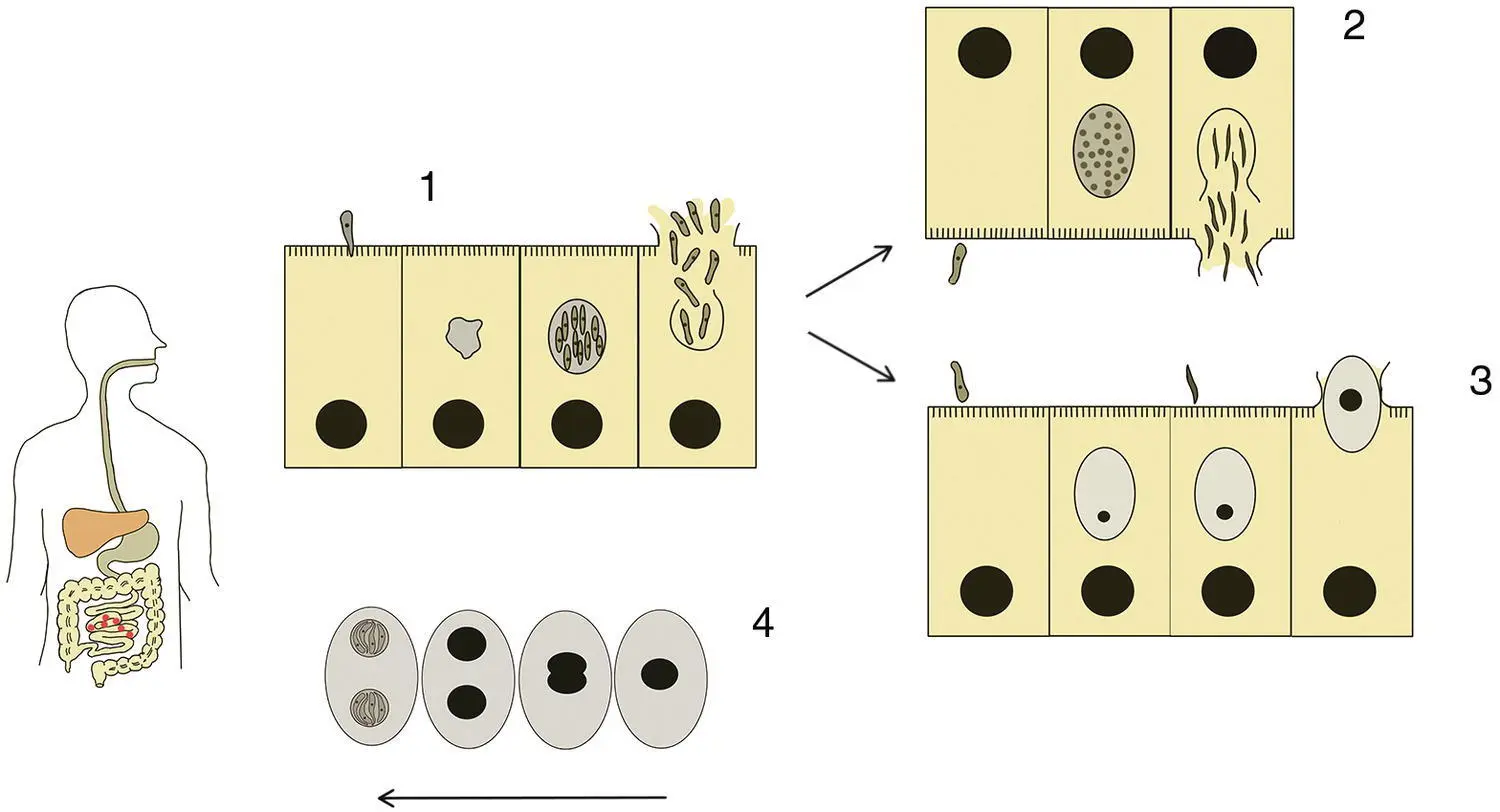
Figure 3.13 Life cycle of Isospora belli . 1: Infection commences following ingestion of a sporulated oocyst. The oocyst releases eight sporozoites when it reaches the small intestine and these invade the gut epithelial cells where they transform into merozoites. The merozoites divide asexually to produce more merozoites. 2: Merozoite forming a multinucleate meront that gives rise to microgametocytes (male). 3: Merozoite giving rise to a macrogametocyte (female). Fusion of a male and a female gamete results in the formation of a zygote, and this develops into an oocyst. 4: Sporulation of the oocyst occurs before it is released in the host’s faeces. Drawings not to scale.
At some point, the merozoites transform into multinucleate meronts and these give rise to microgametes (male) or macrogametes (female). Unless the microgametes and macrogametes occur within the same cell (which is possible), the microgametes leave their host cell to locate a macrogamete and their fusion results in the formation of a zygote that then develops into an oocyst. The death of the host cell releases the oocyst into the gut lumen and leaves the body with the faeces. The oocyst then undergoes sporulation to produce two sporocysts, each of which contains four sporozoites. Transmission is therefore passive by faecal–oral contamination. Environmental conditions probably heavily influence the time taken for sporulation. For example, in some Isospora species, temperatures below 20 °C inhibit sporulation, but it takes less than 16 hours at 30 °C. Nevertheless, transmission is probably through infected food or water rather than direct contact. For example, through not washing one’s hands after defaecation or by contacting an article touched by such a person. This is therefore distinct from Cryptosporidium parvum in which the oocysts are immediately infective after shedding.
3.5.4 Genus Cyclospora
This genus currently contains 13 species that are parasitic in a range of vertebrates (e.g., snakes, rodents), but the most important is Cyclospora cayetanensis that only infects humans.
3.5.4.1 Cyclospora cayetanensis
First described in 1979, this is an emerging parasitic disease with a cosmopolitan distribution (Almeria et al. 2019). There are regular outbreaks in USA and Canada associated with the importation of fresh berries (Dixon et al. 2016). The life cycle begins with the ingestion of oocysts (8–10 μm diameter) containing infective sporozoites. Once the oocysts reach the duodenum and upper small intestine, they release the sporozoites and these invade the gut epithelial cells. They then transform into type I merozoites that then divide to form type II merozoites. The type II merozoites transform into meronts and these give rise to microgametocytes and macrogametocytes. Fusion of a microgametocyte with a macrogametocyte gives rise to zygote, and this develops into an oocyst that is shed with the faeces. The oocyst then undergoes sporulation to produce two sporocysts each containing two sporozoites. Sporulation takes 7–15 days depending upon the environmental conditions and therefore transmission probably occurs through contamination of food and water rather than direct person‐to‐person/contaminated object contact.
The symptoms of infection are non‐specific and resemble those of many other gastrointestinal diseases. Patients suffer from abdominal pain, watery diarrhoea, flatulence, low‐grade fever, anorexia, and weight loss. In endemic regions, the symptoms tend to be worse in young children and in non‐endemic regions most people who become infected express symptoms. Persons who are immunosuppressed and AIDS patients are more seriously affected.
3.5.5 Genus Sarcocystis
Members of this genus are obligate intracellular parasites with a life cycle that involves two hosts – a herbivore intermediate host in which only asexual multiplication occurs and a carnivore definitive host in which sexual reproduction takes place. Most Sarcocystis species are very host‐specific and infect a limited number of closely related intermediate/final hosts ( Table 3.3). You will not be surprised to hear that the taxonomy is confused, and one must take care when using the older literature. For example, some animals once thought to harbour just a single Sarcocystis species actually contain several species. In addition, some species are morphologically identical, and others have synonyms (e.g., Sarcocystis cruzi is also known as Sarcocystis bovicanis – a reflection that the intermediate and definitive hosts are cattle and dogs/ other canines respectively).
Table 3.3 Summary of the most important species of Sarcocystis in human and veterinary medicine.
| Species of Sarcocystis | Synonym | Intermediate host | Definitive host |
|---|---|---|---|
| Sarcocystis bovicanis | Sarcocystis cruzi | Cattle | Dogs and other canines |
| Sarcocystis bovihominis | Sarcocystis hominis | Cattle | Humans |
| Sarcocystis bovifelis | Sarcocystis hirsuta | Cattle | Cats and other felines |
| Sarcocystis Suihominis | Isospora hominis | Pigs | Humans and some primates |
| Sarcocystis ovifelis | Sarcocystis tenella | Sheep | Cats and other felines |
| Sarcocystis hovathi | Sarcocystis gallinarum | Chicken | Dogs and other canines |
The life cycle of S. bovicanis is typical of most Sarcocystis species. In this species, the definitive host is a dog or other canine, whilst the intermediate hosts are cows and other bovids ( Figure 3.14). The life cycle begins when an infected dog sheds free sporocysts or oocysts in its faeces. A cow must then consume the sporocysts/oocysts, and this usually happens through contamination of food or water. When the sporocyts/oocysts reach the cow’s small intestine, they release the sporozoites. The sporozoites then invade the gut epithelial cells and make their way to the blood vessels. The bloodstream then distributes them around the body. The parasites invade the endothelial cells of the blood vessels that serve many of the body’s organ systems. Within the endothelial cells, the parasites transform into merozoites and undergo four cycles of merogony (asexual reproduction). After each cycle, the newly formed merozoites are released, and these infect new endothelial cells downstream of the initial infection. After the last cycle, the merozoites invade skeletal and cardiac muscle cells. Occasionally, smooth muscle, the brain, and spinal cord are also infected. Within these cells, the merozoites transform into metrocytes or ‘mother cells’ each of which divides asexually to form a structure called a sarcocysts ( Figure 3.15). With repeated asexual division, a sarcocyst steadily becomes larger and larger and in some Sarcocystis species may become big enough to be visible to the naked eye. Eventually, the globular metrocytes cease producing new metrocytes and form crescent‐shaped bradyzoites. The time taken for this depends upon the species of Sarcocystis but can be around 2 months. During this time, the sarcocysts are non‐infectious since only bradyzoites can transmit the infection. Completion of the life cycle requires a dog to consume flesh containing the bradyzoites. Digestion of the sarcocyst within the dog’s small intestine releases the bradyzoites, and these become motile. The bradyzoites initially invade the gut epithelial cells and then make their way to the lamina propria region where they transform into either male or female gametes. After gamete fusion, the parasites undergo sporogony to form oocysts that contain two sporocysts. The oocysts are therefore already sporulated when shed and each contains four sporozoites. The oocysts are shed into the lumen of the gut and passed with the faeces. The oocyst has a thin wall and often ruptures when one is preparing faecal samples for microscopy. Consequently, one normally sees sporocysts (16.3 × 10.8 μm) in faecal samples. Sarcocystis seldom causes serious pathology in its definitive hosts.
Читать дальшеИнтервал:
Закладка:
Похожие книги на «Parasitology»
Представляем Вашему вниманию похожие книги на «Parasitology» списком для выбора. Мы отобрали схожую по названию и смыслу литературу в надежде предоставить читателям больше вариантов отыскать новые, интересные, ещё непрочитанные произведения.


Обсуждение, отзывы о книге «Parasitology» и просто собственные мнения читателей. Оставьте ваши комментарии, напишите, что Вы думаете о произведении, его смысле или главных героях. Укажите что конкретно понравилось, а что нет, и почему Вы так считаете.