Pascale Cossart - The New Microbiology
Здесь есть возможность читать онлайн «Pascale Cossart - The New Microbiology» — ознакомительный отрывок электронной книги совершенно бесплатно, а после прочтения отрывка купить полную версию. В некоторых случаях можно слушать аудио, скачать через торрент в формате fb2 и присутствует краткое содержание. Жанр: unrecognised, на английском языке. Описание произведения, (предисловие) а так же отзывы посетителей доступны на портале библиотеки ЛибКат.
- Название:The New Microbiology
- Автор:
- Жанр:
- Год:неизвестен
- ISBN:нет данных
- Рейтинг книги:3 / 5. Голосов: 1
-
Избранное:Добавить в избранное
- Отзывы:
-
Ваша оценка:
The New Microbiology: краткое содержание, описание и аннотация
Предлагаем к чтению аннотацию, описание, краткое содержание или предисловие (зависит от того, что написал сам автор книги «The New Microbiology»). Если вы не нашли необходимую информацию о книге — напишите в комментариях, мы постараемся отыскать её.
, Pascale Cossart tells a splendid story about the revolution in microbiology, especially in bacteriology. This story has wide-ranging implications for human health and medicine, agriculture, environmental science, and our understanding of evolution. The revolution results from the powerful tools of molecular and cellular biology, genomics, and bioinformatics, which have yielded amazing discoveries, from entire genome sequences to video of bacteria invading host cells. This book is for both scientists and especially nonscientists who would like to learn more about the extraordinary world of bacteria.
Dr. Cossart's overview of the field of microbiology research, from infectious disease history to the ongoing scientific revolution resulting from CRISPR technologies, is presented in four parts.
New concepts in microbiology introduces the world of bacteria and some recent discoveries about how they live, such as the role of regulatory RNAs including riboswitches, the CRISPR defense system, and resistance to antibiotics. Sociomicrobiology: the social lives of bacteria helps us see the new paradigm by which scientists view bacteria as highly social creatures that communicate in many ways, for example in the assemblies that reside in our intestine or in the environment. The biology of infections reviews some of history's worst epidemics and describes current and emerging infectious diseases, the organisms that cause them, and how they produce an infection. Bacteria as tools introduces us to molecules derived from microbes that scientists have harnessed in the service of research and medicine, including the CRISPR/Cas9 genome-editing technology.
takes us on a journey through a remarkable revolution in science that is occurring here and now.
The New Microbiology — читать онлайн ознакомительный отрывок
Ниже представлен текст книги, разбитый по страницам. Система сохранения места последней прочитанной страницы, позволяет с удобством читать онлайн бесплатно книгу «The New Microbiology», без необходимости каждый раз заново искать на чём Вы остановились. Поставьте закладку, и сможете в любой момент перейти на страницу, на которой закончили чтение.
Интервал:
Закладка:
Mycoplasma
Mycoplasmas, of the genus Mycoplasma , are bacteria that have cell membranes but lack a cell wall. These relatively small bacteria have historically been difficult to locate and identify because the principal bacterial identification method, the Gram stain, is based on identification of the peptidoglycan in bacterial cell walls. Our inability to detect Mycoplasma has been a problem because many mycoplasmas, which are in most cases commensals (i.e., nonpathogenic) in the respiratory tract or vagina, can be responsible for sexually transmitted infections. Also, because these organisms have no cell walls, they are not affected by antibiotics that target peptidoglycan. Mycoplasmas have some of the smallest bacterial genomes, and the chromosome of Mycoplasma genitalium was the first to be synthesized by means of synthetic biology tools.
Flagella and other appendages
Flagella are found on the surface of bacteria. They are long helical filaments connected to small rotary motors that allow the bacteria to move and spread out in fluid environments. Hairlike appendages known as pili are also found on the surface of some bacteria. Pili allow bacteria to adhere to biological and abiotic surfaces or even to aggregate. Curli , similar to pili, are involved in adherence to surfaces and bacterial aggregation. They closely resemble the aggregating “amyloid” fibers found in the brains of Alzheimer patients.
The shape of bacteria is maintained by their cell wall, but their size depends on the volume of their interior, which expands during bacterial growth. Bacteria have a highly organized internal architecture thanks to molecules that are similar to those found in human or plant cells. It was discovered that bacteria even have skeletons, spiral-shaped cytoskeletons made of actin that are involved in the localization and activation of enzymes that create peptidoglycan and are crucial for cell division.
Bacterial shape and division: proteins similar to actin and tubulin in eukaryotes
Bacillus-type (rod-shaped) bacteria and their cell walls grow depending on the growth medium composition. Growth of the peptidoglycan, and thus also of the cell wall, is controlled by MreB, a protein anchored to the membrane that is similar to the protein actin in eukaryotes. MreB forms a sort of spiral structure that gives bacilli their elongated form. A protein known as crescentin is responsible for the crescent shape of Caulobacter cells. Bacterial growth has a limit. Once a bacterium reaches a certain size, it splits. This is a highly precise process that uses at least two other molecules similar to actin and tubulin, proteins that were once thought to be present only in eukaryotes. In addition, when a bacterium divides, every vital element of the bacterium, including the chromosome (more specifically the DNA), is duplicated and shared between the two resulting daughter cells. At the final phase of division, the key step in the separation of two daughter cells involves another actin-like protein, FtsA, which attaches to the site of division, FtsZ, a protein similar to the tubulin protein in eukaryotic cells. ParM is another protein similar to eukaryotic actin that is involved in the distribution of plasmid DNA between daughter cells.
Most bacteria produce two apparently identical daughter cells. However, some, such as Caulobacter crescentus , do not. Caulobacter is an aquatic bacterium that has become an impressive model for asymmetrical cell division ( Fig. 3). A Caulobacter cell about to divide is immobile, attached by a short stem to a surface such as a rock or the ocean bottom. The unattached end generates a mobile daughter cell that uses a flagellum to propel itself away from the still-attached daughter cell, which will continue to grow and split off more daughters. The mobile cell eventually loses its flagellum and grows a stem that it uses to attach itself to a surface, where it will grow and eventually split off daughter cells of its own.
Are bacteria immortal? Do they have strategies for survival? When stressed by conditions such as desiccation or nutrient deficiency, some bacteria reproduce by forming spores . These are a type of dormant cell that is extremely resistant to heat, cold, dryness, and even some antiseptics. Spores allow bacteria to persist for years or even centuries—and to disseminate ( Fig. 4). When a spore arrives in an appropriate environment, it can germinate and resume a normal binary cell division.
Not all bacteria produce spores, but several that do are considered some of the most dangerous to humans. In late 2001 in the United States, anthrax spores (spores of Bacillus anthracis ) sent through the mail as an act of bioterrorism caused skin, intestinal, and lung infections that led to the deaths of five people. Clostridium tetani is another sporeformer. Its spores can remain dormant in the soil for years, but when they enter the anaerobic environment of an open wound, they can reactivate and cause tetanus.

Figure 3. Caulobacter crescentus is used as a model for the study of differentiated cell division. When these bacteria split, they give birth to two slightly different cells, one with a stem and the other with a flagellum.
Because spores are highly resistant to adverse conditions and can spread easily, it is hard to get rid of them; spores are therefore very dangerous. Take for example the bacterium Clostridium difficile. “C. diff," as it is commonly called, is part of the human intestinal microbiome and is highly resistant to most antibiotics. When a patient is treated with antibiotics, the normal intestinal microbiome changes, leaving the resistant C. difficile to dominate and cause severe colitis and diarrhea. These bacteria are capable of producing spores that can survive almost anywhere for years. Hence, they are becoming a more and more common cause of health care-associated infections, particularly in hospitals.
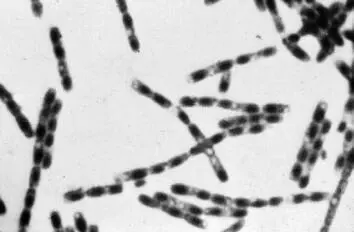
Figure 4. Bacillus anthracis . Under conditions of stress, some bacteria produce spores that contain the bacteria's complete DNA. Spores can survive indefinitely in nature until more favorable conditions trigger them to germinate and replicate normally again.
Bacteria also have lesser-known survival strategies. For example, some bacteria can halt their peptidoglycan synthesis in order to produce progeny that lack peptidoglycan and are not recognized by the immune system. These are called L-form bacteria , from the name of the English surgeon Joseph Lister. Like the mycoplasmas described earlier, they are resistant to many antibiotics and can survive in an infected host for a long time, even during treatment.
It is now possible to watch bacteria divide and to examine the location, the behavior, or the fate of some bacterial proteins. Indeed, new imaging technologies—particularly time-lapse microscopy and superresolution microscopy, both of which use various fluorescent markers—have made it possible to study bacteria in real time. One can observe the precise location of fluorescently linked bacterial proteins (such as the pole or site of division) and see whether they become more intense or disappear during bacterial growth. Combining these imaging techniques with microfluidics—the study of the flow of microquantities of liquids—allows for the real-time observation of bacterial behavior, for example, during changes in cultures or temperature.
Читать дальшеИнтервал:
Закладка:
Похожие книги на «The New Microbiology»
Представляем Вашему вниманию похожие книги на «The New Microbiology» списком для выбора. Мы отобрали схожую по названию и смыслу литературу в надежде предоставить читателям больше вариантов отыскать новые, интересные, ещё непрочитанные произведения.


Обсуждение, отзывы о книге «The New Microbiology» и просто собственные мнения читателей. Оставьте ваши комментарии, напишите, что Вы думаете о произведении, его смысле или главных героях. Укажите что конкретно понравилось, а что нет, и почему Вы так считаете.