S. Jane Flint - Principles of Virology, Volume 2
Здесь есть возможность читать онлайн «S. Jane Flint - Principles of Virology, Volume 2» — ознакомительный отрывок электронной книги совершенно бесплатно, а после прочтения отрывка купить полную версию. В некоторых случаях можно слушать аудио, скачать через торрент в формате fb2 и присутствует краткое содержание. Жанр: unrecognised, на английском языке. Описание произведения, (предисловие) а так же отзывы посетителей доступны на портале библиотеки ЛибКат.
- Название:Principles of Virology, Volume 2
- Автор:
- Жанр:
- Год:неизвестен
- ISBN:нет данных
- Рейтинг книги:3 / 5. Голосов: 1
-
Избранное:Добавить в избранное
- Отзывы:
-
Ваша оценка:
Principles of Virology, Volume 2: краткое содержание, описание и аннотация
Предлагаем к чтению аннотацию, описание, краткое содержание или предисловие (зависит от того, что написал сам автор книги «Principles of Virology, Volume 2»). Если вы не нашли необходимую информацию о книге — напишите в комментариях, мы постараемся отыскать её.
Volume I: Molecular Biology
Volume II: Pathogenesis and Control
Principles of Virology, Fifth Edition
Principles of Virology, Volume 2 — читать онлайн ознакомительный отрывок
Ниже представлен текст книги, разбитый по страницам. Система сохранения места последней прочитанной страницы, позволяет с удобством читать онлайн бесплатно книгу «Principles of Virology, Volume 2», без необходимости каждый раз заново искать на чём Вы остановились. Поставьте закладку, и сможете в любой момент перейти на страницу, на которой закончили чтение.
Интервал:
Закладка:
BOX 2.11
TERMINOLOGY
Which direction: anterograde or retrograde?
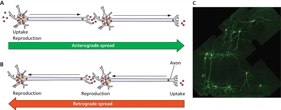
Retrograde and anterograde spread of virus in nerves. (A)Anterograde spread of infection. The virus invades at dendrites or cell bodies and reproduces. Virus particles then spread to axon terminals, where they cross synaptic contacts to invade dendrites or cell bodies of the second neuron. (B)Retrograde spread of infection. The virus invades at axon terminals and spreads to the cell body, where reproduction occurs. Progeny virus particles spread to a neuron at sites of synaptic contact. Particles enter the axon terminal of the second neuron to initiate a second cycle of replication and spread. (C)Identification of a possible micro circuit in the rodent visual cortex (V2) after injection of a green fluorescent protein-expressing strain of pseudorabies virus into the synaptically connected, but distant, V1 region. Infection spread via V1 axons (V1 cell bodies are located far out of the field of view) in a retrograde manner to a subset of V2 cell bodies is seen here. Confocal microscopy and image reconstruction by Botond Roska, Friedrich Miescher Institute, Basel, Switzerland.
Those who study virus spread in the nervous system often use the words retrogradeand anterogradeto describe direction. Confusion can arise because the terms can be used to describe directional movement of virus particles inside a cell, as well as spread between synaptically connected neurons. Spread from the primary neuron to a second-order neuron in the direction of the nerve impulse is called anterograde spread (see figure). Spread in the opposite direction is termed retrograde. Spread inside a neuron is defined by microtubule polarity. Anterograde transport occurs on micro tubules from the cell body toward the axon terminus; retrograde spread occurs from the axon terminus toward the cell body.
Ekstrand MI, Enquist LW, Pomeranz LE. 2008. The alpha-herpesviruses: molecular pathfinders in nervous system circuits. Trends Mol Med 14:134–140.
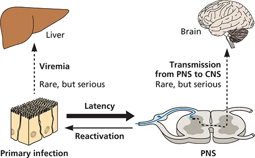
Figure 2.17 Outline of the spread of alphaherpesviruses and relationship to disease. Although herpes simplex virus can infect many cell types, in most infected individuals, it remains restricted to the local site of infection and establishes latency in the ganglia that innervate that site. Under conditions when the host has a weakened immune system, viremia can result in which distal organs become infected and/or the virus may transition from the peripheral nervous system (PNS) to the central nervous system (CNS); again, this is a rare event.
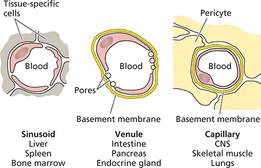
Figure 2.18 Blood-tissue junction in a capillary, venule, and sinusoid. (Left) Sinusoids, lined with macrophages of the reticuloendothelial system, as found in the adrenal glands, liver, spleen, and bone marrow. (Center) Fenestrated endothelium found in the choroid plexus, villi of the intestine, renal glomerulus, pancreas, and endocrine glands. (Right) Continuous endothelium and basement membrane found in the central nervous system, connective tissue, skeletal and cardiac muscle, skin, and lungs. Adapted from Mims CA et al. 1995. Mims’ Pathogenesis of Infectious Disease (Academic Press, Orlando, FL).

Figure 2.19 How viruses gain access to the liver. Two layers of hepatocytes are shown, with the sinusoid at the center, lined with kupffer cells. On the left, transcytosis through the kupffer cells is shown; on the right, direct kupffer cell infection is illustrated, followed by infection of underlying hepatocytes. Viruses not taken up by either route will flow through. Adapted from Mims CA et al. 1995. Mims’ Pathogenesis of Infectious Disease (Academic Press, Orlando, FL).
Organs with Dense Basement Membranes
In the central nervous system, connective tissue, and skeletal and cardiac muscle, capillary endothelial cells are supported by a dense basement membrane, which raises an additional barrier to viral passage into the tissue ( Fig. 2.18and 2.20). In the central nervous system, the basement membrane, formed in part by astrocytic extensions (called “endfeet”) that align with the basolateral surface of the capillary endothelium, is the foundation of the blood-brain barrier ( Fig. 2.21).
Not all capillaries in tissues adhere to one of these three types: for example, in several well-defined parts of the brain, the capillary epithelium is loosely joined together, and the basement membrane is sparse, affording an easier passage for some neurotropic viruses . These highly vascularized sites include the choroid plexus, a sheet of tissue that lies within the brain ventricles and that produces more than 70% of the cerebrospinal fluid that bathes the spinal cord and affords protective cushioning. Some viruses (mumps virus and certain togaviruses) pass through the capillary endothelium and enter the stroma of the choroid plexus, where they may then cross the epithelium into the cerebrospinal fluid either by transcytosis or by directed release following production of progeny virus particles. Once in the cerebrospinal fluid, infection can spread to the ependymal cells lining the ventricles and the underlying brain tissue ( Fig. 2.21). Other viruses (picornaviruses) may infect directly, or be transported across the capillary endothelium. Some viruses (human immunodeficiency virus type 1 and measles virus) cross the endothelium within infected monocytes or lymphocytes (the Trojan Horse approach, described earlier). Increased local permeability of the capillary endothelium, caused, for example, by certain hormones, may also facilitate virus entry into the brain and spinal cord.
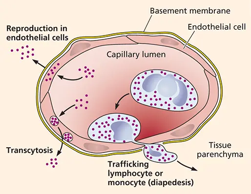
Figure 2.20 How viruses travel from blood to tissues with basement membranes. Schematic of a capillary (similar to Fig. 2.19, right), illustrating different pathways by which viruses may leave the blood and enter underlying tissues. Adapted from Nathanson N (ed). 2007. Viral Pathogenesis and Immunity (Academic Press, London, United Kingdom), with permission.
Skin
In a number of systemic viral infections, rashes are produced when virus particles leave blood vessels and enter the cells that comprise the skin. Viruses that cause rashes include measles virus, rubella virus (German measles), varicella-zoster virus (chicken pox and shingles), some parvoviruses (fifth disease), poxviruses (smallpox), and Coxsackieviruses (hand, foot, and mouth disease). Skin lesions resulting from these infections are notably distinct, distinguished by size, color, frequency, and elevation (an indication of inflammation). Rashes may appear coincident with or subsequent to an infection, although most all appear toward the end of the acute infection. Destruction of cells by virus reproduction and the host immune system are the primary causes of most skin lesions.
Читать дальшеИнтервал:
Закладка:
Похожие книги на «Principles of Virology, Volume 2»
Представляем Вашему вниманию похожие книги на «Principles of Virology, Volume 2» списком для выбора. Мы отобрали схожую по названию и смыслу литературу в надежде предоставить читателям больше вариантов отыскать новые, интересные, ещё непрочитанные произведения.


Обсуждение, отзывы о книге «Principles of Virology, Volume 2» и просто собственные мнения читателей. Оставьте ваши комментарии, напишите, что Вы думаете о произведении, его смысле или главных героях. Укажите что конкретно понравилось, а что нет, и почему Вы так считаете.