M. V. Brian - Ants
Здесь есть возможность читать онлайн «M. V. Brian - Ants» — ознакомительный отрывок электронной книги совершенно бесплатно, а после прочтения отрывка купить полную версию. В некоторых случаях можно слушать аудио, скачать через торрент в формате fb2 и присутствует краткое содержание. Жанр: unrecognised, на английском языке. Описание произведения, (предисловие) а так же отзывы посетителей доступны на портале библиотеки ЛибКат.
- Название:Ants
- Автор:
- Жанр:
- Год:неизвестен
- ISBN:нет данных
- Рейтинг книги:3 / 5. Голосов: 1
-
Избранное:Добавить в избранное
- Отзывы:
-
Ваша оценка:
Ants: краткое содержание, описание и аннотация
Предлагаем к чтению аннотацию, описание, краткое содержание или предисловие (зависит от того, что написал сам автор книги «Ants»). Если вы не нашли необходимую информацию о книге — напишите в комментариях, мы постараемся отыскать её.
Ants — читать онлайн ознакомительный отрывок
Ниже представлен текст книги, разбитый по страницам. Система сохранения места последней прочитанной страницы, позволяет с удобством читать онлайн бесплатно книгу «Ants», без необходимости каждый раз заново искать на чём Вы остановились. Поставьте закладку, и сможете в любой момент перейти на страницу, на которой закончили чтение.
Интервал:
Закладка:
It remains only to mention two glands that open into the sting ( fig. 4). One is the poison gland which consists of two closed tubes feeding a thin-walled storage vesicle and which contains a watery solution of mixed venoms. The other is a smaller gland called after Dufour which contains an oily material secreted by cells which surround the storage space. The poison glands vary quite considerably in different families of ant, depending upon whether they synthesize a thin liquid for squirting or injecting with a sting, or a sticky fluid for smearing.
In general the ant body is neatly divided for separate functions: the head carries the main sense organs and the brain and has in front a mechanism for food capture and food pre-treatment, the mesosoma is specialized for locomotion and the gaster has a region of absorption and food storage but also contains the reproductive organs. Defensive apparatus is disposed both in front and behind but the body is flexible enough for both to act forwards in concert.
LARVAL STRUCTURE
Ants’ eggs are oval and white; each egg weighs only ·00005 gm and is less than a millimetre long. In most of them a legless grub develops. This is of course no bigger than an egg at first and breaks its way out by means of its sharply pointed jaws. It is almost hairless and transparent so that a small, residual blob of egg yolk in its midgut can easily be seen. Though its cuticle is inelastic rather like polythene it is wrinkled and folded so that a good deal of room for expansion is allowed. There are three stages separated by moults, when the old skin is cast off and a new one, folded and wrinkled to allow for growth, is built underneath. Only the head has a firm cuticle that will not expand at all so that its growth only takes place during the moult between successive stages. The second stage has a good many more hairs than the first and the third and last stage is very hairy indeed. It has some hooked hairs as well as the simple ones which enable the grubs to interlock and so be carried about in one group. It is tempting to believe that the workers are able to distinguish the stages by feeling the degree of hairiness with their antennae, but again this has not been tested by experiment.
The head carries jaws with pointed, hardened tips. These are capable of piercing eggs and can masticate soft tissues a little. There are several sense organs of a very simple kind, including minute, unjointed, conical antennae; several other similar ones occur around the mouth and are probably used by the larva to feel and taste what it is eating, as well as to enable it to lock on to the worker’s mouth whilst it is being fed regurgitated liquid. The head, like that of the adult, contains a pharynx innervated by muscles ( fig. 5). It is a pump that only sucks inwards; larvae cannot regurgitate food. The ‘brain’ consists of a dorsal ganglion and a ventral ganglion with nerve trunks running round the oesophagus to connect them. The ventral ganglion is the first of a long, chain-like series that runs down the body near the ventral wall.
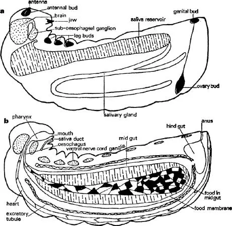
FIG. 5. Larva of Myrmica rubra in third instar, longitudinal sections to show main organs: a. slightly off mid-line, b. on mid-line (sagittal).
Most of the body contents are connected with the digestion and storage of food. A very large, sac-shaped midgut consists of a single layer of large, glandular cells. This is connected to the pharynx by a thin-walled oesophagus and where this and the midgut meet there is a structure for secreting and moulding a thin membrane which encloses the food and is thought to protect the delicate gut cells from abrasion. However, there is no way through from the midgut into the much smaller hindgut and all the food residues collect and condense into a black pellet surrounded by food membranes which are ejected at the end of the larval stage. The hindgut is very thin-walled and collects the liquids extracted by excretory tubules that lie freely in the body fluids. It thus acts as a bladder by storing urine and, if the diet is rich in protein, white insoluble granules of uric acid. The bladder is emptied by a longitudinal contraction of the body; this usually occurs only in response to a light touch on the skin in the region of the anus so that normally there is a worker nearby to collect it up and carry it away. The larva has a single but large pair of tubular digestive glands that start in the front, pass backwards and then enlarge into thin-walled reservoirs which go forwards again well into the first body segment just behind the head before sending tubes that join and run to the lower lip. A great deal of fluid that is rich in protein-splitting enzymes is stored in these reservoirs, ready to be shed on food: as they are thin-walled and quite close to the body wall they are really a considerable hazard for if they are broken in an attack the juices escape into the larval body and digest its internal organs. Saliva may be ejected as a droplet if the larva’s head is pushed backwards.
A long, thin, tubular, valved heart runs mid-dorsally from just above the anus to just behind the brain and circulates body fluid, as in the adult ( fig. 5). There are a number of muscles inside each larva which enable it to make some simple movements, such as bringing its head down towards its abdomen or retracting it into the thorax, moving its jaws and ejecting urine but they also contract in a way that maintains blood pressure against the body wall and hence turgidity. A large part of the body space is filled with storage tissue, called fat-body from the abundance of oil in it; both glycogen and protein are also stored there.
Rudiments of the adult are distributed throughout the larva in appropriate positions ( fig. 5). At first they are simple, hollow sacs surrounded by thin-walled sheaths but as they grow they elongate and split transversely or longitudinally into segments. Thus the antennae are situated in front of the brain and are nearly spherical. The six leg buds are very similar in shape and are arranged in three pairs on either side of the nerve cord in the first three body segments. All of these grow in length and split transversely. The wings are buds, again almost spherical at first, which lie on the side of the second and third segments. They retain a flattened form but develop into a heart-shaped one by the growth of their ventral tips. The ovaries are exceptional. They are paired, solid, carrot-shaped organs fixed by the broad end to the heart not far from the end of the abdomen; their other end passes as a thin filament right down to the genital buds just in front of the anus.
When the chemical signal for metamorphosis is given these buds grow more quickly and begin to join up and assume their adult shape. Even the sheaths spread out and form, at least in the legs and antennae, some of the basal segments. The wings elongate but never split. The ovary, in the case of a queen-forming larva, splits longitudinally into about eight parts which grow along the filament towards the genital buds where they meet the oviduct as it grows upwards. In the worker this is a narrow tube and a broader one in the queen.
All this takes place in the larval skin after the larva has stopped feeding and ejected its food residues through the hindgut which breaks down where it touches the midgut. The membrane enclosing all the residues not only makes this easier but probably prevents gut bacteria getting into the body and starting an infection. The larval head is not big enough to hold the new pupal head and so this is formed in the first body segment; only the tips of the pupal antennae lie in the larval head. The young pupa escapes through a split in the larval skin which starts dorsally just behind the head. Somehow or other it manages to push off the larval skin as far as the tip of its gaster without any help from workers. Simultaneously the petiole is formed by a contraction of the second and third abdominal segments. This together with the inward telescoping of the last body segments seems to reduce the body volume and create enough blood pressure to inflate and smooth out the surface just before it sets.
Читать дальшеИнтервал:
Закладка:
Похожие книги на «Ants»
Представляем Вашему вниманию похожие книги на «Ants» списком для выбора. Мы отобрали схожую по названию и смыслу литературу в надежде предоставить читателям больше вариантов отыскать новые, интересные, ещё непрочитанные произведения.


Обсуждение, отзывы о книге «Ants» и просто собственные мнения читателей. Оставьте ваши комментарии, напишите, что Вы думаете о произведении, его смысле или главных героях. Укажите что конкретно понравилось, а что нет, и почему Вы так считаете.