Library of Congress Cataloging-in-Publication Data
Names: LaDouceur, Elise E., editor.
Title: Invertebrate histology / edited by Elise E LaDouceur.
Description: Hoboken, NJ : Wiley-Blackwell, 2021. | Includes bibliographical references and index.
Identifiers: LCCN 2020024338 (print) | LCCN 2020024339 (ebook) | ISBN 9781119507659 (cloth) | ISBN 9781119507666 (adobe pdf) | ISBN 9781119507604 (epub)
Subjects: MESH: Invertebrates–anatomy & histology
Classification: LCC QL363 (print) | LCC QL363 (ebook) | NLM QL 363 | DDC 592–dc23
LC record available at https://lccn.loc.gov/2020024338LC ebook record available at https://lccn.loc.gov/2020024339
Cover Design: Wiley
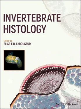
Cover Image: © (cephalopod) Francesco Martini, (histology of an eye) Damien Laudier
Ilze K. BerzinsOne Water, One Health, LLC, Golden Valley, MN, USA
Alvin C. CamusUniversity of Georgia College of Veterinary Medicine, Athens, GA, USA
John E. CooperWildlife Health Services, UK
Michelle M. DennisCenter for Conservation Medicine and Ecosystem Health Department of Biomedical Sciences Ross University School of Veterinary Medicine Basseterre, St Kitts and Nevis Department of Biomedical and Diagnostic Services University of Tennessee College of Veterinary Medicine Knoxville, TN, USA
Jennifer A. Dill-OkuboFlorida Department of Agriculture and Consumer Services, Kissimmee, FL, USA
Alexander EreskovskyInstitut Méditerranéen de Biodiversité et d’Ecologie Marine et Continentale (IMBE), Aix Marseille University, CNRS, IRD, Avignon University, Marseille, France Department of Embryology, Faculty of Biology, Saint-Petersburg State University, Saint-Petersburg, Russia Koltzov Institute of Developmental Biology, Russian Academy of Sciences, Moscow, Russia
Michael M. GarnerNorthwest ZooPath, Monroe, WA, USA
Benjamin KennedyVeterinary Invertebrate Society, Venomtech Ltd, Discovery Park, Sandwich, Kent, UK
György KriskaInstitute of Biology Eötvös Loránd University MTA Centre for Ecological Research, Danube Research Institute, Budapest, Hungary
Elise E.B. LaDouceurJoint Pathology Center, Silver Spring, MD, USA
Damien LaudierLaudier Histology, New York, NY, USA
Andrey LavrovDepartment of Embryology, Faculty of Biology, Saint-Petersburg State University, Saint-Petersburg Pertsov White Sea Biological Station, Biological Faculty, Lomonosov Moscow State University, Moscow, Russia
Péter LőwDepartment of Anatomy, Cell and Developmental Biology Eötvös Loránd University Budapest, Hungary
Kinga MolnárDepartment of Anatomy, Cell and Developmental Biology, Eötvös Loránd University, Budapest, Hungary
Alisa L. NewtonWildlife Conservation Society, Bronx, NY, USA Disney’s Animals, Science and Environment Orlando, FL, USA
Esther C. PetersEnvironmental Science and Policy, George Mason University, Fairfax, VA, USA
Katie J. RoordaJohns Hopkins University, Baltimore, MD, USA
Elemir SimkoWestern College of Veterinary Medicine, University of Saskatchewan, Saskatoon, Canada
Roxanna SmolowitzAquatic Diagnostic Laboratory Roger Williams University Bristol, RI, USA
Steven A. TrimVenomtech Ltd Discovery Park Sandwich, Kent, UK
Sarah C. WoodWestern College of Veterinary Medicine, University of Saskatchewan, Saskatoon, Canada
Roy P.E. YanongTropical Aquaculture Laboratory Fisheries and Aquatic Sciences Program School of Forest Resources and Conservation Institute of Food and Agricultural Sciences University of Florida, Ruskin, FL, USA
Veterinary medicine is a dynamic profession that began over 250 years ago to heal and protect working and warring equids along with livestock for food and other human‐use products. The profession has come a long way since the 1700s, most notably in the breadth of species embraced, and the information that exists and is being explored related to this taxonomic diversity. Increasing human population growth, commerce, technology, and animal welfare are all contributing to this expansion. Our profession is more diverse than ever, and a growing part of that diversity is the inclusion of over 97% of the animal kingdom: the invertebrates.
Dr LaDouceur and her internationally recognized contributors have assembled an organized, easy to navigate, comprehensive, and richly illustrated work focused on the microanatomy and histology of the invertebrates. It is certainly the only book of its kind on the market and one that is long overdue. The text is richly illustrated with beautiful images, drawings, and micrographs, detailing the normal gross and microscopic anatomy of the species covered. Chapters also describe how to properly and efficiently process invertebrate tissues for histology. This is critically important as standard vertebrate tissue‐processing methods frequently do not apply to invertebrates. Anatomic features like chitinous shells, glass spicules, calcium carbonate skeletons, and mesoglea, to name a few, may require specialized fixatives, processing, and staining techniques.
One of the biggest challenges for a clinician or pathologist is being able to recognize and become familiar with what is normal about an animal. This challenge is especially pertinent when dealing with nondomestic species. There is no greater or more diverse animal classification than the invertebrates, estimated to include over 1.3 million described species (and it's likely that the global total could be 10 times this number), representing at least 40 phyla. The editor and authors have wisely focused on the taxa that are the most economically important and/or in need of conservation, protection, and veterinary support. This includes species commonly displayed in zoos and aquaria, taxa that are utilized in the laboratory for research, and animals that are kept as pets.
This detailed and thorough text is a windfall for our profession and anyone working on the health and welfare of these animals. Pathologists, veterinary clinicians, histology technicians, invertebrate zoologists, and students studying in these areas will all find this book highly useful and important for their work. The timing for this book could not be better. I'm sure you, the reader, will agree with me, and find this one of the most important references on your bookshelf, in your laboratory, or digitally on your computer.
Gregory A. Lewbart
Raleigh, NC, USA
Alisa L. Newton1,2 and Michelle M. Dennis3,4
1 Wildlife Conservation Society, Bronx, NY, USA
2 Disney’s Animals, Science and Environment, Orlando, FL, USA
3 Center for Conservation Medicine and Ecosystem Health, Department of Biomedical Sciences, Ross University School of Veterinary Medicine, Basseterre, St Kitts and Nevis
4 Department of Biomedical and Diagnostic Sciences, University of Tennessee College of Veterinary Medicine, Knoxville, TN, USA
Phylum Echinodermata consists of three subphyla (Asterozoa, Echinozoa, and Crinozoa) and five main classes. Subphylum Asterozoa contains two extant classes: Asteroidea (sea stars, sea daisies) and Ophiuroidea (brittle and basket stars). Echinozoa contains two extant classes: Echinoidea (sea urchins, sand dollars) and Holothuroidea (sea cucumbers). Subphylum Crinozoa contains only one extant class: Crinoidea (feather stars, sea lilies). There are 7000 living species of echinoderms (Mulcrone 2005). All are marine and almost exclusively benthic. Some subphyla are mobile (Asterozoa, Echinozoa) and others are sessile (Crinozoa), though some sea lilies have been documented to swim significant distances. Echinoderms do not appear to have near relatives among other invertebrate phyla.
Читать дальше

