Seth Delpachitra - Principles of Dentoalveolar Extractions
Здесь есть возможность читать онлайн «Seth Delpachitra - Principles of Dentoalveolar Extractions» — ознакомительный отрывок электронной книги совершенно бесплатно, а после прочтения отрывка купить полную версию. В некоторых случаях можно слушать аудио, скачать через торрент в формате fb2 и присутствует краткое содержание. Жанр: unrecognised, на английском языке. Описание произведения, (предисловие) а так же отзывы посетителей доступны на портале библиотеки ЛибКат.
- Название:Principles of Dentoalveolar Extractions
- Автор:
- Жанр:
- Год:неизвестен
- ISBN:нет данных
- Рейтинг книги:3 / 5. Голосов: 1
-
Избранное:Добавить в избранное
- Отзывы:
-
Ваша оценка:
Principles of Dentoalveolar Extractions: краткое содержание, описание и аннотация
Предлагаем к чтению аннотацию, описание, краткое содержание или предисловие (зависит от того, что написал сам автор книги «Principles of Dentoalveolar Extractions»). Если вы не нашли необходимую информацию о книге — напишите в комментариях, мы постараемся отыскать её.
Focuses solely on dentoalveolar extractions and surgery Contains full colour photographs and detailed illustrations to aid learning Offers a technical walk-through guide that can be used as a reference prior to surgery Includes a Preface written by Professor Andrew A. C. Heggie, AM Written for dental students, surgical trainees and other dental professionals,
provides a comprehensive and pragmatic review of dentoalveolar extractions.
Principles of Dentoalveolar Extractions — читать онлайн ознакомительный отрывок
Ниже представлен текст книги, разбитый по страницам. Система сохранения места последней прочитанной страницы, позволяет с удобством читать онлайн бесплатно книгу «Principles of Dentoalveolar Extractions», без необходимости каждый раз заново искать на чём Вы остановились. Поставьте закладку, и сможете в любой момент перейти на страницу, на которой закончили чтение.
Интервал:
Закладка:
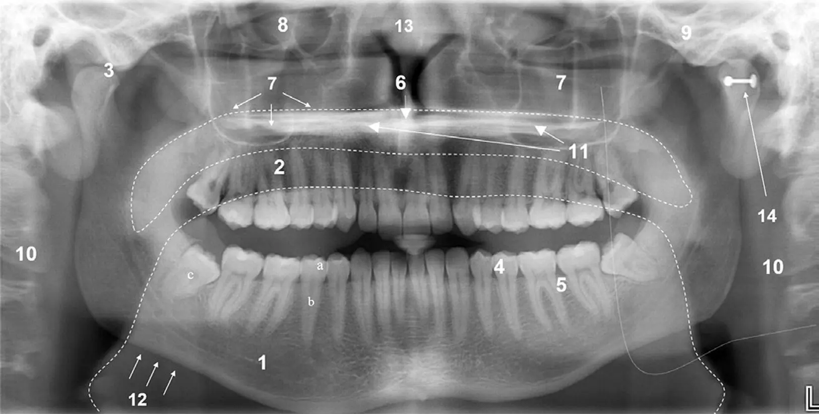
Figure 1.3 Panoramic radiograph: 1. mandible; 2. maxilla; 3. temporomandibular joint; 4. dentition; 5. alveolar process and periodontium; 6. anterior nasal spine; 7. maxillary antrum; 8. orbit; 9. zygoma; 10. cervical spine (double image); 11. hard palate (double image); 12. lower border of mandible; 13. nasal septum; 14. earring causing artifact. Outlines show oro‐ and nasopharyngeal airspaces and double image of mandibular ramus (left side).
Source: Reproduced from The panoramic dental radiograph for emergency physicians by Anton Sklavos, Daniel Beteramia, Seth Navinda Delpachitra, Ricky Kumar, BMJ 36: 565–571. doi:10.1136/emermed‐2018‐208332. Copyright © 2019 with permission from BMJ Publishing Group Ltd.
A procedure should not be undertaken without a diagnostic radiograph, displaying the condition of the tooth, the relevant adjacent structures (inferior alveolar nerve canal, mental foramen, maxillary antrum), and the condition of the adjacent teeth. Generally, a PA radiograph has a limited application and should only be used for emergency single‐tooth extractions or procedures limited to one part of the mouth performed under local anaesthesia. A PA radiograph is not considered an appropriate assessment for proximity of the inferior alveolar canal to the lower third molar teeth; errors in patient or film positioning may alter the radiographic relationship between the canal and associated tooth roots, producing a nondiagnostic image.
The OPG gives a better indication of the overall state of the patient's dentition and of other pathological conditions that may affect the maxillofacial region compared to a PA radiograph. Because the OPG is taken in a standardised manner, it has formed the basis for a number of evidence‐based risk‐assessment tools in the current literature, including assessments of the risk of oroantral communication and inferior alveolar nerve injury.
CBCT is a relatively new and inexpensive method of producing 3D images of maxillomandibular structures ( Figure 1.4). It is indicated when conventional 2D imaging does not produce enough diagnostic information for treatment planning. Most commonly, this is to investigate jaw pathology or the relationship of the inferior alveolar nerve canal to third molar teeth. Whilst CBCT images come in a variety of formats, the serial‐transaxial reformat is useful for the determination of the pathway of the inferior alveolar nerve and its relationship to the mandibular teeth.
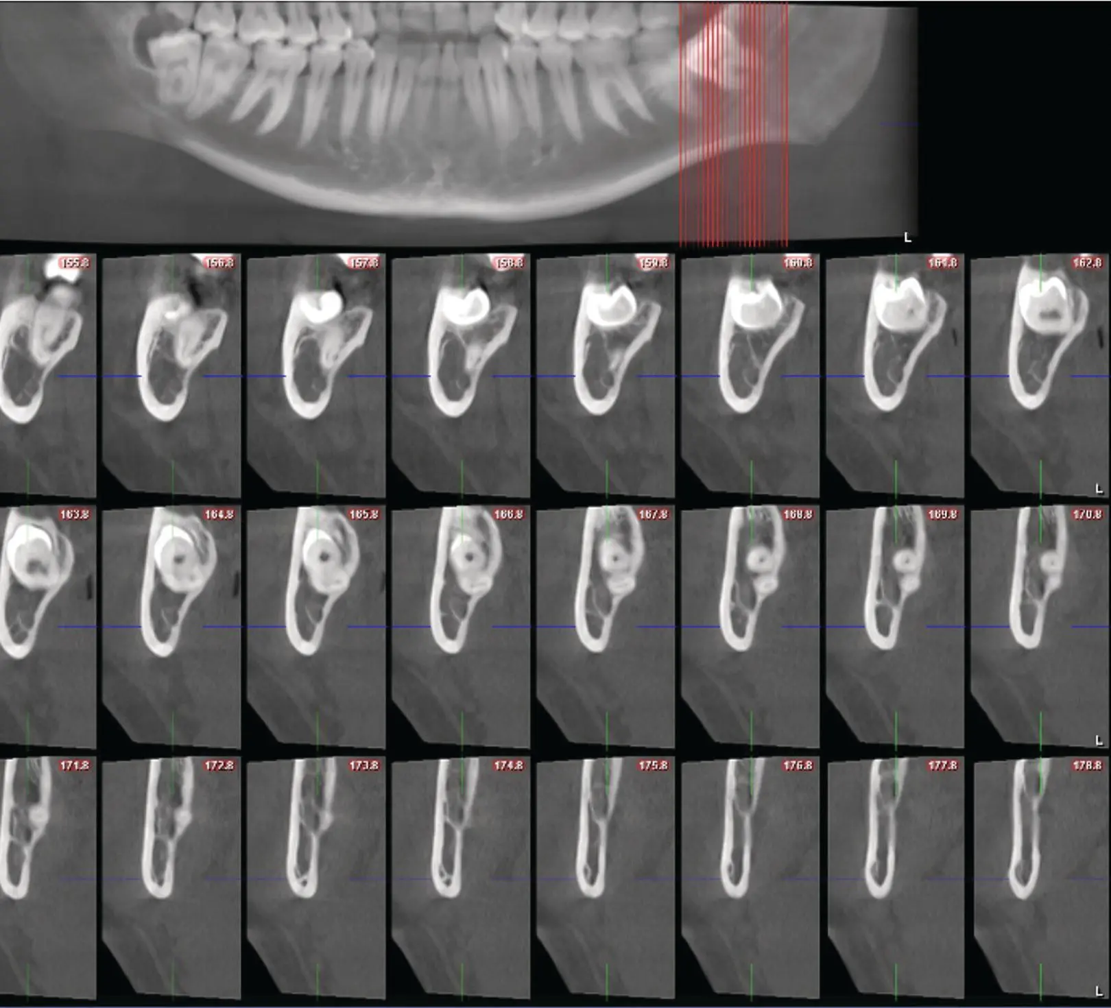
Figure 1.4 Slices of a serial transaxial CBCT.
CBCT images provide a great deal more information than plain‐film imaging, and therefore their interpretation can be challenging. For the surgeon who is not experienced with CBCT images, formal reporting by a specialist radiologist is essential; if they are not interpreted carefully, radiographic signs of nerve risk or bone pathology may be easily missed, likely resulting in a poor outcome for the patient and a litigious scenario for the surgeon.
1.4 Informed Consent
In order to carry out any medical or dental procedure, consent must be obtained from either the patient or their legal guardian. A valid consent may only be given when the patient possesses decision‐making capacity; that is, when they have the ability to weigh up the pros and cons of treatment and come to a sound decision to either undergo or forgo a proposed procedure. This consent must be given voluntarily, without duress or coercion. The decision must be informed, which is to say that the patient must understand the procedure, the expected outcomes, the risks involved, the anticipated recovery time, and the costs of treatment.
For any planned surgical procedure, consent should be both written and verbal, and patients should be provided by the surgeon with a formal document detailing the procedure and its indications, risks, and expected outcomes ( Figure 1.5). When seeking consent from a patient, the surgeon must always provide them sufficient time to ask questions. The patient may request more detailed information about particular risks or outcomes of surgery, and these should be elaborated on and explained in detail.
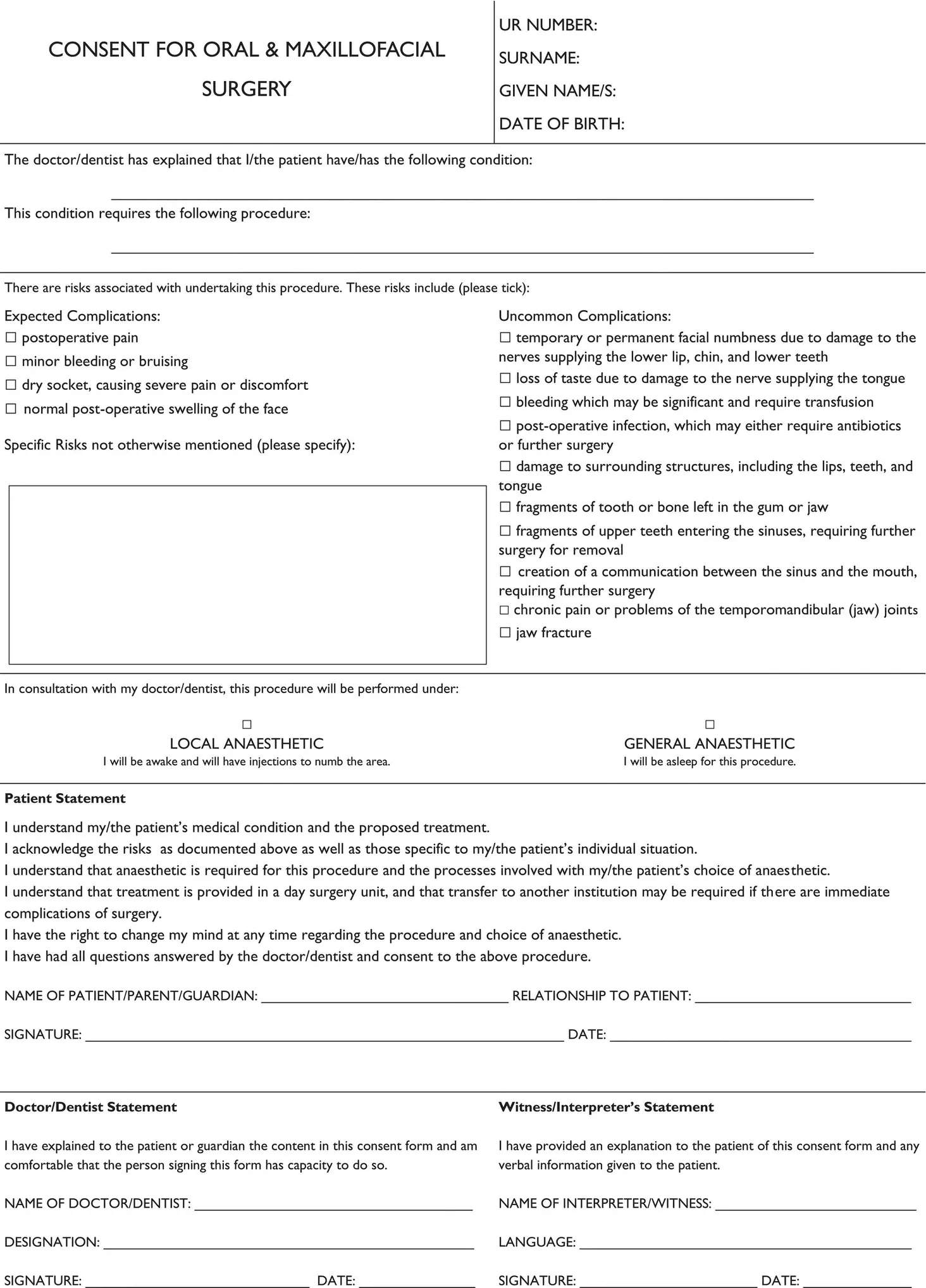
Figure 1.5 Example consent form for dentoalveolar surgery.
In addition, the consent should be specific for the particular patient and their individual circumstances. Alternatives to dentoalveolar extraction should be explained, and the reasons for and against their adoption detailed. In cases where extractions will render the patient without functional teeth, future treatment options for replacement should be discussed; this should include an approximate timeline, who will provide the treatment, and a rough cost estimate.
1.5 Anaesthesia
There are a number of different methods of anaesthesia available for dentoalveolar extractions. These include:
Local anaesthesia only.
Local anaesthesia with relative analgesia.
Local anaesthesia with minor oral sedation.
Local anaesthesia with intravenous (IV) sedation.
Local anaesthesia with general anaesthesia.
Local anaesthesia alone can be administered for in‐chair treatment, and is generally considered the safest option. It is appropriate for most simple dentoalveolar procedures and extractions. The main limitation of local anaesthesia is that it may be unsuitable for complex procedures, when a duration of more than 40 minutes is anticipated, or when the patient is anxious or otherwise uncooperative.
Relative analgesia involves the use of inhaled agents, such as nitrous oxide, to produce conscious sedation. It can be used as an adjunct with local anaesthesia, to improve patient comfort without significant airway risk. Nitrous oxide is usually administered through a nasal hood at concentrations of 50–70%. The advantage of this method is that it can be titrated and rapidly adjusted. However, there is a variable dose–response relationship between individuals, and patients may experience a number of unpleasant side effects if too high a dose is administered. Use of relative analgesia requires additional training, and it is recommended that at least two trained personnel are present in the clinic room when employing this technique.
Oral sedation, when employed effectively, can provide a greater level of sedation than nitrous oxide. Common drug classes used for oral sedation include benzodiazepine and barbiturate medications ( Table 1.1). These drugs have a significant depressant effect on the central nervous system, and hence carry the serious risk of respiratory depression and loss of airway reflexes. Therefore, the use of oral sedation should only be considered when the surgeon and clinic personnel are sufficiently trained in anaesthesia, resuscitation, and airway management.
Table 1.1 Comparisons between commonly used oral benzodiazepine sedatives.
| Drug name | Dose | Onset |
|---|---|---|
| Temazepam | 10–20 mg | 30–120 minutes |
| Diazepam | 10–15 mg | 30–90 minutes |
| Oxazepam | 15–30 mg | 2–3 hours |
IV sedation typically involves more powerful sedative agents, such as midazolam, propofol, or fentanyl. It can only be administered in a setting which is fully prepared for airway management, such as a hospital theatre. As with all methods of anaesthesia where the airway is not secure, there is a risk that the patient will aspirate on any foreign object present in their mouth. This can be particularly dangerous when their reflexes are not protecting the airway. The administration of IV sedation may only be undertaken by a suitably qualified medical specialist or dental specialist with advanced training.
Читать дальшеИнтервал:
Закладка:
Похожие книги на «Principles of Dentoalveolar Extractions»
Представляем Вашему вниманию похожие книги на «Principles of Dentoalveolar Extractions» списком для выбора. Мы отобрали схожую по названию и смыслу литературу в надежде предоставить читателям больше вариантов отыскать новые, интересные, ещё непрочитанные произведения.


Обсуждение, отзывы о книге «Principles of Dentoalveolar Extractions» и просто собственные мнения читателей. Оставьте ваши комментарии, напишите, что Вы думаете о произведении, его смысле или главных героях. Укажите что конкретно понравилось, а что нет, и почему Вы так считаете.