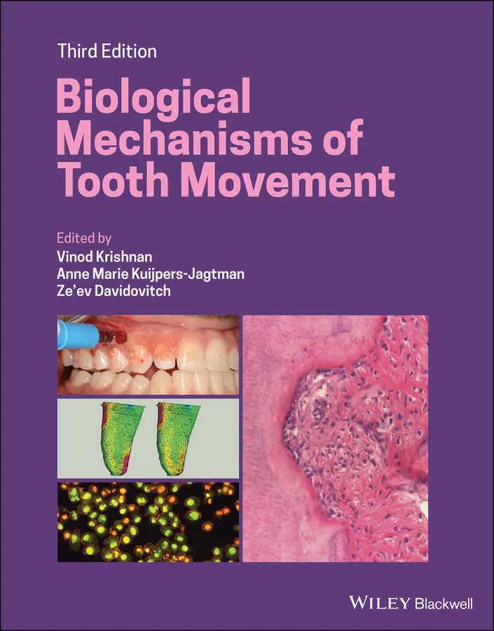
Biological Mechanisms of Tooth Movement
Здесь есть возможность читать онлайн «Biological Mechanisms of Tooth Movement» — ознакомительный отрывок электронной книги совершенно бесплатно, а после прочтения отрывка купить полную версию. В некоторых случаях можно слушать аудио, скачать через торрент в формате fb2 и присутствует краткое содержание. Жанр: unrecognised, на английском языке. Описание произведения, (предисловие) а так же отзывы посетителей доступны на портале библиотеки ЛибКат.
- Название:Biological Mechanisms of Tooth Movement
- Автор:
- Жанр:
- Год:неизвестен
- ISBN:нет данных
- Рейтинг книги:5 / 5. Голосов: 1
-
Избранное:Добавить в избранное
- Отзывы:
-
Ваша оценка:
Biological Mechanisms of Tooth Movement: краткое содержание, описание и аннотация
Предлагаем к чтению аннотацию, описание, краткое содержание или предисловие (зависит от того, что написал сам автор книги «Biological Mechanisms of Tooth Movement»). Если вы не нашли необходимую информацию о книге — напишите в комментариях, мы постараемся отыскать её.
delivers a comprehensive reference for orthodontic trainees and specialists.
It is fully updated to include new chapters on personalized orthodontics as well as the inflammatory process occurring in the dental and paradental tissues. It is heavily illustrated throughout, making it easier for readers to understand and retain the information discussed within. The topics covered range from bone biology, the effects of mechanical loading on tissues and cells, genetics, tissue remodeling, and the effects of diet, drugs, and systemic diseases.
The Third Edition of
features seven sections that cover subjects such as:
The development of biological concepts in orthodontics, including the cellular and molecular biology behind orthodontic tooth movement Mechanics meets biology, including the effects of mechanical loading on hard and soft tissues and cells, and biological reactions to temporary anchorage devices Inflammation and orthodontics, including markers for tissue remodeling in the gingival crevicular fluid and saliva Personalized diagnosis and treatment based on genomic criteria, including the genetic influences on orthodontic tooth movement Rapid orthodontics, including methods to accelerate or decelerate orthodontic tooth movement Perfect for residents and PhD students of orthodontic and periodontal programs,
is also useful to academics, clinicians, bone biologists, and researchers with an interest in the mechanics and biology of tooth movement.
Biological Mechanisms of Tooth Movement — читать онлайн ознакомительный отрывок
Ниже представлен текст книги, разбитый по страницам. Система сохранения места последней прочитанной страницы, позволяет с удобством читать онлайн бесплатно книгу «Biological Mechanisms of Tooth Movement», без необходимости каждый раз заново искать на чём Вы остановились. Поставьте закладку, и сможете в любой момент перейти на страницу, на которой закончили чтение.
Интервал:
Закладка:
Pain during OTM
OTM causes inflammatory reactions in the periodontium and dental pulp, which will stimulate release of various biochemical mediators. The perception of orthodontic pain is the result of a hyperalgesic response elicited by these mediators. Periodontal pain is caused by a process involving the development of pressure, ischemia, inflammation, and edema. Burstone (1964) identified both immediate and delayed pain responses, which begin a few hours after the application of an orthodontic force and last for approximately 5 days (Scheurer et al ., 1996). Krukemeyer et al . (2009) concluded from a survey conducted on 118 patients that 58.5% indicated that they experienced pain for a few days after their appointment, out of which only 26.5% of the patients used pain medication immediately following and 1 day after the last appointment.
Table 4.3Inflammatory responses in dental pulp in response to orthodontic force application.
| In vitro studies (stimulated by substance P) | In vivo studies |
|---|---|
| Prostaglandin E 2(PGE 2) | CGRP and SP |
| IL‐1α and β | Blood flow |
| IL‐6 | Apoptosis |
| TNF‐α | Angiogenesis |
| RANKL | Aspartate amino transferase activity |
Painful sensations result, in part, from the stretching and distortion of tissues by the mechanical loads (Stephenson, 1992), as well as from interactions of multiple inflammatory mediators with local pain receptors (Davies and MacIntyre, 1992). According to a review by Krishnan (2007), the perception of orthodontic pain is due to the changes in blood flow caused by the appliances, correlated with the release and presence of various substances, such as SP, histamine, enkephalin, dopamine, serotonin, glycine, glutamate, gamma‐aminobutyric acid, PGs, leukotrienes, and cytokines (Yamasaki et al ., 1984; Davidovitch et al ., 1988; Davidovitch, 1991; Alhashimi et al ., 2001).
Processing of complex information arising from mechanical force application induces recruitment of neurons, which act by way of chemical mediators as modulators of the effector response to the stimulus. The neurogenic inflammation, which evolves following activation of an orthodontic appliance, starts with the release of neuropeptides from strained afferent nerve endings in and around the treated teeth, as well as in the central nervous system. The simultaneous release of neuropeptides, peripherally and centrally, elicits a painful response (Vandevska‐Radunovic, 1999). Kato et al . (1996) examined the distribution of nerve fibers containing NFP, CGRP, VIP, and NPY in the PDL of the rat first molar after mechanical force application. They observed an increase in the number of NFP‐ and CGRP‐containing nerve fibers at both the stretched and the compressed sides of the PDL after 3 days of force application, which returned to normal after 14 days. They concluded that NFP‐, CGRP‐, VIP‐, and NPY‐containing nerve fibers play an important role in blood flow regulation, tissue remodeling, and modulation of pain perception during tooth movement. This finding was in accordance with earlier reports. Norevall et al . (1995) also agreed about the role of CGRP and SP in tooth movement but contradicted the role of other neuropeptides such as VIP and NPY.
It is imperative that pain control be considered an important aspect of orthodontic mechanotherapy, and that the administration of nonsteroidal anti‐inflammatory drugs (NSAIDs) remains the preferred method for pain control during orthodontic treatment. NSAIDs have been used for the relief of orthodontic pain for decades (Krishnan and Davidovitch, 2006b; Angelopoulou et al ., 2012). It has been well documented that the synthesis of prostaglandin is mediated by COX enzymes and NSAIDs inhibit the activity of COX enzymes. Therefore, NSAIDs could relieve orthodontic pain by inhibiting the release of prostaglandin (Shenoy et al ., 2013). The major concern regarding NSAIDs is the interference with the inflammatory process associated with tooth movement. NSAIDs intake may induce a decrease in levels of prostaglandin, and as a result may inhibit osteoclasts and reduce the rate of tooth movement (Arias and Marquez‐Orozco, 2006). Therefore, their effects on the rate of tooth movement need to be validated in future studies (Long et al ., 2016).
Currently, several treatment modalities have been applied for the relief of orthodontic pain, such as mechanical and behavioural approaches, and low‐level laser therapy (Long et al ., 2016). Mechanical approaches have been proposed to relieve orthodontic pain including vibration, chewing gums, biting wafers, and acupuncture. The proposed mechanism for vibration, chewing gum, and biting wafers lies in the fact that mechanical stimuli activate mechanoreceptors that transmit tactile signals while suppressing the transmission of painful signals (Guyton and Hall, 2000). However, its mechanism remains large unknown.
Behavioural approaches that are applied to relieve orthodontic pain include cognitive behavioral therapy (CBT), physical activity, and music therapy. CBT, a form of psychotherapy, uses several treatment sessions to correct the patient’s negative attitude and decrease their anxiety. As a result, orthodontic pain is relieved in clinical practice (Wang et al., 2015). Music therapy and physical activity distract patient’s attention via the insular cortex‐mediated neural pathway (Xu et al ., 2013; Sandhu and Sandhu 2015). However, the mechanisms are not convincing.
Low‐level laser therapy has been extensively applied for pain relief in both medical and dental practice (Huang et al., 2015; Landucci et al ., 2016). A large body of evidence has confirmed the effectiveness of low‐level laser therapy in alleviating orthodontic pain (Tortamano et al ., 2009; Artés‐Ribas et al ., 2013; Eslamian et al ., 2014). PGE 2and IL‐1β production in stretched human PDL cells was inhibited by laser irradiation in vitro (Shimizu et al ., 1995). Therefore, it is concluded that low‐energy laser irradiation may be useful in reducing the levels of inflammation and pain, as well as decreasing orthodontic treatment time by accelerating the pace of tooth movement, without causing any side effects.
Root resorption and inflammation
Many orthodontists consider external apical root resorption (EARR) to be an unavoidable pathologic consequence of OTM. However, an opposing view is presented in Chapter 17, which describes means to avoid this destructive side effect of orthodontic treatment altogether. The common approach to EARR is to visualize it as an iatrogenic disorder that occurs, unpredictably, after orthodontic treatment, whereby the resorbed apical root portion is replaced with normal bone. This undesirable side effect is described as being the outcome of a sterile, complex inflammatory process that involves various disparate components including mechanical forces, tooth roots, bone, cells, surrounding matrix, and certain known biological messengers (Brezniak and Wasserstein, 2002). Killiany (1999) reported that EARR of >3 mm occurs at a frequency of 30% of a patient population, while 5% of treated individuals were found to have >5 mm of root resorption. Harris et al . (1997, 2001) reported that the sum of the effects of the patients’ sex, age, severity of the malocclusion, and the kind of mechanics used accounts for little of the overall variation in EARR. Orthodontic force applications induce a local process that includes all of the characteristics of inflammation (redness, heat, swelling, pain, and reduced function). This inflammation, which is an essential feature of tooth movement, is actually the fundamental component behind the root‐resorption process (Bosshardt et al ., 1998).
Читать дальшеИнтервал:
Закладка:
Похожие книги на «Biological Mechanisms of Tooth Movement»
Представляем Вашему вниманию похожие книги на «Biological Mechanisms of Tooth Movement» списком для выбора. Мы отобрали схожую по названию и смыслу литературу в надежде предоставить читателям больше вариантов отыскать новые, интересные, ещё непрочитанные произведения.


Обсуждение, отзывы о книге «Biological Mechanisms of Tooth Movement» и просто собственные мнения читателей. Оставьте ваши комментарии, напишите, что Вы думаете о произведении, его смысле или главных героях. Укажите что конкретно понравилось, а что нет, и почему Вы так считаете.