Pascal Ribéreau-Gayon - Handbook of Enology - Volume 1
Здесь есть возможность читать онлайн «Pascal Ribéreau-Gayon - Handbook of Enology - Volume 1» — ознакомительный отрывок электронной книги совершенно бесплатно, а после прочтения отрывка купить полную версию. В некоторых случаях можно слушать аудио, скачать через торрент в формате fb2 и присутствует краткое содержание. Жанр: unrecognised, на английском языке. Описание произведения, (предисловие) а так же отзывы посетителей доступны на портале библиотеки ЛибКат.
- Название:Handbook of Enology: Volume 1
- Автор:
- Жанр:
- Год:неизвестен
- ISBN:нет данных
- Рейтинг книги:4 / 5. Голосов: 1
-
Избранное:Добавить в избранное
- Отзывы:
-
Ваша оценка:
Handbook of Enology: Volume 1: краткое содержание, описание и аннотация
Предлагаем к чтению аннотацию, описание, краткое содержание или предисловие (зависит от того, что написал сам автор книги «Handbook of Enology: Volume 1»). Если вы не нашли необходимую информацию о книге — напишите в комментариях, мы постараемся отыскать её.
is thus the same. It aims to provide practitioners, winemakers, technicians and enology students with foundational knowledge and the most recent research results. This knowledge can be used to contribute to a better definition of the quality of grapes and wine, a greater understanding of chemical and microbiological parameters, with the aim of ensuring satisfactory fermentations and predicting the evolution of wines, an7thd better mastery of wine stabilization processes. As a result, the purpose of this publication is to guide readers in their thought processes with a view to preserving and optimizing the identity and taste of wine and its aging potential.
This third English edition of
, is an enhanced translation from the 7h French 2017 edition, and is published in print as individual themed volumes and as a two-volume set, describing aspects of winemaking using a detailed, scientific approach. The authors, who are highly-respected enologists, examine winemaking processes, theorizing what constitutes a perfect technique and the proper combination of components necessary to produce a quality vintage. They also illustrate methodologies of common problems, revealing the mechanism behind the disorder, thus enabling a diagnosis and solution.
Volume 1:
The Microbiology of
Wine and Vinifications
Coverage includes: Wine microbiology; Yeasts; Yeast metabolism; The conditions for the development of yeasts; Lactic acid bacteria, their metabolism and their development in wine; Acetic bacteria; The use of sulfur dioxide in the treatment of musts and wines; Products and processes acting in addition to sulfur dioxide; Winemaking; The grape and its maturation; Harvesting and processing of grapes after harvest; Vinification in red and white wine making.
The target audience includes advanced viticulture and enology students, professors and researchers, and practicing grape growers and vintners.
Handbook of Enology: Volume 1 — читать онлайн ознакомительный отрывок
Ниже представлен текст книги, разбитый по страницам. Система сохранения места последней прочитанной страницы, позволяет с удобством читать онлайн бесплатно книгу «Handbook of Enology: Volume 1», без необходимости каждый раз заново искать на чём Вы остановились. Поставьте закладку, и сможете в любой момент перейти на страницу, на которой закончили чтение.
Интервал:
Закладка:
Several enzymesare connected to the cell wall or situated in the periplasmic space. One of the most characteristic is invertase or β ‐fructofuranosidase. This enzyme catalyzes the hydrolysis of sucrose into glucose and fructose. It is a thermostable mannoprotein anchored to a β ‐1,6‐glucan of the cell wall. Its molecular weight is 270,000 Da. It contains approximately 50% mannose and 50% protein. Periplasmic acid phosphatase is also a mannoprotein.
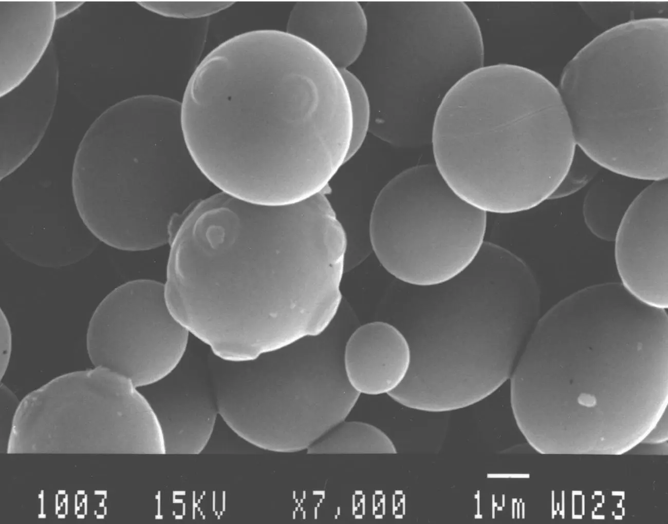
FIGURE 1.3 Scanning electron microscope photograph of proliferating S. cerevisiae cells. The budding scars on the mother cells can be observed.
( Source: Photograph from M. Mercier, Department of Electron Microscopy, Université de Bordeaux I.)
Other periplasmic enzymes that have been noted are β ‐glucosidase, α ‐galactosidase, melibiase, trehalase, aminopeptidase, and esterase. Yeast cell walls also contain endo‐ and exo‐ β ‐glucanases ( β ‐1,3 and β ‐1,6). These enzymes are involved in the reshaping of the cell wall during the growth and budding of cells. Their activity is at a maximum during the exponential growth phase of the population and diminishes notably afterward. However, cells in the stationary phase and even dead yeasts contained in the lees still retain β ‐glucanase activity in their cell walls several months after the completion of fermentation. These endogenous enzymes are involved in the autolysis of the cell wall during the lees aging of wines. This aging method will be covered in the Chapter 13.
1.2.3 General Organization of the Cell Wall and Factors Affecting Its Composition
The cell wall of S. cerevisiae is made up of an outer layer of mannoproteins. These mannoproteins are connected to a matrix of amorphous β ‐1,3‐glucan, which covers an inner layer of fibrous β ‐1,3‐glucan. The inner layer is connected to a small quantity of chitin ( Figure 1.4). β ‐1,6‐glucan probably acts as a cement between the two layers. The rigidity and the shape of the cell wall are due to the internal framework of the fibrous β ‐1,3‐glucan. Its elasticity is due to the outer amorphous layer. The intermolecular structure of the mannoproteins of the outer layer (hydrophobic bonds and disulfide bonds) equally determines cell wall porosity for micromolecules (molecular weights less than 4,500) and impermeability to macromolecules. This impermeability can be affected by treating the cell wall with certain chemical agents, such as β ‐mercaptoethanol. This substance breaks the disulfide bonds, thus destroying the intermolecular network between the mannoprotein chains.
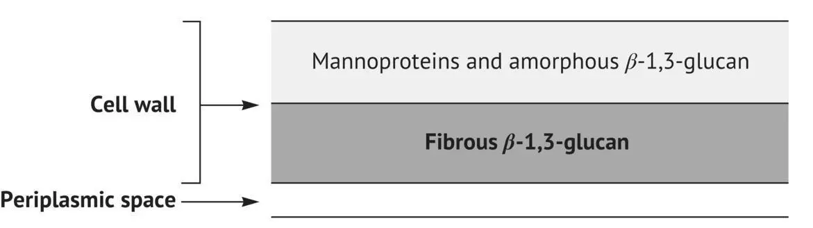
FIGURE 1.4 Cellular organization of the cell wall of S. cerevisiae .
The composition of the cell wall is strongly influenced by nutritive conditions and cell age. The proportion of glucan in the cell wall increases with respect to the amount of sugar in the culture medium. Certain deficiencies (for example, of mesoinositol) also result in an increase in the proportion of glucan compared with mannoproteins. The cell walls of older cells are richer in glucans and in chitin and less rich in mannoproteins than younger ones. For this reason, they are more resistant to physical and enzymatic agents used to break them down. Finally, the composition of cell walls is profoundly modified by morphogenetic alterations (conjugation and sporulation).
1.3 The Plasma Membrane
1.3.1 Chemical Composition and Organization
The plasma membrane is a highly selective barrier controlling exchanges between the living cell and its external environment. This organelle is essential to the life of the yeast.
Like all biological membranes, the yeast plasma membrane is principally made up of lipids and proteins. The plasma membrane of S. cerevisiae contains about 40% lipids and 50% proteins. Glucans and mannans are only present in small quantities (a few percent).
The lipids of the membrane are essentially phospholipids and sterols. They are amphiphilic molecules, i.e. possessing a hydrophilic and a hydrophobic part.
The three principal phospholipids ( Figure 1.5) of the plasma membrane of yeast are phosphatidylethanolamine (PE), phosphatidylcholine (PC), and PI, which represent 70–85% of the total. Phosphatidylserine (PS) and diphosphatidylglycerol or cardiolipin (PG) are less prevalent. Free fatty acids and phosphatidic acid are frequently reported in plasma membrane analysis. They are probably extraction artifacts caused by the activity of certain lipid degradation enzymes.
The fatty acids of the membrane phospholipids contain an even number (14–24) of carbon atoms. The most abundant are C16 and C18 acids. They can be saturated, such as palmitic acid (C16) and stearic acid (C18), or unsaturated, as with oleic acid (C18, double bond in position 9), linoleic acid (C18, two double bonds in positions 9 and 12), and linolenic acid (C18, three double bonds in positions 9, 12, and 15). All membrane phospholipids share a common characteristic: they possess a polar or hydrophilic part made up of a phosphorylated alcohol and a nonpolar or hydrophobic part comprising two more‐or‐less parallel fatty acid chains. Their symbolic representation is shown in Figure 1.6. In an aqueous medium, the phospholipids spontaneously form bimolecular films or a lipid bilayer because of their amphiphilic nature. The lipid bilayers are cooperative but non‐covalent structures. They are maintained in place by mutually reinforced interactions: hydrophobic interactions and van der Waals forces between the hydrocarbon tails, and hydrostatic interactions and hydrogen bonds between the polar heads and water molecules. The examination of cross‐sections of yeast plasma membranes under an electron microscope reveals a classic lipid bilayer structure with a thickness of about 7.5 nm. The membrane surface appears sculpted with creases, especially during the stationary phase. However, the physiological meaning of this anatomical characteristic remains unknown. The plasma membrane also has a depression under the bud scar.
Ergosterol is the primary sterol of the yeast plasma membrane. In addition, 24(28)‐dehydroergosterol and lesser amounts of zymosterol are present ( Figure 1.7). Sterols are exclusively produced in the mitochondria under aerobic conditions during the yeast log phase. As with phospholipids, membrane sterols are amphipathic. The hydrophilic part is composed of the hydroxyl group in the C3 position, while the rest of the molecule is hydrophobic, especially the flexible hydrocarbon tail.
The plasma membrane also contains numerous proteins or glycoproteins presenting a wide range of molecular weights (from 10,000 to 120,000). The available information indicates that the organization of the plasma membrane of a yeast cell resembles the fluid mosaic model. This model, proposed for biological membranes by Singer and Nicolson (1972), consists of two‐dimensional solutions of proteins and oriented lipids. Certain proteins penetrate the membrane; they are called integral proteins ( Figure 1.6). They interact strongly with the nonpolar part of the lipid bilayer. The peripheral proteins are linked to the integral ones by hydrogen bonds. Their location is asymmetrical, at either the inner or the outer side of the plasma membrane. The molecules of proteins and membrane lipids, constantly in lateral motion, are capable of rapidly diffusing in the membrane.
Читать дальшеИнтервал:
Закладка:
Похожие книги на «Handbook of Enology: Volume 1»
Представляем Вашему вниманию похожие книги на «Handbook of Enology: Volume 1» списком для выбора. Мы отобрали схожую по названию и смыслу литературу в надежде предоставить читателям больше вариантов отыскать новые, интересные, ещё непрочитанные произведения.


Обсуждение, отзывы о книге «Handbook of Enology: Volume 1» и просто собственные мнения читателей. Оставьте ваши комментарии, напишите, что Вы думаете о произведении, его смысле или главных героях. Укажите что конкретно понравилось, а что нет, и почему Вы так считаете.