Emergency Management of the Hi-Tech Patient in Acute and Critical Care
Здесь есть возможность читать онлайн «Emergency Management of the Hi-Tech Patient in Acute and Critical Care» — ознакомительный отрывок электронной книги совершенно бесплатно, а после прочтения отрывка купить полную версию. В некоторых случаях можно слушать аудио, скачать через торрент в формате fb2 и присутствует краткое содержание. Жанр: unrecognised, на английском языке. Описание произведения, (предисловие) а так же отзывы посетителей доступны на портале библиотеки ЛибКат.
- Название:Emergency Management of the Hi-Tech Patient in Acute and Critical Care
- Автор:
- Жанр:
- Год:неизвестен
- ISBN:нет данных
- Рейтинг книги:3 / 5. Голосов: 1
-
Избранное:Добавить в избранное
- Отзывы:
-
Ваша оценка:
Emergency Management of the Hi-Tech Patient in Acute and Critical Care: краткое содержание, описание и аннотация
Предлагаем к чтению аннотацию, описание, краткое содержание или предисловие (зависит от того, что написал сам автор книги «Emergency Management of the Hi-Tech Patient in Acute and Critical Care»). Если вы не нашли необходимую информацию о книге — напишите в комментариях, мы постараемся отыскать её.
Provides algorithms for the most common clinical scenarios of device malfunction and related complications Covers management of patients who have undergone major operations such as organ transplantation or complex congenital heart disease repair Presents detailed management plans for a wide range of hardware types and medical conditions Offers expert guidance to practitioners in settings where not all specialties are readily available, such as rural and remote areas or community hospitals Features contributions from a team of experts in various areas of adult and pediatric emergency and critical care medicine
is a must-have clinical reference and guide for pediatric and adult emergency medicine physicians, general pediatricians, internists, general practitioners, critical care specialists, and allied health practitioners.
Emergency Management of the Hi-Tech Patient in Acute and Critical Care — читать онлайн ознакомительный отрывок
Ниже представлен текст книги, разбитый по страницам. Система сохранения места последней прочитанной страницы, позволяет с удобством читать онлайн бесплатно книгу «Emergency Management of the Hi-Tech Patient in Acute and Critical Care», без необходимости каждый раз заново искать на чём Вы остановились. Поставьте закладку, и сможете в любой момент перейти на страницу, на которой закончили чтение.
Интервал:
Закладка:
Buried Bumper Syndrome
Buried bumper syndrome (BBS) is a rare but life‐threatening complication of children with G‐tubes. BBS is defined as the presence of an embedded internal fixation device into the gastric mucosa of the abdominal wall. This is typically caused by securing the external retention device too tightly to the skin surface and thus narrowing the space between the internal and external retention devices and pressing the internal device into the gastric mucosa. Rates of BBS in adults average 1% but can be as high as 5% in pediatric patients. This is seen with both internal balloons and rigid retention devices, but it is more common with the latter. Risk factors for BBS include pediatric age, jejunal extension from the G‐tube, multiple G‐tube placements, and improper home care. Pediatric patients are at a higher risk because of their expected weight gain and compression against the external bolster. Those with GJ tubes are at higher risk because the weight of jejunal extensions is thought to pull the internal bolsters out of their perpendicular placement and cause unequal pressure on the stomach wall. Finally, repeat G‐tube replacements can increase the inflammatory reaction in the stomach wall and thus encourage tissue growth around the internal retention device.
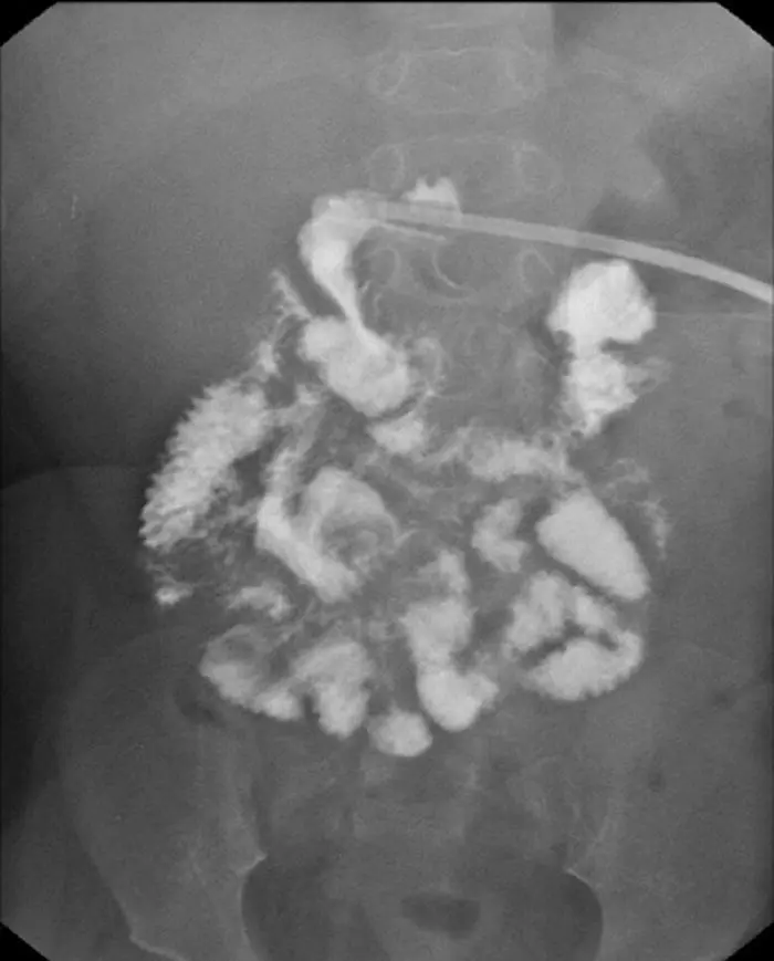
Figure 1.5 Radiograph of a G‐tube dye study shows dye within the small intestine only. This image is consistent with a gastric outlet obstruction whereby the balloon is located in the pylorus blocking dye from filling the stomach.
Patients can be asymptomatic and simply present with inability to feed through the tube. The classic triad for BBS is inability to insert the G‐tube further into the stomach, loss of tube patency (unable to feed or draw back from tubing), and leakage around the tube site. BBS can be complicated by GI bleeding, perforation, and peritonitis, which can be fatal.
BBS is diagnosed by endoscopy. However, abdominal ultrasound and computerized tomography (CT) scan can help identify bumper location if it is not apparent on endoscopy. Depending on the extent of the internal bumper's migration through the gastric mucosa, the bumper may be removed either endoscopically or surgically. Bumpers that have passed through the lamina muscularis propria and are located between the stomach and abdominal wall will need surgical removal.
Intussusception
Intussusception is a well‐described complication of patients with jejunal feeding devices (NJ, GJ, and J‐tubes). Intussusception is defined as one part of the small intestine invaginating or folding into the adjacent portion of small bowel. Complications arise from the pressure placed on the outer layer of small bowel tissue as the inner layer presses against it, thereby decreasing blood flow to the tissue. The pathogenesis of intussusception requires a lead point to pull one section of small bowel into the other. Contrary to classic intussusception where the lead point is either gastric lymphatic tissue or cancerous material, in patients with an enteric feeding device, the extension tubing in the jejunum serves as the lead point.
Patients with intussusception typically present with abdominal pain, bilious emesis, and/or hematemesis. Because of the many comorbidities of patients with enteric feeding devices, the patient may appear asymptomatic. One must have a heightened clinical suspicion. Diagnosis is made by contrast‐enhanced radiography, ultrasound, endoscopy, upper GI, or abdominal CT scan. Tube‐related intussusceptions resolve with tube removal.
Colocutaneous Fistula
Colocutaenous fistula formation is a complication only seen with the percutaneous approaches to gastrostomy. A colocuteneous fistula is caused by trapping a loop of bowel between the abdominal wall and the stomach wall and piercing the G‐tube through all three tissue layers. While, in some cases, patients present with colonic obstruction, this complication may not be detected until the first tube change at which point the tube is replaced into the colonic wall but does not make it to the stomach wall. The feeds are started directly into the colon, and the patient develops diarrhea and dehydration. Treatment includes removing the G‐tube and surgical closure of the fistula.
Consultation
Surgical consultation is needed for surgical emergencies: intussusception, BBS, colocutaneous fistula, peritonitis, and necrotizing fasciitis. Immature tube dislodgement will require replacement by the team responsible for its initial placement, but the emergency department team can initially manage all mature tracts. Consultation is needed if there is significant trauma to the tract, the tube is improperly positioned on dye study, or the patient is unable to tolerate feeds following tube replacement. GJ and J‐tube replacements will typically need interventional radiology consultation. Stomal site bleeding, leakage, or infection may be initially managed by the emergency department and seen in subspecialty clinic for further care. Similarly, gastric outlet obstruction can first be treated with tube repositioning by the emergency department team, but if the obstruction does not resolve, surgical consultation is needed.
Further Reading
1 1 Pearce, C.B. and Duncan, H.D. (2002). Enteral feeding: nasogastric, nasojejunal, percutaneous endoscopic gastrostomy, or jejunostomy: its indications and limitations. Postgrad. Med. J. 78: 198–204.
2 2 Prabhakaran, S., Doraiswamy, V.A., Nagaraja, V. et al. (2012). Nasoenteric tube complications. Scand. J. Surg. 101: 147–155.
3 3 Taheri, M.R., Singh, H., and Duerken, D.R. (2011). Peritonitis after gastrostomy tube replacement: a case series and review of literature. J. Parenter. Enteral Nutr. 35: 56–60.
4 4 Ibegbu, E., Relan, M., and Vega, K.J. (2007). Retrograde jejunoduodenogastric intussusception due to a replacement percutaneous gastrostomy tube presenting as upper gastrointestinal bleeding. World J. Gastroenterol. 13: 5285–5284.
5 5 Jamil, Y., Idris, M., Kashif, N. et al. (2012). Jejunoduodenogastric intussusception secondary to percutaneous gastrostomy tube in an adult patient. Jpn. J. Radiol. 30: 277–280.
6 6 Cyrany, J., Rejchrt, S., Kopacova, M., and Bures, J. (2016). Buried bumper syndrome: a complication of percutaneous endoscopic gastrostomy. World J. Gastroenterol. 22: 618–627.
7 7 Stewart, C.E., Mutalib, M., Pradhan, A. et al. (2016). Buried bumper syndrome in children: incidence and risk factors. Eur. J. Gastroenterol. Hepatol. 29: 181–184.
8 8 Goldin, A.B., Heiss, K.F., Hall, M. et al. (2016). Emergency department visits and readmissions among children after gastrostomy tube placement. J. Pediatr. 174: 139–145.
9 9 Powers, J., Chance, R., Bortenschalger, L. et al. (2003). Bedside placement of small‐bowel feeding tubes in the intensive care unit. Crit. Care Nurse 23: 16–24.
10 10 Tiancha, H., Jiyong, J., and Min, Y. (2015). How to promote bedside placement of postpyloric feeding tube: a network meta‐analysis of randomized controlled trials. J. Parenter. Enteral Nutr. 39: 521–530.
11 11 Gallagher, E.J. (2004). Nasogastric tubes: hard to swallow. Ann. Emerg. Med. 44: 138–141.
12 12 Cirgin Ellett, M.L.C., Cohen, M.D., Perkins, S.M. et al. (2012). Comparing methods of determining insertion length for placing gastric tubes in children 1 month to 17 years of age. J. Spec. Pediatr. Nurs. 17: 19–32.
13 13 Stepter, C.R. (2012). Maintaining placement of temporary enteral feeding tubes in adults: a critical appraisal of the evidence. Medsurg Nurs. 21: 61–69.
Читать дальшеИнтервал:
Закладка:
Похожие книги на «Emergency Management of the Hi-Tech Patient in Acute and Critical Care»
Представляем Вашему вниманию похожие книги на «Emergency Management of the Hi-Tech Patient in Acute and Critical Care» списком для выбора. Мы отобрали схожую по названию и смыслу литературу в надежде предоставить читателям больше вариантов отыскать новые, интересные, ещё непрочитанные произведения.


Обсуждение, отзывы о книге «Emergency Management of the Hi-Tech Patient in Acute and Critical Care» и просто собственные мнения читателей. Оставьте ваши комментарии, напишите, что Вы думаете о произведении, его смысле или главных героях. Укажите что конкретно понравилось, а что нет, и почему Вы так считаете.