Anne M. Zajac - Veterinary Clinical Parasitology
Здесь есть возможность читать онлайн «Anne M. Zajac - Veterinary Clinical Parasitology» — ознакомительный отрывок электронной книги совершенно бесплатно, а после прочтения отрывка купить полную версию. В некоторых случаях можно слушать аудио, скачать через торрент в формате fb2 и присутствует краткое содержание. Жанр: unrecognised, на английском языке. Описание произведения, (предисловие) а так же отзывы посетителей доступны на портале библиотеки ЛибКат.
- Название:Veterinary Clinical Parasitology
- Автор:
- Жанр:
- Год:неизвестен
- ISBN:нет данных
- Рейтинг книги:5 / 5. Голосов: 1
-
Избранное:Добавить в избранное
- Отзывы:
-
Ваша оценка:
Veterinary Clinical Parasitology: краткое содержание, описание и аннотация
Предлагаем к чтению аннотацию, описание, краткое содержание или предисловие (зависит от того, что написал сам автор книги «Veterinary Clinical Parasitology»). Если вы не нашли необходимую информацию о книге — напишите в комментариях, мы постараемся отыскать её.
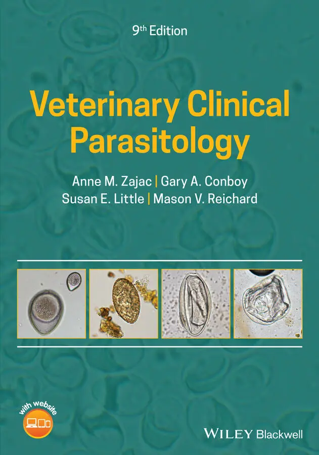
Describes accurate and cost-effective techniques for diagnosing parasitic infections in animals Presents clear and concise information on the distribution, life cycle, and significance of each parasite covered Contains hundreds of color clinical images to enable rapid picture-matching and aid in diagnosis Offers a spiral-bound format that allows the book to lie flat on the benchtop, ideal for regular use in daily practice Features revised content throughout, including new chapters and images, an improved format, an enhanced color scheme, and an updated companion website
is an essential resource for practicing veterinarians, veterinary technicians, diagnosticians, researchers, and students in need of a timely and efficient reference on morphologic identification of parasites in different animal species.
Veterinary Clinical Parasitology — читать онлайн ознакомительный отрывок
Ниже представлен текст книги, разбитый по страницам. Система сохранения места последней прочитанной страницы, позволяет с удобством читать онлайн бесплатно книгу «Veterinary Clinical Parasitology», без необходимости каждый раз заново искать на чём Вы остановились. Поставьте закладку, и сможете в любой момент перейти на страницу, на которой закончили чтение.
Интервал:
Закладка:
2 Chapter 2Fig. 2.1 Dioctophyme eggs are larger than those of Pearsonema and have a thi...Fig. 2.2 Pearsonema feliscati egg in urine sediment. Methylene blue stain is...Fig. 2.3 If the microscope is focused on the shell wall of the Pearsonema eg...Fig. 2.4 Stephanurus dentatus adults produce typical strongylid eggs that ca...Fig. 2.5 The eggs of Trichosomoides have bipolar plugs and are embryonated w...Fig. 2.6 Tritrichomonas foetus organisms from culture. The undulating membra...Fig. 2.7 Microfilariae of Onchocerca gutterosa ( arrow ) in bovine skin. Diagn...Fig. 2.8 Horn flies ( Haematobia irritans ) feeding on a Stephanofilaria stile ...Fig. 2.9 Microfilariae of Onchocerca lupi from the uterus of an adult worm. ...Fig. 2.10 First‐stage larvae of Dracunculus insignis . These distinctive larv...Fig. 2.11 Third‐stage larva of Pelodera strongyloides recovered from a canin...Fig. 2.12 (A) Anterior end of Pelodera third‐stage larva. The bulb of the rh...Fig. 2.13 Thelazia in the eye of a cow.Fig. 2.14 Extensive skin thickening and wrinkling caused by Besnoitia besnoi ...Fig. 2.15 Scleral “pearls,” or cysts, of Besnoitia bennetti in the eye of a ...
3 Chapter 3Fig. 3.1 Technique for making a blood smear. (A) Bring a spreader slide back...Fig. 3.2 Results of a hematocrit test using a blood sample containing D. imm ...Fig. 3.3 Hepatozoon gamont in a polymorphonucleocyte. The parasite is sausag...Fig. 3.4 Erythrocyte containing pear‐shaped Babesia canis piroplasms. The pa...Fig. 3.5 Babesia gibsoni ( arrow ) is a smaller organism than B. canis . The pe...Fig. 3.6 Small Cytauxzoon felis merozoites in infected erythrocytes ( arrow ) ...Fig. 3.7 Mononuclear cell containing a Cytauxzoon felis schizont.Fig. 3.8 Leishmania sp. amastigotes in a lymph node impression smear.Fig. 3.9 Leishmania amastigote ( arrow ) in a macrophage from a canine lymph n...Fig. 3.10 Stained trypomastigote of Trypanosoma cruzi in a blood smear from ...Fig. 3.11 Microfilariae of Dirofilaria immitis recovered from a blood sample...Fig. 3.12 Microfilariae of Dirofilaria immitis have gently tapered heads (A)...Fig. 3.13 Microfilariae are at an earlier developmental stage than first‐sta...Fig. 3.14 Microfilariae of Acanthocheilonema ( Dipetalonema ) reconditum in a ...Fig. 3.15 The tails of some individual microfilariae of Acanthocheilonema re ...Fig. 3.16 Dirofilaria repens microfilaria. This species is expanding its ran...Fig. 3.17 The head of Dirofilaria repens is blunt in comparison to the taper...Fig. 3.18 Babesia bigemina can be seen in this bovine blood smear. The teard...Fig. 3.19 Composite photo showing Theileria equi in equine red blood cells. ...Fig. 3.20 Composite photo of Babesia caballi . The definitive diagnostic form...Fig. 3.21 Theileria parva multinucleated schizont in a lymphocyte. The speci...Fig. 3.22 Theileria orientalis in a bovine blood smear ( arrows ).Fig. 3.23 Trypanosoma brucei and T. congolense are found in domestic mammals...Fig. 3.24 Trypanosoma congolense has a subterminal kinetoplast that is on th...Fig. 3.25 Trypanosoma vivax is found in Africa and other parts of the world....Fig. 3.26 Trypanosoma theileri is found worldwide in cattle, and a similar p...Fig. 3.27 Trypanosoma evansi is an important pathogen of horses and camels i...Fig. 3.28 Trypanosoma simiae is a parasite of African warthogs that is trans...Fig. 3.29 Microfilariae of Setaria spp. may occasionally be seen in ruminant...Fig. 3.30 Host leukocytes and erythrocytes containing the sausage‐shaped Leu ...Fig. 3.31 Birds may be infected with more than one species of protozoa. Both...Fig. 3.32 Gamonts of Haemoproteus in a Swainson’s hawk. The gamonts are ofte...Fig. 3.33 The appearance of multiple stages of the parasite (signet‐ring sta...
4 Chapter 4Fig. 4.1 Schematic ELISA antigen detection procedures: (A) test surface: pol...Fig. 4.2 Schematic of antigen detection using an immunochromatographic later...Fig. 4.3 Comparison of common antibody detection procedures: indirect fluore...Fig. 4.4 Schematic of a conventional PCR assay. PCR is a process used to sel...Fig. 4.5 Simplified illustration of how a qPCR assay differs from a conventi...Fig. 4.6 Simple schematic of fluorescence plot of results from a multiplex q...
5 Chapter 5Fig. 5.1 Important characteristics for identification of a number of common ...Fig. 5.2 Pruritic mite infestations may stimulate intense grooming by the ho...Fig. 5.3 Sarcoptes and related mites are typically round bodied. The third a...Fig. 5.4 Sarcoptes scabiei var canis causes “scabies” or “sarcoptic mange” i...Fig. 5.5 Sarcoptic mange in an alpaca. In chronic sarcoptic mange, affected ...Fig. 5.6 Notoedres mites are similar in appearance to Sarcoptes . However, th...Fig. 5.7 Knemidokoptes is a round‐bodied mite, generally similar in appearan...Fig. 5.8 Budgerigar with a deformed beak resulting from the proliferative le...Fig. 5.9 Like other sarcoptiform mites, Trixacarus is a round‐bodied mite wi...Fig. 5.10 Chorioptes mites are more elongated, with longer legs than the sar...Fig. 5.11 Female Chorioptes mites with eggs present ( arrows ). Mite eggs are ...Fig. 5.12 Psoroptic mange or “scab” can be a serious infestation in ruminant...Fig. 5.13 Psoroptic ear mange in a rabbit.Fig. 5.14 Psoroptes sp. mites have a more oval shape and longer legs than ro...Fig. 5.15 Psoroptes may be up to 800 μm in length. Shown here are specimens ...Fig. 5.16 Otodectes cynotis infestation in a cat. The mites cause the produc...Fig. 5.17 Mating Otodectes cynotis mites from a ferret. Otodectes is another...Fig. 5.18 Gravid female of Otodectes cynotis with egg present ( arrow ).Fig. 5.19 Male Otodectes cynotis . The pair of distinct circular structures e...Fig. 5.20 Demodex is most often seen as a clinical problem in dogs. Lesions ...Fig. 5.21 In goats and cattle, clinical demodecosis is usually associated wi...Fig. 5.22 Demodex spp. mites (A) have a distinct, elongated appearance and a...Fig. 5.23 Many animals are parasitized by Demodex spp. Shown here is Demodex Fig. 5.24 Cheyletiella is a surface mite that can be collected by brushing t...Fig. 5.25 Cheyletiella spp. are readily identified microscopically by the pr...Fig. 5.26 Psorobia simplex from a mouse. Note the rounded body, short legs, ...Fig. 5.27 The body of the Lynxacarus mite is laterally compressed like that ...Fig. 5.28 Male Leporacarus mites have a brown anterior shield that projects ...Fig. 5.29 Adult Leporacarus on the hair of a rabbit. The female mites also b...Fig. 5.30 Chirodiscoides from a guinea pig. The first two pairs of legs are ...Fig. 5.31 Mycoptes musculinus from the hair coat of a mouse. In males, the f...Fig. 5.32 Radfordia is found at the base of the hairs. The first pair of leg...Fig. 5.33 Feather mite from a chicken. Feather mite species show great varia...Fig. 5.34 Megninia , a feather mite from a finch. Feather mites infest variou...Fig. 5.35 Ornithonyssus spp. belong to the mesostigmatid order of mites. The...Fig. 5.36 Another characteristic used to differentiate Ornithonyssus from th...Fig. 5.37 Dermanyssus gallinae infests both domestic and wild birds. The anu...Fig. 5.38 Pneumonyssoides caninum , the nasal mite of dogs. A related mite, P ...Fig. 5.39 Ophionyssus mites are the most common external parasite on captive...Fig. 5.40 Ophionyssus , like other mesostigmatid mites, has claws on the tips...Fig. 5.41 Only the six‐legged larvae of chiggers are parasitic, which is hel...Fig. 5.42 Specimen of trombiculid larvae that cause mammalian chigger infestation.Fig. 5.43 Typical chigger lesions on the leg of a parasitologist. Chiggers m...Fig. 5.44 Ticks are often found attached on parts of the body that are diffi...Fig. 5.45 Comparison of the basis capituli and mouthparts of females of the ...Fig. 5.46 Larval ticks are often called “seed ticks” because of their small ...Fig. 5.47 Female ticks of highest veterinary medical importance in the Unite...Fig. 5.48 Key to adult tick genera found in North America. Examination of ti...Fig. 5.49 Dorsal (top row) and ventral (bottom row) aspects of all motile st...Fig. 5.50 Dorsal ( left ) and ventral ( right ) views of Amblyomma americanum ny...Fig. 5.51 Amblyomma maculatum : ( left ) male; ( right ) female. The Gulf Coast t...Fig. 5.52 Amblyomma cajennense : ( left ) male; female ( right ). The Cayenne tic...Fig. 5.53 Amblyomma variegatum : ( left ) male; ( right ) female. The tropical bo...Fig. 5.54 Amblyomma spp. are common in the tropics and subtropics. They are ...Fig. 5.55 Hyalomma spp. ticks are important disease vectors in Africa, Asia,...Fig. 5.56 Engorged nymph and engorged adult female Ixodes scapularis , the de...Fig. 5.57 Unfed adult female ( left ) and male ( right ) Ixodes scapularis viewe...Fig. 5.58 A distinctive morphologic detail of the Ixodes ticks is the groove...Fig. 5.59 Dermacentor variabilis : female ( left ); male ( right ). Like many mem...Fig. 5.60 Dermacentor andersoni : ( left ) male; ( right ) female; the Rocky Moun...Fig. 5.61 Dermacentor albipictus , the winter or moose tick, is a one‐host ti...Fig. 5.62 Dermacentor ( Anocentor ) nitens engorged female ( left ) and male ( ri ...Fig. 5.63 Engorged female Rhipicephalus sanguineus (brown dog tick). This sp...Fig. 5.64 Rhipicephalus sanguineus male ( left ) and engorged female ( right ). ...Fig. 5.65 The brown dog tick, Rhipicephalus sanguineus . Members of this tick...Fig. 5.66 Rhipicephalus ( Boophilus ) microplus female ( left ) and male ( right )...Fig. 5.67 From left to right, engorged Ixodes , Haemaphysalis , and Rhipicepha ...Fig. 5.68 Haemaphysalis longicornis , the longhorned tick, was recently disco...Fig. 5.69 Haemaphysalis leporispalustris , the rabbit tick, is found in North...Fig. 5.70 The palps of Haemaphysalis ticks are wider than they are long, and...Fig. 5.71 Otobius , the spinose ear tick, and Dermacentor andersoni . This pic...Fig. 5.72 Partially engorged larva of Otobius megnini , the spinose ear tick....Fig. 5.73 This closer view of Otobius megnini nymphs shows the spines that c...Fig. 5.74 Argas sp., the fowl tick, is a soft tick. The ventral location of ...Fig. 5.75 The surface of the soft tick Ornithodoros is covered with mammilla...Fig. 5.76 Chewing lice of domestic animals are usually smaller than sucking ...Fig. 5.77 Sucking louse infestation on a calf. Note the reddish‐brown color ...Fig. 5.78 Bovicola ovis is a small white or tan chewing louse that can be ve...Fig. 5.79 Section of bovine skin with louse eggs (nits) attached to the hair...Fig. 5.80 Haematopinus spp. have prominent ocular points ( arrow , partially o...Fig. 5.81 Linognathus spp. have no ocular points. Unlike Haematopinus , the s...Fig. 5.82 Each sucking louse leg ends in a prominent claw. Shown are the cla...Fig. 5.83 Solenopotes capillatus , the little blue cattle louse, is less comm...Fig. 5.84 Pediculus humanus has well‐developed eyes, no ocular points, and t...Fig. 5.85 The human crab louse, Pthirus pubis , has a distinctive crab‐shaped...Fig. 5.86 Sucking lice ( Polyplax ) species from a rat. Typical of sucking lic...Fig. 5.87 Polyplax egg glued to a rat hair. The presence of lice eggs (“nits...Fig. 5.88 Gliricola porcelli , a chewing louse of guinea pigs, is one of thre...Fig. 5.89 Like other chewing lice, the head of Bovicola spp. is broader than...Fig. 5.90 Chewing lice ( Werneckiella equi ) in the hairs of a horse.Fig. 5.91 Lesions on the shoulder and neck of a horse with a heavy burden of...Fig. 5.92 White louse eggs (nits) can be seen attached to the hairs of this ...Fig. 5.93 Trichodectes canis is the canine chewing louse.Fig. 5.94 The head of Felicola subrostratus , the feline chewing louse, is no...Fig. 5.95 Eggs (nits) of Felicola subrostratus adhered to cat hair.Fig. 5.96 There are more than 700 species of avian lice, all of which are ch...Fig. 5.97 Lipeurus caponis , the wing louse of poultry.Fig. 5.98 Laemobothrion sp. from an eagle.Fig. 5.99 Columbicula columbae , the slender pigeon louse, on the flight feat...Fig. 5.100 Columbicula columbae , the slender pigeon louse.Fig. 5.101 Puppy infested with fleas. This severe level of infestation cause...Fig. 5.102 Key to common flea species in the United States.Fig. 5.103 Female ( left ) and male ( right ) Ctenocephalides felis . The cat fle...Fig. 5.104 Ctenocephalides spp. eggs ( arrow ) are about 0.5 mm long. Larvae a...Fig. 5.105 Pet owners may find larvae of the cat flea, C. felis felis , in th...Fig. 5.106 The human flea, Pulex irritans , is less common on people in indus...Fig. 5.107 Echidnophaga gallinacea , the sticktight flea, has no combs and a ...Fig. 5.108 Xenopsylla cheopsis , the oriental rat flea. Genal and pronotal co...Fig. 5.109 Cediopsylla simplex , a rabbit flea.Fig. 5.110 Posterior spiracles of bot fly larvae. Row 1: Cuterebra spp. spir...Fig. 5.111 Paired spiracle plates on the posterior end of a bot larvae ( Cute ...Fig. 5.112 The hairy body of adult warble (bot) flies makes them look more l...Fig. 5.113 Bot larvae ( Cuterebra sp.) spiracles visible through patent openi...Fig. 5.114 Cuterebra larvae are about 2.5 cm in length and covered with spin...Fig. 5.115 Veterinary practitioners occasionally remove young Cuterebra larv...Fig. 5.116 Dermatobia hominis bots are often seen in the second‐instar larva...Fig. 5.117 Eggs (nits) of Gasterophilus adhered to horse hair.Fig. 5.118 Eggs of Gasterophilus intestinalis , the most common equine bot sp...Fig. 5.119 Second‐instar Gasterophilus larvae. Following treatment with a ma...Fig. 5.120 Equine stomach bot, Gasterophilus . Species can be distinguished b...Fig. 5.121 Early third‐instar cattle grub, Hypoderma lineatum . Grubs or warb...Fig. 5.122 Hypoderma sp. grub emerging from its subcutaneous location on the...Fig. 5.123 Ovine nasal bot, Oestrus ovis . These bots are occasionally seen b...Fig. 5.124 Posterior spiracles of maggots associated with fly strike.Fig. 5.125 A case of ovine “fly strike” or “fly blow” in which an animal has...Fig. 5.126 Posterior spiracles of Lucilia spp. larva.Fig. 5.127 Lucilia spp. is one of the genera of blow flies that cause facult...Fig. 5.128 Screwworm infestation on the ear of a calf. If untreated, these i...Fig. 5.129 Screwworm maggots. If screwworm infestation is suspected in the U...Fig. 5.130 Cochliomyia sp. larvae showing pigmented tracheal trunks ( arrow )....Fig. 5.131 Typical hippoboscid louse flies showing the flattened appearance ...Fig. 5.132 Adult and pupal stages of the sheep ked, Melophagus . Although som...Fig. 5.133 Adult Lipoptena cervi from a moose. Alopecia is reported in moose...Fig. 5.134 Tabanid flies. These large biting flies are familiar worldwide. H...Fig. 5.135 Deer flies, Chrysops , also belong to the tabanid group but are sm...Fig. 5.136 Head and mouthparts of Stomoxys calcitrans , the stable fly ( left )...Fig. 5.137 Horn flies, Haematobia irritans , are a major pest of cattle throu...Fig. 5.138 The stable fly, Stomoxys calcitrans , is approximately the size of...Fig. 5.139 Glossina , the tsetse fly, is the vector of trypanosomiasis in dom...Fig. 5.140 Mosquitoes are common in many regions. Mosquito larvae develop in...Fig. 5.141 Culicoides (midges or no‐see‐ums) are very small biting flies (ra...Fig. 5.142 Adult triatomine bug, Triatoma sanguisuga , an important vector of...Fig. 5.143 Adult bedbug, Cimex lectularis . Photo courtesy of Megan Lineberry...Fig. 5.144 Dorsal ( top row ) and ventral ( bottom row ) adult and nymphal bedbu...Fig. 5.145 Cimex adjunctus , the eastern bat bug, can be mistaken for C. lect ...
Читать дальшеИнтервал:
Закладка:
Похожие книги на «Veterinary Clinical Parasitology»
Представляем Вашему вниманию похожие книги на «Veterinary Clinical Parasitology» списком для выбора. Мы отобрали схожую по названию и смыслу литературу в надежде предоставить читателям больше вариантов отыскать новые, интересные, ещё непрочитанные произведения.


Обсуждение, отзывы о книге «Veterinary Clinical Parasitology» и просто собственные мнения читателей. Оставьте ваши комментарии, напишите, что Вы думаете о произведении, его смысле или главных героях. Укажите что конкретно понравилось, а что нет, и почему Вы так считаете.