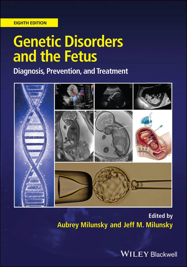
Genetic Disorders and the Fetus
Здесь есть возможность читать онлайн «Genetic Disorders and the Fetus» — ознакомительный отрывок электронной книги совершенно бесплатно, а после прочтения отрывка купить полную версию. В некоторых случаях можно слушать аудио, скачать через торрент в формате fb2 и присутствует краткое содержание. Жанр: unrecognised, на английском языке. Описание произведения, (предисловие) а так же отзывы посетителей доступны на портале библиотеки ЛибКат.
- Название:Genetic Disorders and the Fetus
- Автор:
- Жанр:
- Год:неизвестен
- ISBN:нет данных
- Рейтинг книги:4 / 5. Голосов: 1
-
Избранное:Добавить в избранное
- Отзывы:
-
Ваша оценка:
Genetic Disorders and the Fetus: краткое содержание, описание и аннотация
Предлагаем к чтению аннотацию, описание, краткое содержание или предисловие (зависит от того, что написал сам автор книги «Genetic Disorders and the Fetus»). Если вы не нашли необходимую информацию о книге — напишите в комментариях, мы постараемся отыскать её.
Genetic Disorders and the Fetus — читать онлайн ознакомительный отрывок
Ниже представлен текст книги, разбитый по страницам. Система сохранения места последней прочитанной страницы, позволяет с удобством читать онлайн бесплатно книгу «Genetic Disorders and the Fetus», без необходимости каждый раз заново искать на чём Вы остановились. Поставьте закладку, и сможете в любой момент перейти на страницу, на которой закончили чтение.
Интервал:
Закладка:
643 643. Kunsela P, Seppala MB, Brock DJH, et al. Amniotic fluid fibronectin in normal pregnancy and in pregnancies with anencephalic fetus. Biomedicine 1978; 29:296.
644 644. Vlodavsky I, Voss R, Yarkoni S, et al. Stimulation of human amniotic fluid cell proliferation and colony formation by cell plating on a naturally produced extracellular matrix. Prenat Diagn 1982; 2:13.
645 645. Crickard K, Golbus MS. Influence of extracellular matrix on the proliferation of human amniotic fluid cells in vitro. Prenat Diagn 1982; 2:89.
646 646. Brackertz M, Kubbies M, Feige A, et al. Decreased oxygen supply enhances growth in culture of human midtrimester amniotic fluid cells. Hum Genet 1983; 64:334.
647 647. Held KR, Sönnichsen S. The effect of oxygen tension on colony formation and cell proliferation of amniotic fluid cells in vitro. Prenat Diagn 1984; 4:171.
648 648. Barnes D, Sato G. Serum‐free cell culture: a unifying approach. Cell 1980; 22:649.
649 649. Gospodarowicz D, Moran JS, Owashi ND. Effects of fibroblast growth factor and epidermal growth factor on the rate of growth of amniotic fluid‐derived cells. J Clin Endocrinol Metab 1977; 44:651.
650 650. Porreco RP, Bradshaw C, Sarkar S, et al. Enhanced growth of amniotic fluid cells in presence of fibroblast growth factor. Obstet Gynecol 1980; 55:55.
651 651. Art to Science. Freezing and thawing serum and other biological materials: optimal procedures minimize damage and maximize shelf‐life. HyClone Laboratories, Inc, 1992; 11:1.
652 652. Chang HC, Jones OW. A new growth medium for human amniotic fluid cells. Karyogram 1981; 7:54.
653 653. Chang HC, Jones OW, Masui H. Human amniotic fluid cells grown in a hormone‐supplemented medium: suitability for prenatal diagnosis. Proc Natl Acad Sci U S A 1982; 79:4795.
654 654. Biddle WC, Kuligowski S, Lockwood DH, et al. AmnioMAXTM‐C100: a new specialized cell culture medium for the propagation of human amniocytes. Focus 1992; 14:80.
655 655. Claussen U. The pipette method: a new rapid technique for chromosome analysis in prenatal diagnosis. Hum Genet 1980; 54:277.
656 656. Salk D, Disteche C, Stenchever MR, et al. Routine use of Chang medium for prenatal diagnosis: improved growth and increased chromosomal breakage. Am J Hum Genet 1983; 35:151A.
657 657. Masia A, Jenkins EC, Duncan C. Chromosomal abnormalities in cultures with Chang medium. Am J Hum Genet 1986; 39:260.
658 658. Krawczun MS, Jenkins EC, Masia A, et al. Chromosomal abnormalities in amniotic fluid cell cultures: a comparison of apparent pseudomosaicism in Chang and RPMI‐1640 media. Clin Genet 1989; 35:139.
659 659. Eiben B, Goebel R, Hansen S, et al. Early amniocentesis – a cytogenetic evaluation of over 1500 cases. Prenat Diagn 1994; 14:497.
660 660. Assel BG, Lewis SM, Dickerman LH, et al. Single operator comparison of early and mid‐second‐trimester amniocentesis. Obstet Gynecol 1992; 79:940.
661 661. Corning guide for identifying and correcting common cell growth problems. http://www.bioprocessonline.com/doc/identifying‐correcting‐cell‐growthproblems‐0001 (accessed May 19, 2015).
662 662. Persutte WH, Lenke RR. Failure of amniotic‐fluid cell growth: is it related to fetal aneuploidy? Lancet 1995; 345:96.
663 663. Elejalde BR, de Elejalde MM, Soto A. Amniotic‐fluid cell growth and fetal aneuploidy. Lancet 1995; 345:924.
664 664. Lam YH, Tang MH, Sin SY, et al. Clinical significance of amniotic‐fluid‐cell culture failure. Prenat Diagn 1998; 18:343.
665 665. American College of Medical Genetics Standards and Guidelines for Clinical Genetics Laboratories. https://www.acmg.net/ACMG/Medical-Genetics-Practice-Resources/Technical_Standards_and_Guidelines.aspx (accessed July 8, 2020).
666 666. Milunsky A, Bender CS. Failure of amniotic‐fluid cell growth with toxic tubes. N Engl J Med 1979; 301:47.
667 667. Kohn G. Failure of amniotic fluid cell cultures due to syringe toxicity. Prenat Diagn 1981; 1:233.
668 668. Chiesa J, Bureau JP. Nothing ventured, nothing gained! Prenat Diagn 1999; 19:894.
669 669. Seguin LR, Palmer CG. Variables influencing growth and morphology of colonies of cells from human amniotic fluid. Prenat Diagn 1983; 3:107.
670 670. Felix JS, Doherty RA. Amniotic fluid cell culture II. Evaluation of a red blood cell lysis procedure for culture of cells from blood‐contaminated amniotic fluid. Clin Genet 1979; 15:215.
671 671. Johansson KE, Bolske G. Evaluation and practical aspects of the use of a commercial DNA probe for detection of mycoplasma infections in cell cultures. J Biochem Biophys Methods 1989; 19:185.
672 672. Knutsen T. Laboratory safety, quality control and regulations. In: Barch MJ, ed. The AGT cytogenetics laboratory manual, 3rd edn. Philadelphia: Lippincott Williams & Wilkins, 1997:597.
673 673. Holtge GA. Laboratory safety. In: McClatchey KD, ed. Clinical laboratory medicine, 2nd edn. Philadelphia: Lippincott Williams & Wilkins, 2002:78.
674 674. Travers EM. Basic laboratory management. In: McClatchey KD, ed. Clinical laboratory medicine, 2nd edn. Philadelphia: Lippincott Williams & Wilkins, 2002:3.
675 675. Tsai MS, Lee JL, Chang YJ, et al. Isolation of human multipotent mesenchymal stem cells from second‐trimester amniotic fluid using a novel two‐stage culture protocol. Hum Reprod 2004; 19:1450.
676 676. De Coppi P, Bartsch G, Jr., Siddiqui MM, et al. Isolation of amniotic stem cell lines with potential for therapy. Nat Biotechnol 2007; 25:100.
677 677. Guillot PV, Gotherstrom C, Chan J, et al. Human first trimester fetal MSC express pluripotency markers and grow faster and have longer telomeres than adult MSC. Stem Cells 2007; 25:646.
678 678. Roubelakis MG, Pappa KI, Bitsika V, et al. Molecular and proteomic characterization of human mesenchymal stem cells derived from amniotic fluid: comparison to bone marrow mesenchymal stem cells. Stem Cells Dev 2007; 16:931.
679 679. Zhou J,Wang D, Liang T, et al. Amniotic fluid‐derived mesenchymal stem cells: characteristics and therapeutic applications. Arch Gynecol Obstet 2014; 290:223.
680 680. In't Anker PS, Scherjon SA, Kleijburg‐van der Keur C, et al. Amniotic fluid as a novel source of mesenchymal stem cells for therapeutic transplantation. Blood 2003; 102:1548.
681 681. Trounson A. A fluid means of stem cell generation. Nat Biotechnol 2007; 25:62.
682 682. Liu YW, Roan JN, Wang SP, et al. Xenografted human amniotic fluid‐derived stem cell as a cell source in therapeutic angiogenesis. Int J Cardiol 2013; 168:66.
683 683. Petsche Connell J, Camci‐Unal G, Khademhosseini A, et al. Amniotic fluid‐derived stem cells for cardiovascular tissue engineering applications. Tissue Eng Part B Rev 2013; 19:368.
684 684. Kang NH, Hwang KA, Kim SU, et al. Potential antitumor therapeutic strategies of human amniotic membrane and amniotic fluid‐derived stem cells. Cancer Gene Ther 2012; 19:517.
685 685. Kaviani A, Perry TE, Dzakovic A, et al. The amniotic fluid as a source of cells for fetal tissue engineering. J Pediatr Surg 2001; 36:1662.
686 686. Gucciardo L, Lories R, Ochsenbein‐Kolble N, et al. Fetal mesenchymal stem cells: isolation, properties and potential use in perinatology and regenerative medicine. BJOG 2009; 116:166.
687 687. Murphy SV, Atala A. Amniotic fluid and placental membranes: unexpected sources of highly multipotent cells. Semin Reprod Med 2013; 31:62.
4 Molecular Aspects of Placental Development
Wendy P. Robinson 1,2 and Deborah E. McFadden 1,2
1BC Children's Hospital Research Institute, Vancouver, BC, Canada
2University of British Columbia, Vancouver, BC, Canada
Overview
The placenta is a fetal organ that is discarded after birth, but is essential to ensuring normal development in utero . It regulates fetal growth, protects the fetus from infection and other adverse exposures, as well as generally programming the fetus for good health after birth. Screening for placental disease is an important component to the assessment of the fetus in pregnancy. Reduced placental efficiency can lead to fetal growth restriction (FGR) and/or maternal preeclampsia (PE). This can be caused by genetic changes within the placenta or by environmental influences, such as maternal stress or drug exposure. In this chapter, causes of placental disease and the role of the placenta in diagnosis of fetal health will be reviewed with a focus on genetic associations.
Читать дальшеИнтервал:
Закладка:
Похожие книги на «Genetic Disorders and the Fetus»
Представляем Вашему вниманию похожие книги на «Genetic Disorders and the Fetus» списком для выбора. Мы отобрали схожую по названию и смыслу литературу в надежде предоставить читателям больше вариантов отыскать новые, интересные, ещё непрочитанные произведения.


Обсуждение, отзывы о книге «Genetic Disorders and the Fetus» и просто собственные мнения читателей. Оставьте ваши комментарии, напишите, что Вы думаете о произведении, его смысле или главных героях. Укажите что конкретно понравилось, а что нет, и почему Вы так считаете.