Pubudu N. Pathirana - Human Motion Capture and Identification for Assistive Systems Design in Rehabilitation
Здесь есть возможность читать онлайн «Pubudu N. Pathirana - Human Motion Capture and Identification for Assistive Systems Design in Rehabilitation» — ознакомительный отрывок электронной книги совершенно бесплатно, а после прочтения отрывка купить полную версию. В некоторых случаях можно слушать аудио, скачать через торрент в формате fb2 и присутствует краткое содержание. Жанр: unrecognised, на английском языке. Описание произведения, (предисловие) а так же отзывы посетителей доступны на портале библиотеки ЛибКат.
- Название:Human Motion Capture and Identification for Assistive Systems Design in Rehabilitation
- Автор:
- Жанр:
- Год:неизвестен
- ISBN:нет данных
- Рейтинг книги:3 / 5. Голосов: 1
-
Избранное:Добавить в избранное
- Отзывы:
-
Ваша оценка:
Human Motion Capture and Identification for Assistive Systems Design in Rehabilitation: краткое содержание, описание и аннотация
Предлагаем к чтению аннотацию, описание, краткое содержание или предисловие (зависит от того, что написал сам автор книги «Human Motion Capture and Identification for Assistive Systems Design in Rehabilitation»). Если вы не нашли необходимую информацию о книге — напишите в комментариях, мы постараемся отыскать её.
A guide to the core ideas of human motion capture in a rapidly changing technological landscape Human Motion Capture and Identification for Assistive Systems Design in Rehabilitation
Human Motion Capture and Identification for Assistive Systems Design in Rehabilitation
Human Motion Capture and Identification for Assistive Systems Design in Rehabilitation — читать онлайн ознакомительный отрывок
Ниже представлен текст книги, разбитый по страницам. Система сохранения места последней прочитанной страницы, позволяет с удобством читать онлайн бесплатно книгу «Human Motion Capture and Identification for Assistive Systems Design in Rehabilitation», без необходимости каждый раз заново искать на чём Вы остановились. Поставьте закладку, и сможете в любой момент перейти на страницу, на которой закончили чтение.
Интервал:
Закладка:
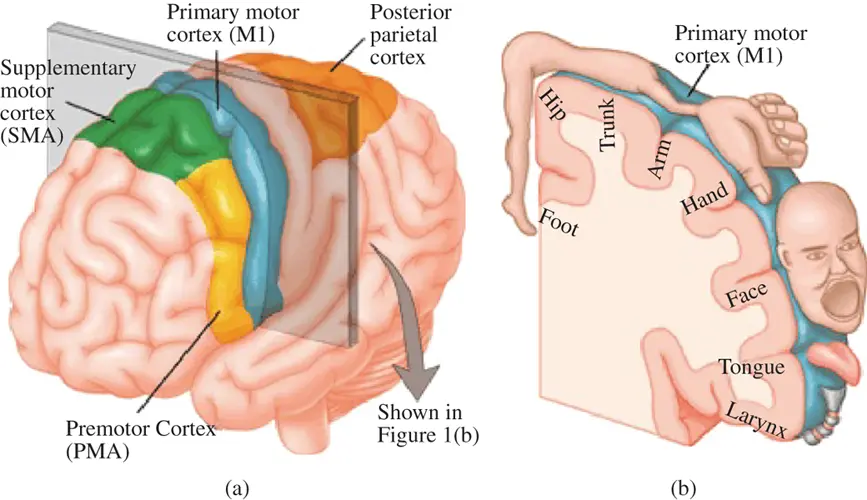
Figure 1.2 Functional description of the brain motor cortex.
1.2 Musculoskeletal Injuries and Neurological Movement Disorders
Movement difficulties can be due to a number of causes and generally are classified as neuromuscular disorders. The causes of these abnormal movements can be classified into two main categories: musculoskeletal injuries and neurological movement disorders.
1.2.1 Musculoskeletal injuries
These injuries are normally observed in joints associated with certain degrees of movement, such as shoulder, elbow and wrists in the upper extremities and hips, knees and ankles in lower ones, which may eventually lead to abnormal movements or event disabilities. Some examples of disorders are shown in Tables 1.1and 1.2.
1.2.2 Neuromuscular disorders
The disorders that can be associated with the nervous and muscular systems affect the movements and can sometimes exhibit characteristic movement patterns associated with certain conditions. Neuromuscular disorders affect the nerves that control the voluntary muscles – muscles that can normally be controlled by the individual. Such disorders include motor neurone diseases, neuropathies, muscular dystrophies and neurodegenerative disorders. These disorders can be classified according to the area of the neuromuscular system that is affected.
Table 1.1 Examples of musculoskeletal injuries in joints of the upper extremities. Inj is for injury and ST is for studies about using physical rehabilitation to treat the conditions or stimulate recovery (the same as that in Table 1.2).
| Shoulder | Elbow | Wrist | |
|---|---|---|---|
| Movements | Flexion, extension, abduction, adduction, internal and external rotation [84] | “Flexion and extension at the ulnohumeral and radiocapitellar articulations, while pronation and supination at the proximal radioulnar joint” [56] | Flexion, extension, radial deviation and ulnar deviation [271] |
| Inj1 | Shoulder impingement | Tennis elbow | Carpal tunnel syndrome |
| Description | It “occurs against the anterior edge and undersurface of the anterior third of the acromion, the coracoacromial ligament, and, at times, the acromioclavicular joint” [255] and deemed as one of the factors that lead to shoulder disability [254]. | Although it is not perfectly understood, it negatively influences “the attachment of the extensors of the forearm at the lateral side of the elbow”, thereby leading to pain [365]. | It usually is caused by the pressure on the median nerve on a wrist and leads to various conditions, such as pain, paraesthesiae, hypoaesthesia and so on [287]. |
| ST | [238] | [366] | [251] |
| Inj2 | Adhesive capsulitis | Scaphoid | |
| Description | The general cause leading to this condition is described as “progressive fibrosis and ultimate contracture of the glenohumeral joint capsule” [258]. | It is usually caused by a hyperextended and radially deviated wrist and seen in patients aged between 15 and 40 [167]. | |
| ST | [343] | [299] |
Upper motor neurone disorders–
Conditions such as a cerebrovascular accident (stroke), Parkinson's disease, multiple sclerosis, Huntington's disease (Huntington's chorea) and Creutzfeldt‐Jakob disease are examples of upper motor neurone diseases.
Lower motor neurone disorders – spinal muscular atrophies
Lower motor neurones are located in either the anterior grey column, anterior nerve roots (spinal lower motor neruones) or the cranial nerve nuclei of the brain stem and cranial nerves with a motor function (cranial nerve lower motor neurones) [1]. All voluntary movement relies on spinal lower motor neurones, which innervate skeletal muscle fibres and act as a link between the upper motor neurons and muscles [2, 3]. Cranial nerve lower motor neurones control movements of the eyes and tongue, and contribute to chewing, swallowing and vocalisation [4]. Damage to the lower motor neurone can lead to flaccid paralysis.
Table 1.2 Examples of musculoskeletal injuries in joints of the lower extremities.
| Hip | Knee | Ankle | |
|---|---|---|---|
| Movements | Flexion, extension, abduction, adduction, internal and external rotation [149] | There are two ways to describe the degree of freedom (DOF) in a knee. One is with two DOFs (flexion‐extension and axial rotation) [234] and the other is with six DOFs (flexion‐extension, varus‐valgus, internal‐external rotation and mediolateral, anteroposterior and superoinferior translation around mediolateral, anteroposterior and superoinferior axis) [122]. | Extension, flexion, valgus and varus [301] |
| Inj1 | Hamstring strain | Patellofemoral pain syndrome | Achilles tendonitis |
| Description | It usually associated with lower extremity activities, like football, soccer, dancing and so on, while this condition occurs in different phases of motions in various types of activity [149]. | It is an anterior knee pain and mainly resulted from “aberrant motion of the patella in the trochlear groove” [123]. | The physical findings of this condition include soft tissue swelling, local tenderness and sometimes crepitus [256]. |
| ST | [326] | [214] | [222] |
| Inj2 | Groin pain | Anterior cruciate ligament (ACL) injury | Lateral sprain |
| Description | It contributes 2–5% of all sport injuries [243]. Vincent et al . [243] also mentioned that the diagnosis of this pain is hard because of its complex anatomy in the affected region, as well as the coexistence of multiple injuries. | The causes are in two major categories, including non‐contact (usually resulted from sudden deceleration before changing direction or a landing motion) and contact (valgus collapse) [45]. | It can be deemed as the most common injury in ankles [114], which is usually cause by inversion of the foot [114]. |
| ST | [302] | [74] | [103] |
Neuropathies
Neuropathies involve dysfunction of the peripheral nerves, which consist of: motor neurones, that carry the electrical signals directly from the spinal cord and brain stem to activate muscle movement; the sensory neurones, which convey sensory information such as pain, temperature, light touch, vibration and position to the brain; and the autonomic neurons, which go to the internal organs and control blood vessel reflexes.
Neuromuscular junction disorders
Myasthenia gravis and Lambert-Eaton syndrome are examples of neuromuscular junction disorders. Muscular dystrophies and inflammatory myopathies such as polymyositis are examples of primary muscular (myopathic) disorders.
Neurodegenarative classification encapsulates the progressive loss of structure or function of the neurone and a number of conditions exhibit this form of progression. Hence, amyotrophic lateral sclerosis (ALS), Parkinson's and Huntington's are classified as neurodegenerative diseases that affect movement. These conditions exhibit characteristically slower movement compared to healthy people – hypokinesis – or excessive and abnormal involuntary movements – hyperkinesis [156]. Some common examples of hypokinesis include bradykinesia, freezing, rigidity and stiff muscles, while those belonging to hyperkinesis are chorea, dyskinesia, myoclonus, tics and tremor [242].
Читать дальшеИнтервал:
Закладка:
Похожие книги на «Human Motion Capture and Identification for Assistive Systems Design in Rehabilitation»
Представляем Вашему вниманию похожие книги на «Human Motion Capture and Identification for Assistive Systems Design in Rehabilitation» списком для выбора. Мы отобрали схожую по названию и смыслу литературу в надежде предоставить читателям больше вариантов отыскать новые, интересные, ещё непрочитанные произведения.


Обсуждение, отзывы о книге «Human Motion Capture and Identification for Assistive Systems Design in Rehabilitation» и просто собственные мнения читателей. Оставьте ваши комментарии, напишите, что Вы думаете о произведении, его смысле или главных героях. Укажите что конкретно понравилось, а что нет, и почему Вы так считаете.