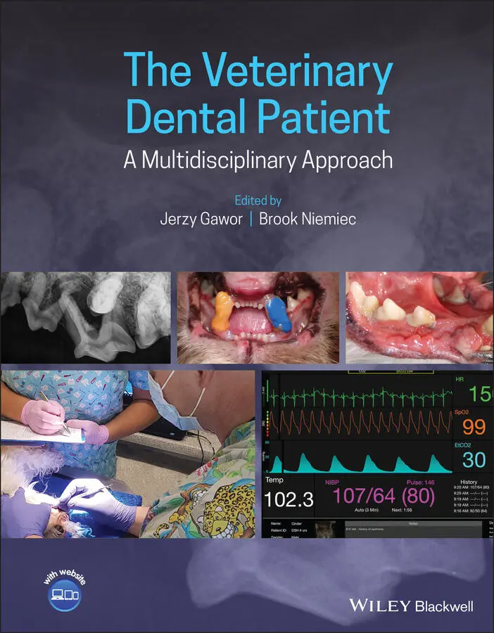
The Veterinary Dental Patient - A Multidisciplinary Approach
Здесь есть возможность читать онлайн «The Veterinary Dental Patient - A Multidisciplinary Approach» — ознакомительный отрывок электронной книги совершенно бесплатно, а после прочтения отрывка купить полную версию. В некоторых случаях можно слушать аудио, скачать через торрент в формате fb2 и присутствует краткое содержание. Жанр: unrecognised, на английском языке. Описание произведения, (предисловие) а так же отзывы посетителей доступны на портале библиотеки ЛибКат.
- Название:The Veterinary Dental Patient: A Multidisciplinary Approach
- Автор:
- Жанр:
- Год:неизвестен
- ISBN:нет данных
- Рейтинг книги:3 / 5. Голосов: 1
-
Избранное:Добавить в избранное
- Отзывы:
-
Ваша оценка:
The Veterinary Dental Patient: A Multidisciplinary Approach: краткое содержание, описание и аннотация
Предлагаем к чтению аннотацию, описание, краткое содержание или предисловие (зависит от того, что написал сам автор книги «The Veterinary Dental Patient: A Multidisciplinary Approach»). Если вы не нашли необходимую информацию о книге — напишите в комментариях, мы постараемся отыскать её.
The Veterinary Dental Patient: A Multidisciplinary Approach
The Veterinary Dental Patient: A Multidisciplinary Approach — читать онлайн ознакомительный отрывок
Ниже представлен текст книги, разбитый по страницам. Система сохранения места последней прочитанной страницы, позволяет с удобством читать онлайн бесплатно книгу «The Veterinary Dental Patient: A Multidisciplinary Approach», без необходимости каждый раз заново искать на чём Вы остановились. Поставьте закладку, и сможете в любой момент перейти на страницу, на которой закончили чтение.
Интервал:
Закладка:
18 Index
19 End User License Agreement
List of Tables
1 Chapter 1 Table 1.1 Suggested business plan for the equipment of the dental part of a p...
2 Chapter 2 Table 2.1 Verbal behaviors significantly associated with positive or negative... Table 2.2 Nonverbal behaviors significantly associated with positive or negat...
3 Chapter 5 Table 5.1 Oral health parameters assessed during patient examination and inte...
4 Chapter 7Table 7.1 American Heart Association recommendations for antimicrobial prophy...Table 7.2 Common dental antimicrobials.Table 7.3 Potential criteria for identification of veterinary patients that r...Table 7.4 Basic measures to reduce the incidence of needlestick injuries.Table 7.5 Basic goals of cleaning, disinfection, and sterilization.Table 7.6 Required cleaning and disinfection practices for different types of...
5 Chapter 8Table 8.1 Comparison of selected parameters characterizing pet oral care in P...
6 Chapter 10Table 10.1 Description of the tests required to diagnose hereditary diseases ...Table 10.2 Scale for the negative influence of hereditary disease on patient ...
7 Chapter 11Table 11.1 Nonsteroidal anti‐inflammatory drugs (NSAIDs): canine and feline d...Table 11.2 Common local anesthetics used in veterinary anesthesia and pain ma...
8 Chapter 12Table 12.1 Drugs used in pre‐anesthetic combinations. aTable 12.2 AAHA recommended intravenous fluid rates during general anesthesia...Table 12.3 Anesthesia induction protocols for dogs and cats.Table 12.4 Single injections of sedative or analgesic drugs used to decrease ...Table 12.5 Drugs used as CRIs during anesthesia to reduce MAC.Table 12.6 Anesthetic management for obese patients.
9 Chapter 13Table 13.1 Cardiovascular effects of important anesthetic drugs in physiologi...
10 Chapter 15Table 15.1 Bacterial genera cultured from both blood and plaque in different ...Table 15.2 Incidence of bacteria by procedure group in human dentistry.Table 15.3 Expected healing times for different tissues.Table 15.4 Recommendations when dental (class A or B) and other surgeries (clas...
11 Chapter 18Table 18.1 Dental formulae for cats and dogs. I (incisors); C (canines); P (p...Table 18.2 Estimated timing of eruption of different teeth in different speci...
12 Chapter 22Table 22.1 Amount of alveolar bone to be removed in different clinical situat...
List of Illustrations
1 Chapter 1 Figure 1.1 Waiting room TV screen with informational dental presentation. Figure 1.2 Feline cabinet, following the standards of a cat‐friendly clinic.... Figure 1.3 Dental models and instruments as well as a small whiteboard can b... Figure 1.4 White board drawings explaining a crown height‐reduction procedur... Figure 1.5 Educational posters avaliable at www.vetdentalrad.com. Figure 1.6 Educational video shown on a tablet in a waiting room. Figure 1.7 Finger protection during feline oral cavity assessment. Figure 1.8 Dental operatory. Figure 1.9 Protection of the patient during sedation. Figure 1.10 Central distribution of (a) water in preparation and postoperati... Figure 1.11 Handheld X‐ray generator. Figure 1.12 Wall‐mounted anesthesia machine. Figure 1.13 Hazardous materials have to be stored in dedicated containers an... Figure 1.14 An adjustable table is helpful when working on with larger patie... Figure 1.15 Veterinary dentist demonstrating the pathology to the pet owner.... Figure 1.16 Discussion in the consulting room is easier when a high‐quality ... Figure 1.17 Presentation displayed in the waiting room explaining the safety... Figure 1.18 An object (e.g., a treated tooth) can be radiographically assess... Figure 1.19 Advantages of 3D versus 2D imaging. (a) The sequestrum revealed ... Figure 1.20 Device combining a scaler and a polisher. Figure 1.21 Dental unit main board, including (from left) suction, three‐way... Figure 1.22 (a) Combination of a UNC probe and explorer in a single instrume... Figure 1.23 Series of combined probes and explorers. (a) Probes. From left: ... Figure 1.24 (a) Mouth props extending a dog's jaws. (b) Selection of props i... Figure 1.25 Mirror (a) reflecting light into the caudal part of the oral cav... Figure 1.26 Magnification helps greatly with dental procedures. Figure 1.27 Charting: filling in (a) a paper dental chart and (b) an electro... Figure 1.28 The oral surgical general kit includes, from left: periosteal el... Figure 1.29 Extraction kit: luxators (left) and elevators (right), as well a... Figure 1.30 Rescue kit ‐ From left: four superslim luxators and elevators, m... Figure 1.31 Selection of burs for extraction. Figure 1.32 Periodontal kit. From bottom: mirror, three curettes, periodonta... Figure 1.33 Area‐specific curettes. Figure 1.34 Use of CHX solution during extraction of a badly infected mouth.... Figure 1.35 Plaque‐disclosing solution. Figure 1.36 Disposable custom‐made pharyngeal packs made of gauze or sponge....
2 Chapter 2 Figure 2.1 Scheme illustrating problems associated with selling veterinary d... Figure 2.2 Client–pet and client–veterinarian bonds. Figure 2.3 Example of a human–dog bond, with humanization efforts. Figure 2.4 Diplomas in the waiting room. Figure 2.5 Array of thank‐you pictures. Figure 2.6 Nonverbal communication: (a) smile versus (b) non‐smile. Figure 2.7 A centralized position of the patient is necessary to display emp...
3 Chapter 3 Figure 3.1 Framework for clinical assessment. Figure 3.2 Webinar in dentistry. Figure 3.3 Face‐to‐face learning. Figure 3.4 Interactive session. Figure 3.5 WSAVA Dental guidelines team From left: Paulo Stegall DACVAA (Can...
4 Chapter 4 Figure 4.1 The first impression when a client enters the clinic: North Downs... Figure 4.2 The kennel assistant, a first‐line dental team member. Figure 4.3 (a) This patient came to boarding with a fractured 304. (b) An un... Figure 4.4 Veterinary care assistant packing a dental kit. Figure 4.5 Four‐handed dentistry: examination and charting. Figure 4.6 Preparation of the operatory. Figure 4.7 (a, b) Seven‐year‐old Yorkshire terrier that has received annual Figure 4.8 Concious animal assessment. Figure 4.9 Client estimate, prepared based on a filled‐in dental chart. Figure 4.10 Anaesthetised animal assessment. Figure 4.11 Modified pen grasp of a scaling handpiece. Figure 4.12 Color coding of patients for follow‐up visits. Figure 4.13 Informational brochures in a waiting room. Figure 4.14 Before and after smile book examples.
5 Chapter 5 Figure 5.1 Dental Index app. Figure 5.2 Dental plaque identification is part of pet owner education, and ... Figure 5.3 Double‐headed toothbrushes have certain benefits in plaque contro... Figure 5.4 Mechanized toothbrush used in dogs. Figure 5.5 Selection of toothpastes. Figure 5.6 Teaching tooth brushing should be gradual and gentle. Figure 5.7 Avoid wide opening of the mouth when teaching tooth brushing. Figure 5.8 Indirect (with the use of fingerbrush) application of Maxiguard i... Figure 5.9 Application of the waxy barrier sealant. Figure 5.10 Kibbles intended for oral hygiene (right) are larger than normal... Figure 5.11 Hill’s Dental Care T/D diet for dogs and cats. Figure 5.12 Dental treats/chews must be attractive for pets. Figure 5.13 With bones or very hard chews, there is danger of (a) breaking t... Figure 5.14 The new version of Vet Aquadent without xylitol. Figure 5.15 Water additives will not work when the animal prefers tap water.... Figure 5.16 Plaque Off series.
6 Chapter 6Figure 6.1 Comparison of the dentition of a dog that regularly received a de...Figure 6.2 Entrapment of a large piece of bone in a dog's mandible.Figure 6.3 VOHC website.Figure 6.4 CBCT image showing the relations of occlusion of teeth in differe...
7 Chapter 7Figure 7.1 Anaerobic transport medium for sample collection (top). Sterile g...Figure 7.2 (a) Deep periodontal pocket in the left mandibular molar area of ...Figure 7.3 Odontogenic abscess in a cat: (a) deep periodontal pocket (circle...Figure 7.4 Antimicrobial prophylaxis for routine descaling is the most commo...Figure 7.5 Cultured bacteria from a periodontal abscess with antibiotic susc...Figure 7.6 Presence of dental plaque demonstrated with the use of (a) ultrav...Figure 7.7 Antiseptics used in veterinary dentistry: (a) Vet aquadent, conta...Figure 7.8 Personal protective equipment: gown, gloves, mask, and eye protec...Figure 7.9 Conjunctivitis as a result of wearing contact lenses during a den...Figure 7.10 Skin wound caused by lack of protection of sharp working tips.Figure 7.11 Autoclaved instruments are a necessary condition for performing ...
Читать дальшеИнтервал:
Закладка:
Похожие книги на «The Veterinary Dental Patient: A Multidisciplinary Approach»
Представляем Вашему вниманию похожие книги на «The Veterinary Dental Patient: A Multidisciplinary Approach» списком для выбора. Мы отобрали схожую по названию и смыслу литературу в надежде предоставить читателям больше вариантов отыскать новые, интересные, ещё непрочитанные произведения.


Обсуждение, отзывы о книге «The Veterinary Dental Patient: A Multidisciplinary Approach» и просто собственные мнения читателей. Оставьте ваши комментарии, напишите, что Вы думаете о произведении, его смысле или главных героях. Укажите что конкретно понравилось, а что нет, и почему Вы так считаете.