The Veterinary Dental Patient - A Multidisciplinary Approach
Здесь есть возможность читать онлайн «The Veterinary Dental Patient - A Multidisciplinary Approach» — ознакомительный отрывок электронной книги совершенно бесплатно, а после прочтения отрывка купить полную версию. В некоторых случаях можно слушать аудио, скачать через торрент в формате fb2 и присутствует краткое содержание. Жанр: unrecognised, на английском языке. Описание произведения, (предисловие) а так же отзывы посетителей доступны на портале библиотеки ЛибКат.
- Название:The Veterinary Dental Patient: A Multidisciplinary Approach
- Автор:
- Жанр:
- Год:неизвестен
- ISBN:нет данных
- Рейтинг книги:3 / 5. Голосов: 1
-
Избранное:Добавить в избранное
- Отзывы:
-
Ваша оценка:
The Veterinary Dental Patient: A Multidisciplinary Approach: краткое содержание, описание и аннотация
Предлагаем к чтению аннотацию, описание, краткое содержание или предисловие (зависит от того, что написал сам автор книги «The Veterinary Dental Patient: A Multidisciplinary Approach»). Если вы не нашли необходимую информацию о книге — напишите в комментариях, мы постараемся отыскать её.
The Veterinary Dental Patient: A Multidisciplinary Approach
The Veterinary Dental Patient: A Multidisciplinary Approach — читать онлайн ознакомительный отрывок
Ниже представлен текст книги, разбитый по страницам. Система сохранения места последней прочитанной страницы, позволяет с удобством читать онлайн бесплатно книгу «The Veterinary Dental Patient: A Multidisciplinary Approach», без необходимости каждый раз заново искать на чём Вы остановились. Поставьте закладку, и сможете в любой момент перейти на страницу, на которой закончили чтение.
Интервал:
Закладка:
Progress in technology and continuous cooperation between manufacturers and specialists are producing better and better solutions for veterinary dentistry. Selection of the optimal supplier is an individual decision. Before that decision is made, a trial is strongly recommended.
1.10 Dental Instrumentation
Specific instruments are necessary to perform oral surgery and dentistry. One cannot compromise patient care by using instruments inappropriately or using nonmedical instruments. The decision about which types or brands should be purchased is based on individual preferences. However, the author can recommend some based on experience and personal needs. Most instruments can be organized in groups (kits) dedicated to specific procedures or specific species or sizes of animal. A very important part of the correct use of any instrument is having a proper grasp. Also important is maintaining the correct shape of working surface or tip, which is associated with sharpening and conservation. Ensuring that instruments are always clean, ready, and sharp is a must, not only in the dental world, but in all of medicine.
This section presents the absolute basic instruments that every general practice offering any form of dentistry should be equipped with.
1.10.1 Diagnostic Kit
An explorer and periodontal probe are very often combined, having one side of the instrument being an explorer and the other being a probe. For dogs, a combination of a UNC 17 periodontal probe with a shepherd hook explorer is this author's preference ( Figure 1.22). In cats, a finer explorer ODU or Orban explorer combined with a Michigan probe with Williams markings will better adapt to the feline gingival sulcus and smaller oral cavity. Additionally, evaluation of areas suspected of tooth resorption is more convenient with this explorer ( Figure 1.23).
Mouth props are preferred to gags as they do not apply additional force to the temporomandibular joint area, and in cats they diminish the risk of complications of excessive jaws opening (Stiles et al. 2012) ( Figure 1.24).
For conscious patient examination, finger protectors are very useful where there is danger of the patient hurting the assessor (see Figure 1.7).
Mirrors can be used to visualize the palatal and lingual surfaces of teeth, the caudal part of the oral cavity, the pharynx, and the choanae. The most caudal areas can be illuminated by light reflection from the mirror ( Figure 1.25).
Revealing small lesions or details during oral examination is easier with the use of magnification combined with lightening ( Figure 1.26). For beginners, 2.5 dental loupes are a good choice. With time, 3.5× magnification may be preferred. It is important to understand that using magnification will not immediately improve the quality of a procedure. It takes experience and training, but magnification will become an invaluable support for the operator, help to avoid errors, and expose small details during operations.
The next step in magnifying the operating area is the use of an operating microscope, which further improves the assessment of surgical tissues.
During oral assessment, it is important to record all observations on a dental chart appropriate to the species and age of the patient. Regardless of whether two‐ or four‐handed dentistry (i.e., with or without an assistant) is performed, filling in the chart is obligatory ( Figure 1.27).
High‐quality photography can further augment the diagnostic process. It can be used for presentation to the owner, for communication with a referring veterinarian, for comparison at distant follow‐up, and for illustration of publications or presentations. This is a matter of personal preference, but the camera should preserve natural colors and provide sufficient magnification and focal distance. The latter can be difficult in compact cameras, which require very small distances for macro mode, where lenses and external optics may get foggy from the humid oral environment.
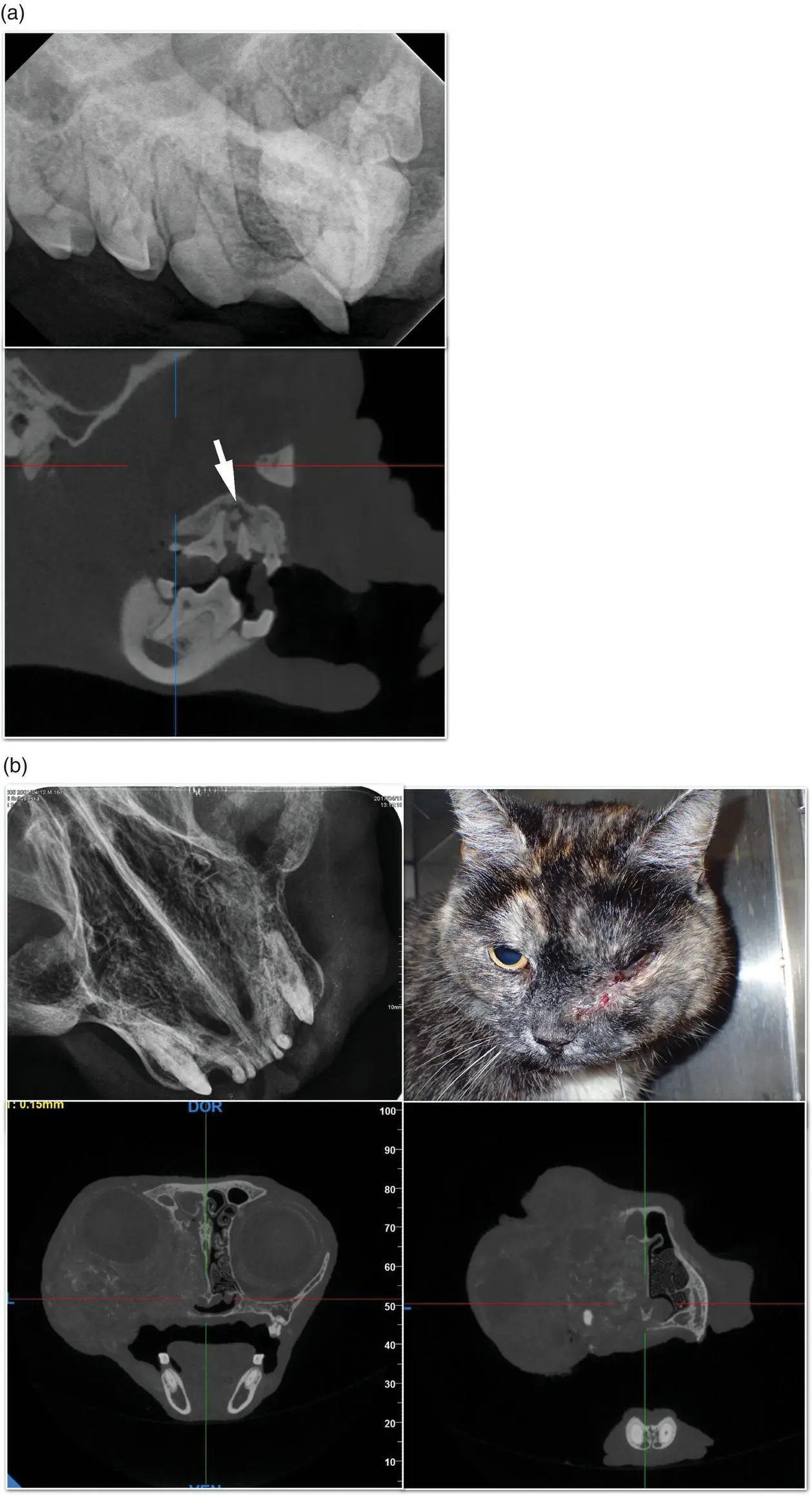
Figure 1.19 Advantages of 3D versus 2D imaging. (a) The sequestrum revealed next to the infected root of the 208 (arrow) would not be identified in a standard radiograph. (b) The extent of neoplastic growth is better assessed in 3D than in intraoral radiograph.
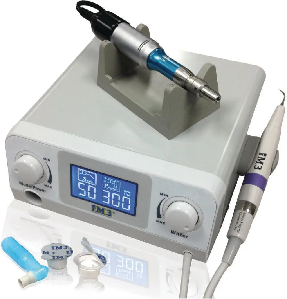
Figure 1.20 Device combining a scaler and a polisher.
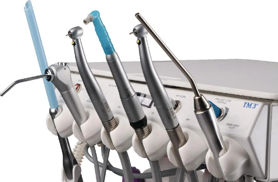
Figure 1.21 Dental unit main board, including (from left) suction, three‐way syringe, high‐speed handpiece, low‐speed handpiece, second high‐speed handpiece, and illuminating wand.
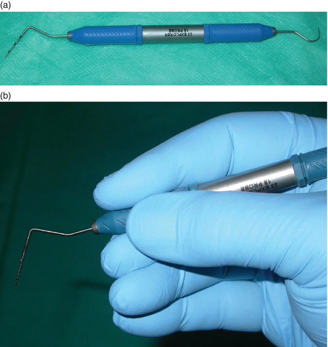
Figure 1.22 (a) Combination of a UNC probe and explorer in a single instrument. (b) and its pen grasp.
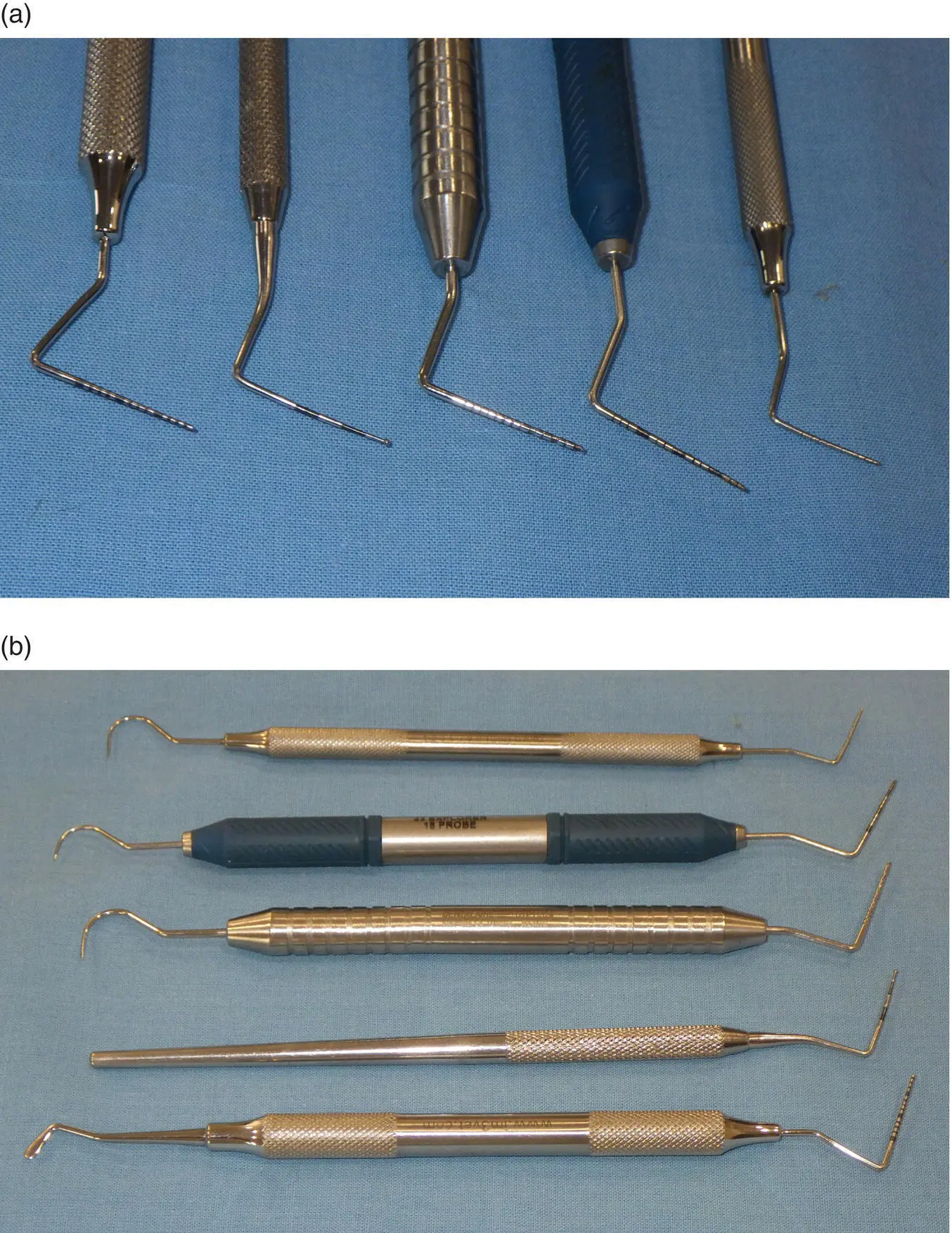
Figure 1.23 Series of combined probes and explorers. (a) Probes. From left: UNC 15, Michigan, Niemiec, UNC 17, Williams. (b) Whole instruments.
1.10.2 Surgical Kit
In most cases, oral surgery will mean extraction. More information on this topic is provided in Chapter 22. The oral surgery kit has been developed and presented in textbooks by several specialists (Niemiec, Verstraete, Reiter), and oral surgeons have their own preferences in terms of type, size, and brand. Nevertheless, all agree that the following should always be included: blade holder, tissue forceps, periosteal elevator, tissue scissors, suture scissors, and needle holder ( Figure 1.28). The part of the needle holder that grasps the suture should be delicate or it can weaken the material, causing it to break after a few sutures. Oral needle holders must not be used for materials larger than 4/0 or they will immediately lose good attachment to sutures and needle.
Tissue scissors with serrated cutting edges provide a better margin of the cut mucosa and gums, but they should never be used for sutures. Suture scissors should preferably be blunt‐ended in order not to harm the tissue while cutting the sutures.
The surgical kit may have medium/large canine and cats/small dogs variations and be packed together with diagnostic instruments. For surgery in the caudal area of the mouth or oropharynx, instruments must be long enough for comfortable operation.
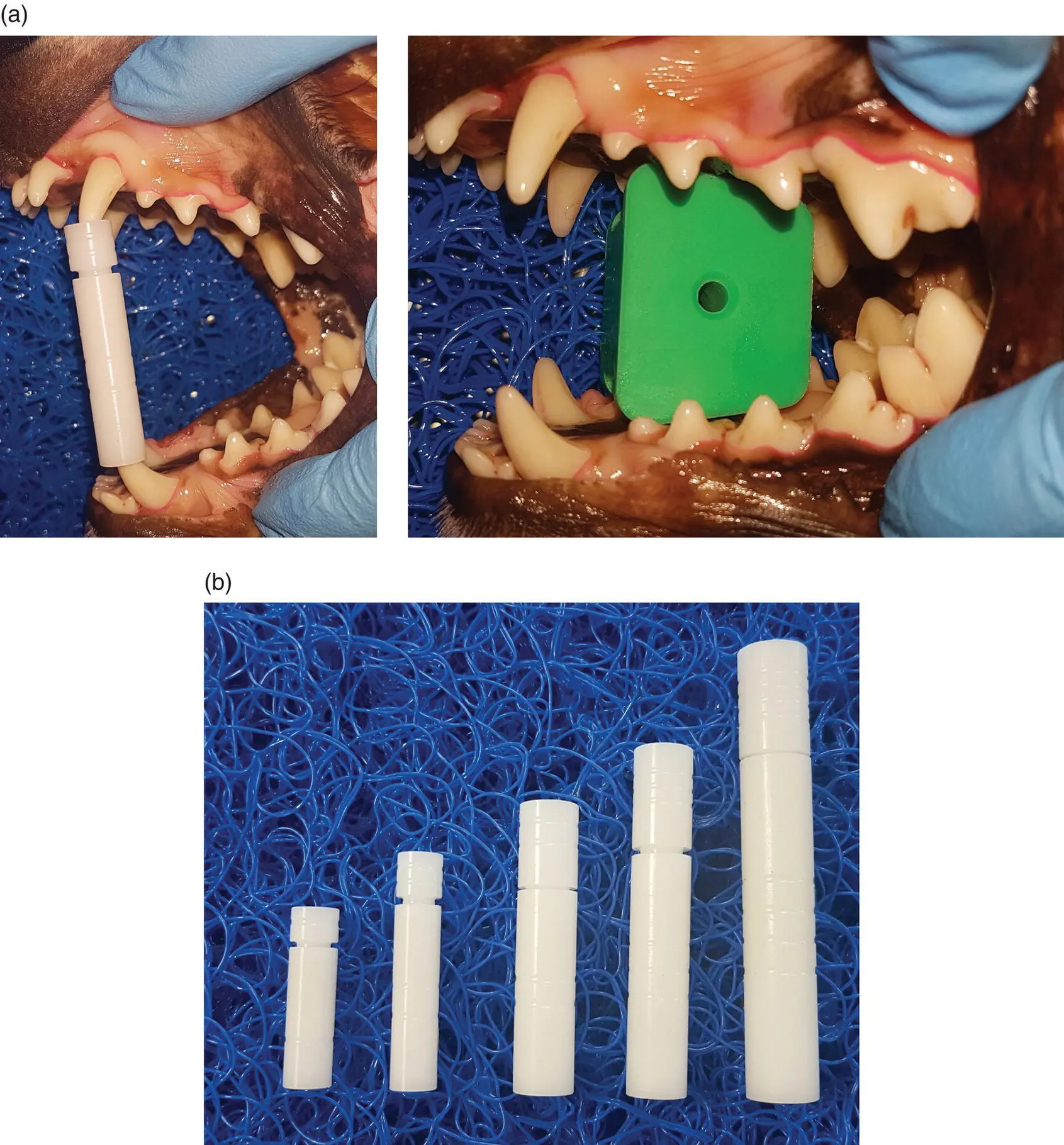
Figure 1.24 (a) Mouth props extending a dog's jaws. (b) Selection of props in different sizes for dogs and cats.
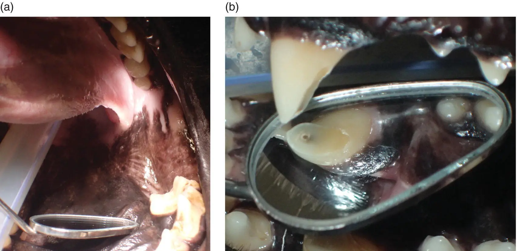
Figure 1.25 Mirror (a) reflecting light into the caudal part of the oral cavity and (b) showing the other side of a tooth.
Читать дальшеИнтервал:
Закладка:
Похожие книги на «The Veterinary Dental Patient: A Multidisciplinary Approach»
Представляем Вашему вниманию похожие книги на «The Veterinary Dental Patient: A Multidisciplinary Approach» списком для выбора. Мы отобрали схожую по названию и смыслу литературу в надежде предоставить читателям больше вариантов отыскать новые, интересные, ещё непрочитанные произведения.


Обсуждение, отзывы о книге «The Veterinary Dental Patient: A Multidisciplinary Approach» и просто собственные мнения читателей. Оставьте ваши комментарии, напишите, что Вы думаете о произведении, его смысле или главных героях. Укажите что конкретно понравилось, а что нет, и почему Вы так считаете.