Saeid Sanei - EEG Signal Processing and Machine Learning
Здесь есть возможность читать онлайн «Saeid Sanei - EEG Signal Processing and Machine Learning» — ознакомительный отрывок электронной книги совершенно бесплатно, а после прочтения отрывка купить полную версию. В некоторых случаях можно слушать аудио, скачать через торрент в формате fb2 и присутствует краткое содержание. Жанр: unrecognised, на английском языке. Описание произведения, (предисловие) а так же отзывы посетителей доступны на портале библиотеки ЛибКат.
- Название:EEG Signal Processing and Machine Learning
- Автор:
- Жанр:
- Год:неизвестен
- ISBN:нет данных
- Рейтинг книги:3 / 5. Голосов: 1
-
Избранное:Добавить в избранное
- Отзывы:
-
Ваша оценка:
EEG Signal Processing and Machine Learning: краткое содержание, описание и аннотация
Предлагаем к чтению аннотацию, описание, краткое содержание или предисловие (зависит от того, что написал сам автор книги «EEG Signal Processing and Machine Learning»). Если вы не нашли необходимую информацию о книге — напишите в комментариях, мы постараемся отыскать её.
EEG Signal Processing and Machine Learning — читать онлайн ознакомительный отрывок
Ниже представлен текст книги, разбитый по страницам. Система сохранения места последней прочитанной страницы, позволяет с удобством читать онлайн бесплатно книгу «EEG Signal Processing and Machine Learning», без необходимости каждый раз заново искать на чём Вы остановились. Поставьте закладку, и сможете в любой момент перейти на страницу, на которой закончили чтение.
Интервал:
Закладка:
Following the generation of an IPSP, there is an overflow of cations from the nerve cell or an inflow of anions into the nerve cell. This flow ultimately causes a change in potential along the nerve cell membrane. Primary transmembranous currents generate secondary ional currents along the cell membranes in the intracellular and extracellular space. The portion of these currents that flow through the extracellular space is directly responsible for the generation of field potentials. These field potentials, usually with less than 100 Hz frequency, are called EEGs when there are no changes in the signal average and called DC potential if there are slow drifts in the average signals, which may mask the actual EEG signals. A combination of EEG and DC potentials is often observed for some abnormalities in the brain such as seizure (induced by pentylenetetrazol), hypercapnia, and asphyxia [22]. We next focus on the nature of APs.
1.4 Action Potentials
AP is actually the information transmitted by a nerve. These potentials are caused by an exchange of ions across the neuron membrane and an AP is a temporary change in the membrane potential that is transmitted along the axon. It is usually initiated in the cell body and normally travels in one direction. The membrane potential depolarizes (becomes more positive) producing a spike. After the peak of the spike the membrane repolarizes (becomes more negative). The potential becomes more negative than the resting potential and then returns to normal. The APs of most nerves last between 5 and 10 ms. Figure 1.4shows an example of an AP.
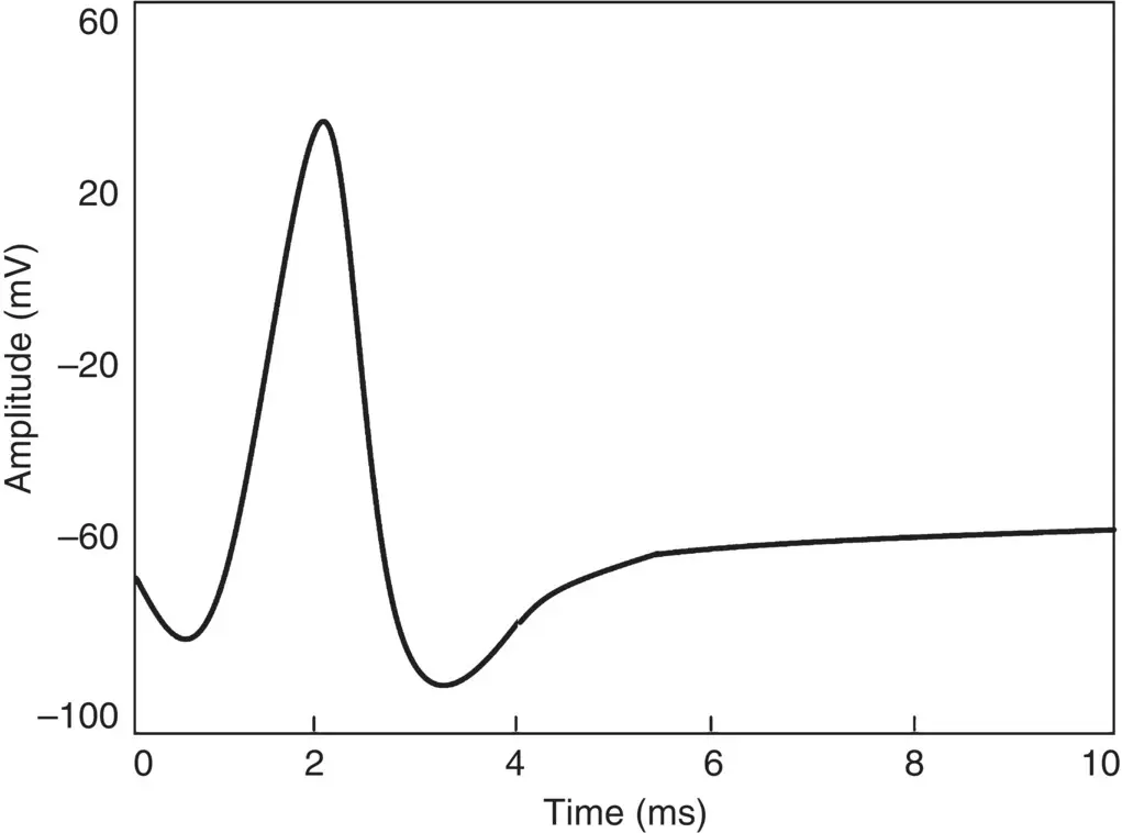
Figure 1.4 An example of an AP.
The conduction velocity of APs lies between 1 and 100 m s −1. APs are initiated by many different types of stimuli; sensory nerves respond to many types of stimuli, such as: chemical, light, electricity, pressure, touch, and stretching. Conversely, the nerves within the CNS (brain and spinal cord) are mostly stimulated by chemical activity at synapses.
A stimulus must be above a threshold level to set off an AP. Very weak stimuli cause a small local electrical disturbance, but do not produce a transmitted AP. As soon as the stimulus strength goes above the threshold, an AP appears and travels down the nerve.
The spike of the AP is mainly caused by opening of Na (sodium) channels. The Na pump produces gradients of both Na and K (potassium) ions, both are used to produce the AP; Na is high outside the cell and low inside. Excitable cells have special Na and K channels with gates that open and close in response to the membrane voltage (voltage‐gated channels). Opening the gates of Na channels allows Na to rush into the cell, carrying a +ve charge. This makes the membrane potential positive (depolarization), producing the spike. Figure 1.5shows the stages of the process during evolution of an AP for a giant squid.
For a human being the amplitude of the AP ranges between approximately −60 to 10 mV. During this process [23]:
1 When the dendrites of a nerve cell receive the stimulus the Na+ channels will open.
2 If the opening is sufficient to drive the interior potential from −70 mV up to −55 mV, the process continues.
3 As soon as the action threshold is reached, additional Na+ channels (sometimes called voltage‐gated channels) open. The Na+ influx drives the interior of the cell membrane up to about +30 mV. The process to this point is called depolarization.
4 Then Na+ channels close and the K+ channels open. Since the K+ channels are much slower to open, the depolarization has time to be completed. Having both Na+ and K+ channels open at the same time would drive the system towards neutrality and prevent the creation of the AP. Figure 1.5 Changing the membrane potential for a giant squid by closing the Na channels and opening K channels.(Source: adapted from Ka Xiong Charand [23].)
5 Having the K+ channels open, the membrane begins to repolarize back towards its rest potential.
6 The repolarization typically overshoots the rest potential to a level of approximately −90 mV. This is called hyperpolarization, and would seem to be counterproductive, but it is actually important in the transmission of information. Hyperpolarization prevents the neuron from receiving another stimulus during this time, or at least raises the threshold for any new stimulus. Part of the importance of hyperpolarization is in preventing any stimulus already sent up an axon from triggering another AP in the opposite direction. In other words, hyperpolarization assures that the signal is proceeding in one direction.
After hyperpolarization, the Na +/K +pumps eventually bring the membrane back to its resting state of −70 mV.
The nerve requires approximately two milliseconds before another stimulus is presented. During this time no AP can be generated. This is called the refractory period. The generation of EEG signals is next described.
1.5 EEG Generation
An EEG signal is an indirect measurement of currents that flow during synaptic excitations of the dendrites of many pyramidal neurons in the cerebral cortex. When the brain cells (neurons) are activated, the synaptic currents are produced and propagate through the dendrites. This current generates a magnetic field measurable by EMG machines and a secondary electrical field over the scalp measurable by EEG systems.
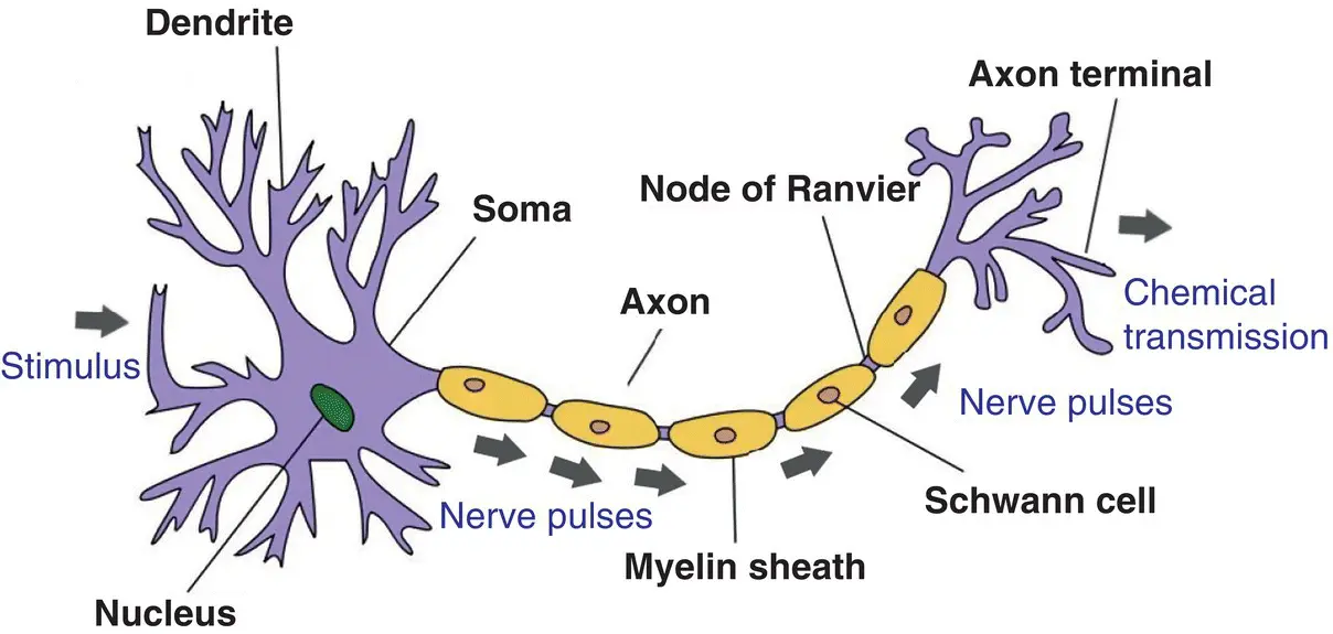
Figure 1.6 Structure of a neuron.
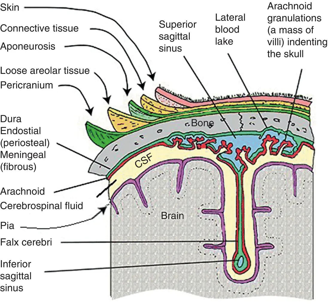
Figure 1.7 The head layers from brain to scalp.
Differences of electrical potentials are caused by summed post‐synaptic graded potentials from pyramidal cells that create electrical dipoles between the soma (body of a neuron) and apical dendrites which branch from neurons ( Figure 1.6). The current in the brain is generated mostly due to pumping the positive ions of sodium, Na +, potassium, K +, calcium, or Ca ++, and the negative ion of Cl −, through the neuron membranes in the direction governed by the membrane potential [24].
The human head consists of three main layers of scalp, skull, brain ( Figure 1.7) including many other thin layers in‐between. In addition, the scalp consists of different layers such as skin, connective tissue, which is a thin layer of fat and fibrous tissue lying beneath the skin, the loose areolar connective tissue, and the pericranium, which is the periosteum of the skull bones and provides nutrition to bone and capacity for repair. Conversely, the brain is covered by a thin layer of cortex, which encompasses various brain tissues. The cortex includes arachnoid, meninges, dura, epidural, and subarachnoid space. The skull attenuates the signals approximately one hundred times more than the soft tissue. Conversely, most of the noise is generated either within the brain (internal noise) or over the scalp (system noise or external noise). Therefore, only large populations of active neurons can generate enough potential to be recordable using the scalp electrodes. These signals are later amplified greatly for display purposes. Approximately 10 11neurons are developed at birth when the CNS becomes complete and functional [25]. This makes an average of 10 4neurons per cubic millimetre. Neurons are interconnected into neural nets through synapses. Adults have approximately 5.10 14synapses. The number of synapses per neuron increases with age, whereas the number of neurons decreases with age.
Читать дальшеИнтервал:
Закладка:
Похожие книги на «EEG Signal Processing and Machine Learning»
Представляем Вашему вниманию похожие книги на «EEG Signal Processing and Machine Learning» списком для выбора. Мы отобрали схожую по названию и смыслу литературу в надежде предоставить читателям больше вариантов отыскать новые, интересные, ещё непрочитанные произведения.


Обсуждение, отзывы о книге «EEG Signal Processing and Machine Learning» и просто собственные мнения читателей. Оставьте ваши комментарии, напишите, что Вы думаете о произведении, его смысле или главных героях. Укажите что конкретно понравилось, а что нет, и почему Вы так считаете.