Chia-shu Lin - Dental Neuroimaging
Здесь есть возможность читать онлайн «Chia-shu Lin - Dental Neuroimaging» — ознакомительный отрывок электронной книги совершенно бесплатно, а после прочтения отрывка купить полную версию. В некоторых случаях можно слушать аудио, скачать через торрент в формате fb2 и присутствует краткое содержание. Жанр: unrecognised, на английском языке. Описание произведения, (предисловие) а так же отзывы посетителей доступны на портале библиотеки ЛибКат.
- Название:Dental Neuroimaging
- Автор:
- Жанр:
- Год:неизвестен
- ISBN:нет данных
- Рейтинг книги:4 / 5. Голосов: 1
-
Избранное:Добавить в избранное
- Отзывы:
-
Ваша оценка:
Dental Neuroimaging: краткое содержание, описание и аннотация
Предлагаем к чтению аннотацию, описание, краткое содержание или предисловие (зависит от того, что написал сам автор книги «Dental Neuroimaging»). Если вы не нашли необходимую информацию о книге — напишите в комментариях, мы постараемся отыскать её.
Provides the latest neuroimaging-based evidence on the brain mechanisms of oral functions Dental Neuroimaging: The Role of the Brain in Oral Functions
Dental Neuroimaging: The Role of the Brain in Oral Functions
Dental Neuroimaging — читать онлайн ознакомительный отрывок
Ниже представлен текст книги, разбитый по страницам. Система сохранения места последней прочитанной страницы, позволяет с удобством читать онлайн бесплатно книгу «Dental Neuroimaging», без необходимости каждый раз заново искать на чём Вы остановились. Поставьте закладку, и сможете в любой момент перейти на страницу, на которой закончили чтение.
Интервал:
Закладка:
Library of Congress Cataloging‐in‐Publication Data
Names: Lin, Chia‐Shu, 1976– author.
Title: Dental neuroimaging : the role of the brain in oral functions / Chia‐Shu Lin.
Description: Hoboken, NJ : Wiley‐Blackwell, 2022. | Includes bibliographical references and index.
Identifiers: LCCN 2021047940 (print) | LCCN 2021047941 (ebook) | ISBN 9781119724209 (paperback) | ISBN 9781119724254 (Adobe PDF) | ISBN 9781119724230 (epub)
Subjects: MESH: Stomatognathic System–diagnostic imaging | Neuroimaging–methods | Brain Mapping–methods
Classification: LCC RK308 (print) | LCC RK308 (ebook) | NLM WU 102 | DDC 617.5/22–dc23
LC record available at https://lccn.loc.gov/2021047940LC ebook record available at https://lccn.loc.gov/2021047941
Cover Design: Wiley
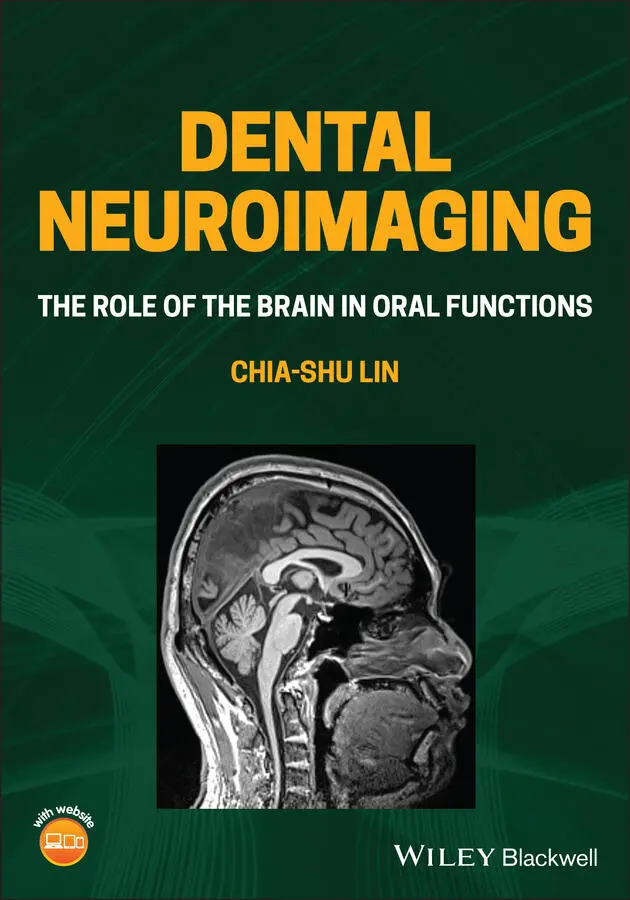
Cover Images: Courtesy of Chia‐Shu Lin; © KJ_Photography/Shutterstock
This book is dedicated to my parents, for their love and caring, my wife, I‐ting, and our children, Yuan‐han and Yi‐hsien, who are my inspiration.
List of Figures
| 1.1 | The association between the brain and the stomatognathic system. The traditional perspective highlights the brain as a ‘systemic factor’ associated with oral health, just like the factors related to other body systems. The functional perspective highlights that the brain and mental functions guided by the brain play an essential role in stomatognathic functions. |
| 1.2 | A general view of the neural circuitries of the brain mechanisms of orofacial functions. The circuitries between the central and peripheral sites (i.e. pathways labelled in blue and red) are investigated primarily via animal models. Notably, the circuitries within the brain (i.e. the intracortical pathways labelled in black) have not been fully elucidated. Source: Avivi-Arber and Sessle (2018).Reproduced with permission of John Wiley and Sons. |
| 1.3 | Theoretical frameworks of the association between the brain, oral functions and behaviour. (a) The oral-to-behaviour (OB) framework, (b) the oral-brain-behaviour (OBB) framework and (c) the brain–stomatognathic axis (BSA). |
| 2.1 | The general concept of the blood-oxygen-level-dependent (BOLD) mechanism. (a) Transportation of oxygenated haemoglobin during a resting condition, when neural activity is low. (b) Transportation of oxygenated haemoglobin when neural activity increases. The neurons demand more energy by consuming oxygen provided by oxygenated haemoglobin. Via a complex haemodynamic process (e.g. an increasing rate and volume of cerebral flow), the amount of oxygenated haemoglobin increases (relatively to the amount of deoxygenated haemoglobin), leading to an over-supply or compensation of the oxygen demand from neurons. |
| 2.2 | Examples of functional magnetic resonance imaging (fMRI) investigation of chewing movement. (a) The first-level analysis. In a chewing experiment, the task conditions (i.e. when subjects are chewing) are contrasted to the baseline conditions (i.e. when subjects are resting). Brain activation reflects the difference in blood-oxygen-level-dependent (BOLD) signals in the task vs. the baseline condition. The first-level analysis focuses on the pattern of brain activation at the individual subject. (b) The second-level analysis. The second-level analysis focuses on the association between brain activation and individual variability. The association can be explored by investigating the correlation between brain activation and individual performance or comparing brain activation between different clinical groups. |
| 2.3 | Methodological considerations of a functional magnetic resonance imaging study. (a) Subjects may show great inter-individual variability in their general conditions, such as sex, age and general physical/psychological conditions. (b) Subjects may show great inter-individual variability in their personal trait and performance (e.g. pain ratings) related to an experimental task. (c) Subjects differ in brain morphology. When individual brains are compared, the individual images are normalized to a template image, using linear transformation (i.e. translation, rotation, resizing and shearing) and nonlinear transformation approaches. |
| 2.4 | Statistical analysis at the individual level and the group level. (a) The analysis at the individual level focuses on the association between task progression and the blood-oxygen-level-dependent (BOLD) time series, as shown in the left panel. For each voxel, a strong association indicates that the BOLD signals of the voxel can be predicted by the task condition, in contrast to the baseline condition (e.g. Voxel A), as shown in the right panel. (b) The analysis at the group level focuses on the association between brain features (e.g. brain activation of grey matter volume) and group factors. The association may reflect the difference in brain features between patient and control groups (the left panel) or the correlation between brain features and clinical factors (the right panel). (c) A typical image result consists of the statistical values (e.g. the t-score) from multiple voxels (represented as the grid), which are visualized by a colour scale, as shown in the left panel. The result can be thresholded according to intensity (i.e. the t-score). For example, only the voxels with a t-score >6 are preserved after thresholding, as shown in the middle panel. The result can be thresholded according to the size of a cluster of voxels. For example, only the clusters with a size larger than 100 voxels will be preserved after thresholding, as shown in the right panel. |
| 2.5 | Experimental design of functional neuroimaging research. (a) Under the assumption of pure insertion, the difference of brain activation between two experimental conditions only reflects the mental function contrasted by the conditions (e.g. perception of pain intensity). However, the contrast may be associated with more than one mental function (e.g. perception of pain intensity and attention to noxious stimuli). (b) A factorial design helps to delineate the association between two mental functions. For example, the light grey area denotes the effect of increased pain on brain activation, and the dark grey area denotes the effect of increased attention. (c) A conjunction design focuses on the pattern of brain activation common to two experimental conditions (e.g. a clenching task and a chewing task). The activation may reflect the brain mechanisms of a mental function common to both task conditions. |
| 2.6 | Conceptual differences between functional specialization, functional segregation and functional integration. (a) Functional specialization highlights the association between a brain region and a specific mental function. For example, activation at the occipital lobe is considered mainly for the processing of visual perception. (b) Functional segregation highlights that a mental function is associated with multiple brain regions that are functionally connected within a module. For example, visual cognition is associated with the module consisting of the yellow regions, and motor control is associated with the module consisting of the blue regions. (c) Functional integration highlights the pattern of global communication between multiple brain regions. For example, individual variability in mental functions may be associated with the efficiency of how information is distributed in a network. |
| 2.7 | Analysis of resting-state functional connectivity. (a) The spontaneous blood-oxygen-level-dependent (BOLD) activity is acquired using resting-state functional magnetic resonance imaging. Subjects fix their eyesight on a crosshair without additional external stimuli. (b) Brain images are segmented into multiple regions according to a brain atlas. (c) To each brain region, the mean BOLD time series is extracted by averaging the time series from all the voxels within a region. (d) Association between the regional time series is quantified with correlation coefficients. (e) The correlation coefficient represents the strength of the connection between each pair of brain regions. (f) In the seed-based approach, a brain region of interest (i.e. the ‘seed’ region) is pre-selected. Functional connectivity is calculated between the seed region and all the other voxels to explore the brain regions that have a strong connection with the seed region. |
Интервал:
Закладка:
Похожие книги на «Dental Neuroimaging»
Представляем Вашему вниманию похожие книги на «Dental Neuroimaging» списком для выбора. Мы отобрали схожую по названию и смыслу литературу в надежде предоставить читателям больше вариантов отыскать новые, интересные, ещё непрочитанные произведения.


Обсуждение, отзывы о книге «Dental Neuroimaging» и просто собственные мнения читателей. Оставьте ваши комментарии, напишите, что Вы думаете о произведении, его смысле или главных героях. Укажите что конкретно понравилось, а что нет, и почему Вы так считаете.