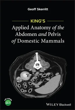
Geoff Skerritt - King's Applied Anatomy of the Abdomen and Pelvis of Domestic Mammals
Здесь есть возможность читать онлайн «Geoff Skerritt - King's Applied Anatomy of the Abdomen and Pelvis of Domestic Mammals» — ознакомительный отрывок электронной книги совершенно бесплатно, а после прочтения отрывка купить полную версию. В некоторых случаях можно слушать аудио, скачать через торрент в формате fb2 и присутствует краткое содержание. Жанр: unrecognised, на английском языке. Описание произведения, (предисловие) а так же отзывы посетителей доступны на портале библиотеки ЛибКат.
- Название:King's Applied Anatomy of the Abdomen and Pelvis of Domestic Mammals
- Автор:
- Жанр:
- Год:неизвестен
- ISBN:нет данных
- Рейтинг книги:5 / 5. Голосов: 1
-
Избранное:Добавить в избранное
- Отзывы:
-
Ваша оценка:
- 100
- 1
- 2
- 3
- 4
- 5
King's Applied Anatomy of the Abdomen and Pelvis of Domestic Mammals: краткое содержание, описание и аннотация
Предлагаем к чтению аннотацию, описание, краткое содержание или предисловие (зависит от того, что написал сам автор книги «King's Applied Anatomy of the Abdomen and Pelvis of Domestic Mammals»). Если вы не нашли необходимую информацию о книге — напишите в комментариях, мы постараемся отыскать её.
King’s Applied Anatomy of the Abdomen and Pelvis of the Domestic Mammals King’s Applied Anatomy of the Abdomen and Pelvis of the Domestic Mammals
King's Applied Anatomy of the Abdomen and Pelvis of Domestic Mammals — читать онлайн ознакомительный отрывок
Ниже представлен текст книги, разбитый по страницам. Система сохранения места последней прочитанной страницы, позволяет с удобством читать онлайн бесплатно книгу «King's Applied Anatomy of the Abdomen and Pelvis of Domestic Mammals», без необходимости каждый раз заново искать на чём Вы остановились. Поставьте закладку, и сможете в любой момент перейти на страницу, на которой закончили чтение.
Интервал:
Закладка:
Table of Contents
1 Cover
2 Title Page
3 Copyright Page
4 Dedication Page
5 Foreword
6 Preface
7 Acknowledgements
8 About the Author
9 About the Contributors
10 About the Companion Website
11 1 The Boundaries of the Abdomen1.1 Introduction 1.2 The Diaphragm (Figure 8.3) 1.3 The Layers of the Abdominal Wall 1.4 The Sheath of the Rectus Abdominis Muscle (Figures 1.10a–c) 1.5 Clinical Importance of the Ventral Body Wall 1.6 The Inguinal Canal (Figures 1.11 and 1.12) 1.7 Hernias
12 2 Gastrointestinal Function2.1 Introduction 2.2 Functions of the Alimentary Tract 2.3 Regions of the Alimentary Tract (Figure 2.1) 2.4 Clinical Conditions Affecting Gastrointestinal Function
13 3 The Mesenteries, Ligaments and Omenta 3.1 The Greater Omentum (Figure 3.1) 3.2 The Clinical Significance of the Greater Omentum 3.3 The Lesser Omentum 3.4 Ligaments
14 4 The Stomach (Figures 4.1–4.4) 4.1 Overview of the Mammalian Stomach 4.2 Species Variations 4.3 Clinical Conditions
15 5 The Small Intestines 5.1 Duodenum, Jejunum and Ileum 5.2 Species Variations 5.3 Clinical Conditions
16 6 The Large Intestine6.1 Overview 6.2 Species Variations 6.3 Clinical Conditions
17 7 The Liver and Pancreas7.1 The Liver (Figure 7.1) 7.2 Anatomy of the Liver 7.3 Histology of the Liver 7.4 The Gall Bladder 7.5 Species Variations 7.6 The Pancreas 7.7 Clinical Conditions of the Liver and Pancreas
18 8 Arteries of the Abdomen and Pelvis (Figures 8.1–8.3) 8.1 The Branches of the Abdominal Aorta 8.2 Species Variations
19 9 Veins of the Abdomen and Pelvis (Figure 9.1) 9.1 Tributaries of the Caudal Vena Cava 9.2 The Hepatic Portal Vein 9.3 The Mammary Glands
20 10 Lymphatics and the Spleen10.1 The Lymphatic System (Figure 10.1) 10.2 The Spleen 10.3 Species Variations 10.4 Clinical Conditions Affecting the Lymphatic System
21 11 The Nerves of the Abdomen and Pelvis 11.1 General Somatic Afferent Neurons 11.2 General Visceral Afferent Neurons 11.3 General Somatic Efferent Neurons 11.4 General Visceral Efferent Neurons 11.5 Clinical Conditions 11.6 Regional Anaesthesia
22 12 The Kidneys (Figures 12.1 and 12.2) 12.1 Nitrogenous Excretion 12.2 Gross Anatomy of the Kidneys 12.3 Species Variations (Figure 12.2) 12.4 Clinical Conditions of the Kidneys of the Domestic Mammals 12.5 Urinary Bladder and Urethra 12.6 Adrenal Gland
23 13 The Ovaries and Ovarian Bursae 13.1 The Ovary (Figures 13.1 and 13.2) 13.2 Species Variations 13.3 The Ovarian Bursa (Figure 13.1) 13.4 The Uterine Tube, also called the Fallopian Tube or the Oviduct (Figure 13.2)
24 14 The Uterus, Uterine Tube, Vestibule and Vagina14.1 The Uterine Cornu 14.2 The Body of the Uterus (Figures 14.1–14.4) 14.3 The Pregnant Uterus 14.4 Placentation 14.5 The Cervix Uteri (Figures 14.1–14.4) 14.6 The Vagina (Figures 14.1–14.4) 14.7 The Vestibule (Figures 14.5–14.8) 14.8 Clinical Conditions
25 15 The Mammalian Penis 15.1 The Penis 15.2 Erectile Tissue 15.3 The Muscles of the Penis 15.4 The Blood Supply and Venous Drainage of the Penis 15.5 Species Variations of the Penis 15.6 The Prepuce (Figure 15.1)
26 16 The Testes16.1 The Anatomy of the Testes 16.2 Species Variations (see Figure 16.1) 16.3 The Scrotum 16.4 The Tissue Layers of the Testes and Scrotum (Figure 16.2) 16.5 The Blood Supply and Drainage of the Testes 16.6 The Epididymis 16.7 Species Variations (Figure 16.5) 16.8 The Descent of the Testes (Figure 16.6) 16.9 Induction of Testicular Descent
27 17 The Accessory Sex Glands17.1 The Accessory Glands 17.2 Prostate Gland 17.3 Vesicular Glands 17.4 Ampulla of the Ductus Deferens 17.5 Bulbourethral Glands 17.6 Clinical Conditions of the Accessory Glands 17.7 Anal Glands
28 18 Diagnostic Imaging of the Abdomen (Figures 18.1–18.4) 18.1 Radiographic Anatomy 18.2 Specific Organs 18.3 Magnetic Resonance Imaging (MRI) 18.4 Computed Tomography 18.5 Ultrasonography 18.6 Diagnostic Imaging in Equine Patients 18.7 Diagnostic Imaging in Farm Animals 18.8 Laparoscopy in Dogs and Cats
29 Appendix Questionnaire Answers to Questionnaire
30 Bibliography
31 Index
32 End User License Agreement
List of Illustrations
1 Chapter 1 Figure 1.1 Lateral view of inguinal region of horse showing the rectus abdom... Figure 1.2 Ventral view of the inguinal region of horse showing the rectus a... Figure 1.3 Lateral view of the inguinal region of the horse left external ob... Figure 1.4 Ventral view of inguinal region of the horse showing the left and... Figure 1.5 Lateral view of the abdomen of the ox showing the left external a... Figure 1.6 Lateral view of inguinal area of horse showing the internal abdom... Figure 1.7 Ventral view of inguinal region showing internal abdominal obliqu... Figure 1.8 Lateral view of abdomen of ox showing left abdominal oblique musc... Figure 1.9 Medial view of abdomen of ox showing the transverse abdominal mus... Figure 1.10 Transverse sections through the ventral body wall to show the sp... Figure 1.11 Ventral view of inguinal canal of the pig. The left side of the ... Figure 1.12 Lateral view of the inguinal canal of the horse. A window has be...
2 Chapter 2 Figure 2.1 The general arrangement of the intestines of the domestic mammals...
3 Chapter 3 Figure 3.1 Transverse diagram of the abdomen to show the greater omentum, om...
4 Chapter 4Figure 4.1 The stomachs of the domestic animals. The areas in blue are those...Figure 4.2 Right lateral aspect of the stomach of the ox.Figure 4.3 Left lateral aspect of the stomach of the ox.Figure 4.4 The interior of the right half of the rumen and reticulum of the ...Figure 4.5 Left lateral aspect of the gastrointestinal tract of the ox, in s...
5 Chapter 5Figure 5.1 Right aspect of the gastrointestinal tract of a mare in situ.Figure 5.2 Left aspect of the gastrointestinal tract of the horse, in situ....Figure 5.3 Right lateral aspect of the gastrointestinal tract of the ox, in ...Figure 5.4 Left lateral aspect of the gastrointestinal tract of the pig, in ...Figure 5.5 The right lateral view of the abdomen of the bitch.Figure 5.6 The left lateral view of the abdomen of the dog.
6 Chapter 6Figure 6.1 Caecum and ascending colon of the horse. The organs are viewed fr...Figure 6.2 Semi‐diagrammatic right lateral view of the intestines of the ox ...Figure 6.3 Diagram of the intestines of the pig as viewed from the right sid...
7 Chapter 7Figure 7.1 Diagram of the caudal view showing the visceral surfaces of a typ...Figure 7.2 The liver of the horse. The left lobe is subdivided and the papil...Figure 7.3 The liver of ruminants. The liver is almost entirely displaced to...Figure 7.4 Diagram of the liver of the dog, cat and pig. The right and left ...
8 Chapter 8Figure 8.1 Ventral view diagram of the main arteries of the abdomen of the d...Figure 8.2 Diagram of the arteries of the pelvis of the dog. 1 = right testi...Figure 8.3 Diagram of the cranial view of the mammalian diaphragm. This exam...
9 Chapter 9Figure 9.1 Diagram of the abdominal veins of the dog. Note that the right ve...
10 Chapter 10Figure 10.1 Visceral surface of the spleen of the domestic species.
11 Chapter 11Figure 11.1 Summary of the afferent pathways from the viscera, excluding pai...Figure 11.2 Diagram to show the sympathetic motor pathway of the somatic reg...Figure 11.3 Diagram of the sympathetic motor pathway to the viscera. Refer t...Figure 11.4 Summary of the motor outflow of the sympathetic system. The diag...Figure 11.5 Summary of the pain pathways from the viscera. The great majorit...Figure 11.6 Diagram of the sympathetic and parasympathetic motor pathways to...Figure 11.7 Diagram of the reflexive innervation of the anal sphincters of t...
12 Chapter 12Figure 12.1 Diagram of a nephron showing a filtration arrangement unit for t...Figure 12.2 Diagrams showing the degree of lobation of the mammalian kidney....
Читать дальшеИнтервал:
Закладка:
Похожие книги на «King's Applied Anatomy of the Abdomen and Pelvis of Domestic Mammals»
Представляем Вашему вниманию похожие книги на «King's Applied Anatomy of the Abdomen and Pelvis of Domestic Mammals» списком для выбора. Мы отобрали схожую по названию и смыслу литературу в надежде предоставить читателям больше вариантов отыскать новые, интересные, ещё непрочитанные произведения.
Обсуждение, отзывы о книге «King's Applied Anatomy of the Abdomen and Pelvis of Domestic Mammals» и просто собственные мнения читателей. Оставьте ваши комментарии, напишите, что Вы думаете о произведении, его смысле или главных героях. Укажите что конкретно понравилось, а что нет, и почему Вы так считаете.


