Geoff Skerritt - King's Applied Anatomy of the Abdomen and Pelvis of Domestic Mammals
Здесь есть возможность читать онлайн «Geoff Skerritt - King's Applied Anatomy of the Abdomen and Pelvis of Domestic Mammals» — ознакомительный отрывок электронной книги совершенно бесплатно, а после прочтения отрывка купить полную версию. В некоторых случаях можно слушать аудио, скачать через торрент в формате fb2 и присутствует краткое содержание. Жанр: unrecognised, на английском языке. Описание произведения, (предисловие) а так же отзывы посетителей доступны на портале библиотеки ЛибКат.
- Название:King's Applied Anatomy of the Abdomen and Pelvis of Domestic Mammals
- Автор:
- Жанр:
- Год:неизвестен
- ISBN:нет данных
- Рейтинг книги:5 / 5. Голосов: 1
-
Избранное:Добавить в избранное
- Отзывы:
-
Ваша оценка:
King's Applied Anatomy of the Abdomen and Pelvis of Domestic Mammals: краткое содержание, описание и аннотация
Предлагаем к чтению аннотацию, описание, краткое содержание или предисловие (зависит от того, что написал сам автор книги «King's Applied Anatomy of the Abdomen and Pelvis of Domestic Mammals»). Если вы не нашли необходимую информацию о книге — напишите в комментариях, мы постараемся отыскать её.
King’s Applied Anatomy of the Abdomen and Pelvis of the Domestic Mammals King’s Applied Anatomy of the Abdomen and Pelvis of the Domestic Mammals
King's Applied Anatomy of the Abdomen and Pelvis of Domestic Mammals — читать онлайн ознакомительный отрывок
Ниже представлен текст книги, разбитый по страницам. Система сохранения места последней прочитанной страницы, позволяет с удобством читать онлайн бесплатно книгу «King's Applied Anatomy of the Abdomen and Pelvis of Domestic Mammals», без необходимости каждый раз заново искать на чём Вы остановились. Поставьте закладку, и сможете в любой момент перейти на страницу, на которой закончили чтение.
Интервал:
Закладка:
Insertion: The linea alba and prepubic tendon.
Structure: Most of the muscle fibres run caudoventrally. At its origin it consists of muscle fibres but towards its insertion caudoventrally it becomes a tendinous aponeurosis. Towards its insertion in the prepubic tendon there is a slit in the aponeurosis; this is the superficial inguinal ring. The slit divides the tendon into an abdominal part cranially and a pelvic part caudally. The caudal edge of the pelvic part of the tendon is the inguinal ligament.
Species variations:The external abdominal oblique muscle of the dogand pigis mainly muscular almost to the dorsal edge of the rectus abdominis muscle. In ruminantsthere is no origin from the lumbodorsal fascia, but there is an insertion on the tuber coxae. In the ox the aponeurosis of this muscle is extensive. In the horsethe external abdominal oblique muscle is very large and inserts onto the femoral fascia, linea alba, tuber coxae and the prepubic tendon.
1.3.5 Internal abdominal oblique muscle ( Figures 1.6– 1.8)
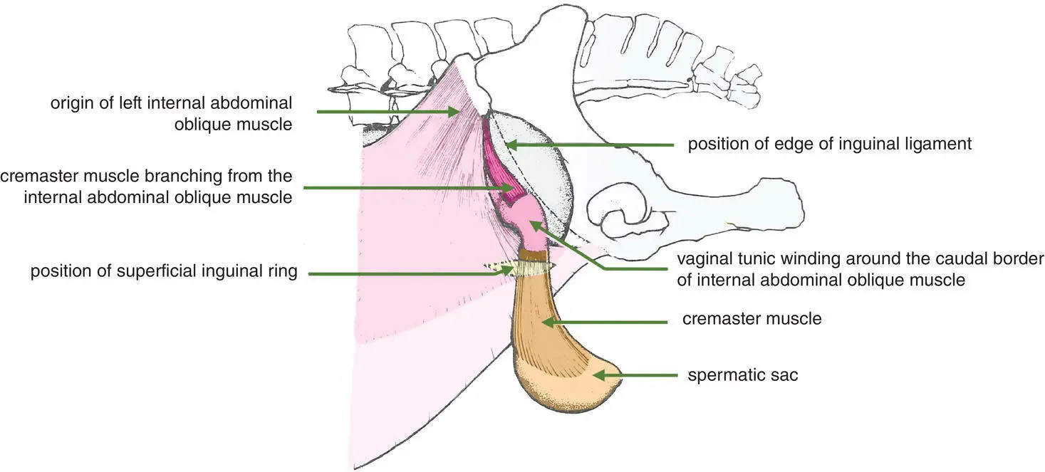
Figure 1.6 Lateral view of inguinal area of horse showing the internal abdominal oblique muscle. The left external abdominal oblique muscle has been removed although the position of the left superficial inguinal ring is shown. The mid‐section of the left cremaster muscle has been excised to expose the vaginal tunic.
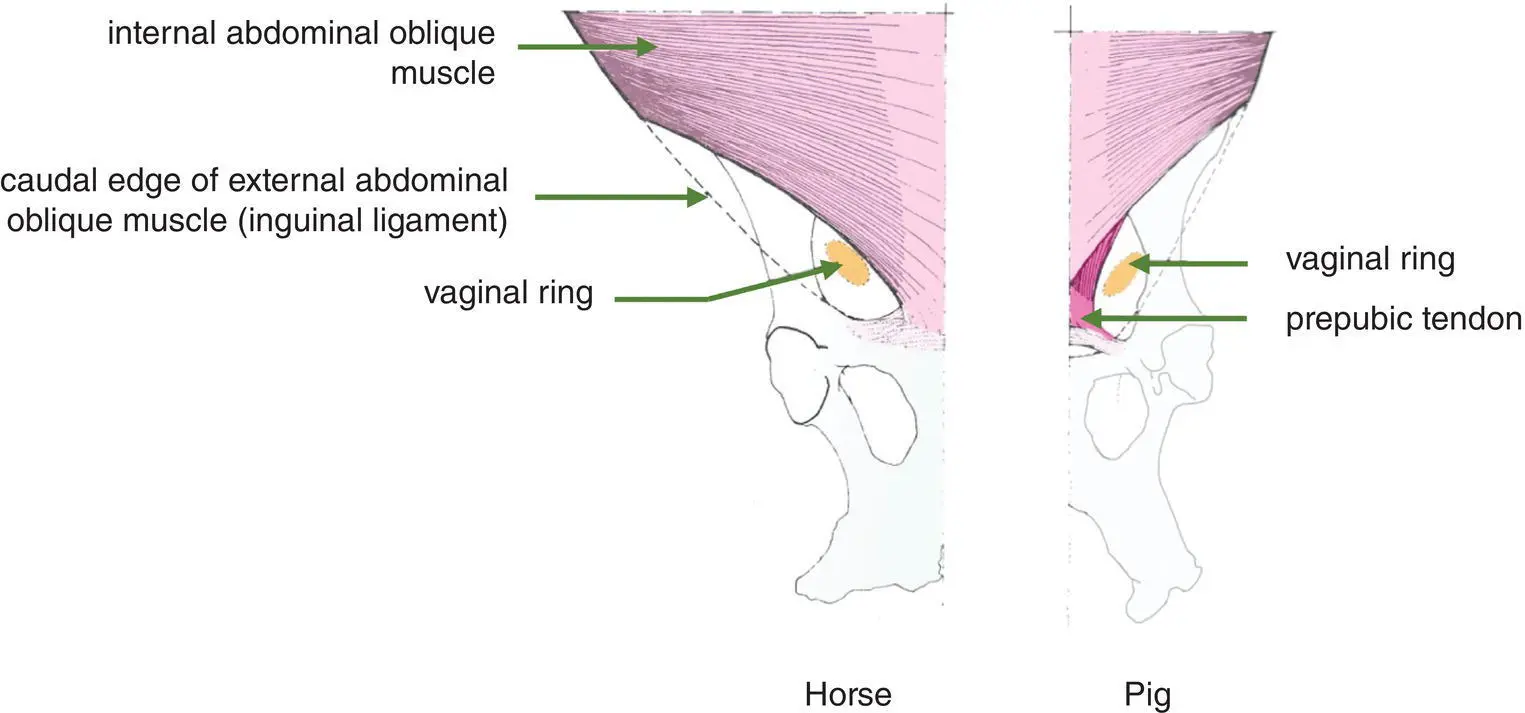
Figure 1.7Ventral view of inguinal region showing internal abdominal oblique muscle.
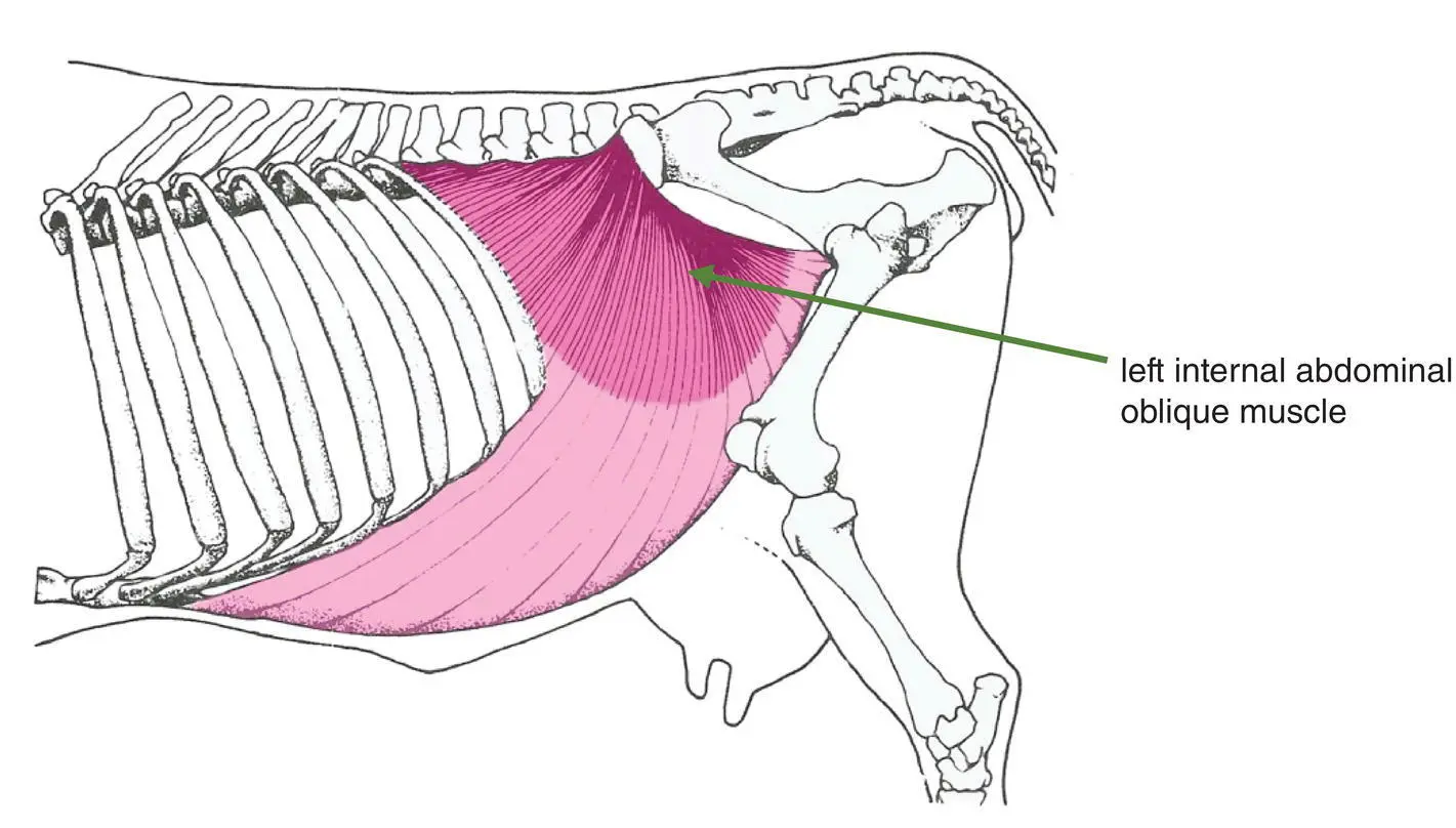
Figure 1.8 Lateral view of abdomen of ox showing left abdominal oblique muscle. The external abdominal oblique has been removed.
Origin: Tuber coxae and lumbodorsal fascia.
Insertion: Linea alba (except for the most caudal part), last rib and cartilages of the caudal ribs.
Structure: This is a sheet of muscle and tendon with the fibres running cranioventrally. It is muscular at its origin and becomes tendinous ventrally. In the male a slip of the internal abdominal oblique muscle passes through the inguinal canal on the lateral aspect of the vaginal process and becomes the cremaster muscle (see Section 16.4).
Species variations: The fibres of this muscle run almost ventrally in the dog. In carnivores the tendinous portion divides to pass dorsally and ventrally to the rectus abdominis muscle in the cranial third of the abdomen; it passes only ventrally in the caudal two‐thirds of the abdomen. In the oxthe internal abdominal oblique is quite substantial, being the largest flank muscle in this species; its tendon passes both ventrally and dorsally to the rectus abdominis. In the horsethe internal abdominal oblique muscle originates only from the tuber coxae, and its tendon passes ventrally to the rectus abdominis. See Figures 1.10a–c for a summary of the species variation of the sheath of the rectus abdominis.
1.3.6 Transverse abdominal muscle ( Figure 1.9)
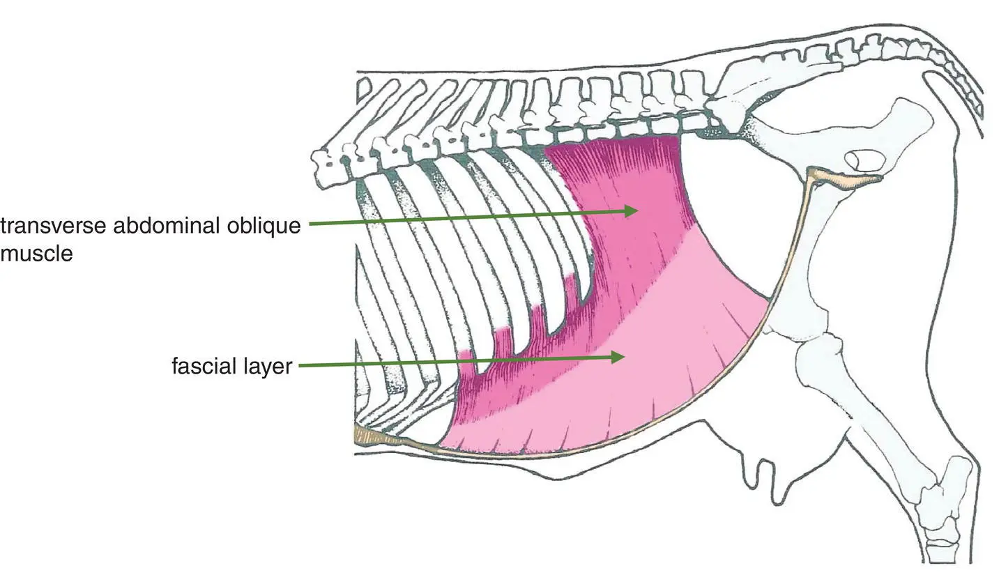
Figure 1.9 Medial view of abdomen of ox showing the transverse abdominal muscle.
Origin: The medial surfaces of the ventral parts of the caudal ribs and the deep layers of the lumbodorsal fascia.
Insertion: The linea alba.
Structure: Again this muscle is sheet‐like, although its fibres run ventrally and transversely to the longitudinal axis. Caudally the muscle thins out to only a fascial layer.
Species variations:In the dogthe cranial two‐thirds of the tendon pass dorsally to the rectus abdominis with the caudal third passing ventrally.
1.3.7 Retroperitoneal fascia
This tissue layer is equivalent to the superficial fascia but less defined. Its significance is due to its large fat content in the adult pig, fat ponies and beef breeds of cattle. Where the fascia is minimal the peritoneum is closely applied to the transverse abdominal muscle. The falciformligament (see Sections 3.3and 3.4) is a fold of peritoneum attached to the liver. It is a remnant of the peritoneum that contained the umbilical vein of the foetus; it attaches to the abdominal wall at the umbilicus.
1.3.8 Parietal peritoneum
This peritoneal layer lines the whole abdominal wall. It is a largely transparent and delicate layer that is reflected as mesenteries that are continuous with the visceral peritoneum that covers the abdominal viscera. The peritoneum comprises an outer layer of simple squamous epithelium called the mesothelium and is supported by a layer of loose connective tissue.
1.4 The Sheath of the Rectus Abdominis Muscle ( Figures 1.10a–c)
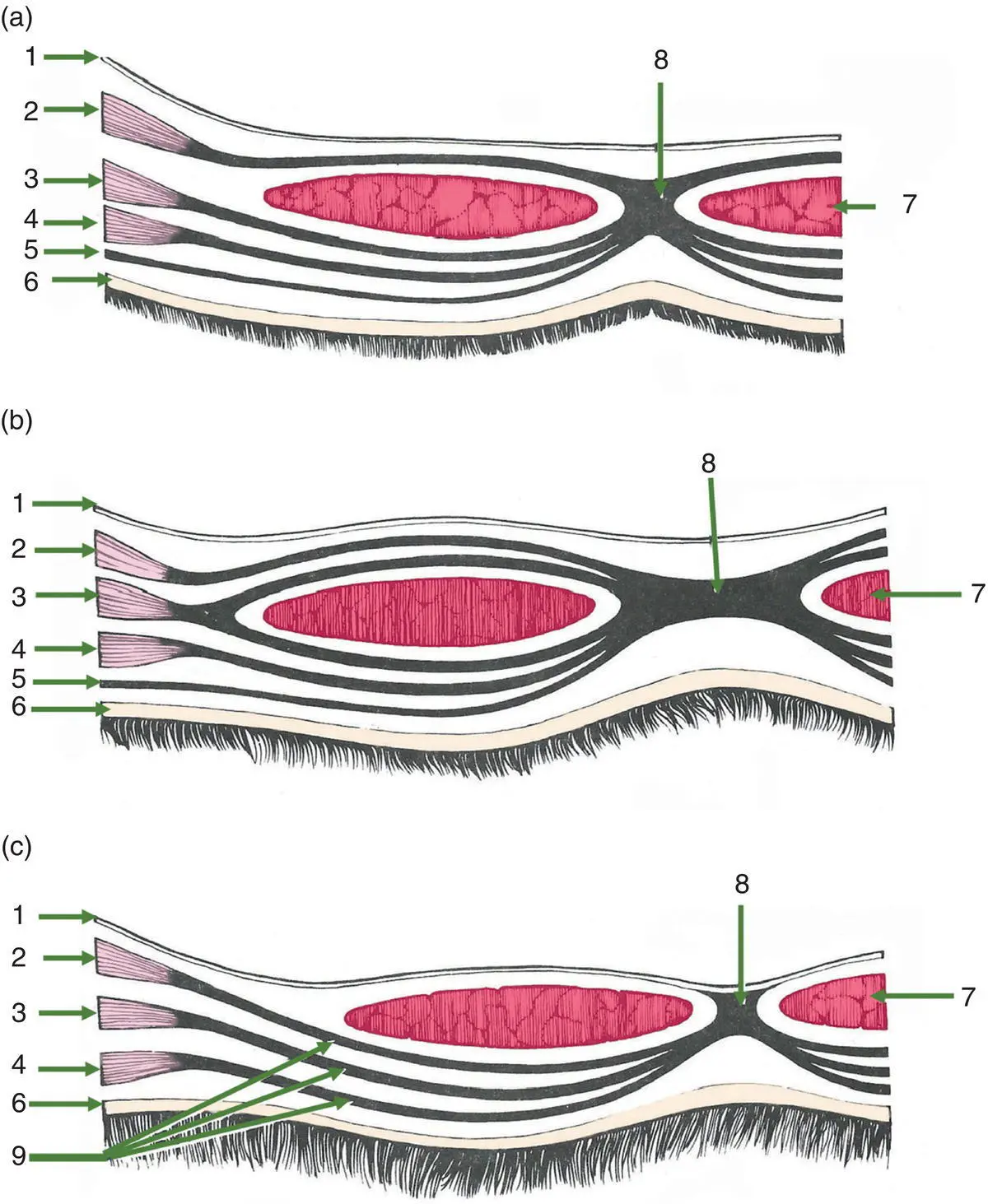
Figure 1.10 Transverse sections through the ventral body wall to show the species variation in the sheath of the rectus abdominis. (a) The horse, (b) the ox and (c) the dog (caudal third of abdomen only).1 = parietal peritoneum; 2 = transverse abdominis muscle; 3 = interior oblique abdominis muscle; 4 = exterior oblique abdominis muscle; 5 = yellow abdominis tunic; 6 = skin; 7 = rectus abdominis muscle; 8 = linea alba; 9 = ventral sheath of rectus abdominis muscle
The aponeuroses of the external, internal and transverse abdominal oblique muscles together form a sheath that encloses the rectus abdominis muscle either side of the midline of the abdominal wall. There are species differences and, in the dog, variations in the craniocaudal location.
Species variations:In the caudal third of the abdomen of the dogthe tendon of the transverse abdominal muscle lies ventral to the rectus abdominis muscle. In the middle third of the abdomen the transverse abdominal tendon lies dorsal, and that of the internal abdominal oblique muscle passes ventral to the rectus abdominis (as in the horse). In the cranial third of the abdomen the tendon of the transverse abdominal muscle lies dorsal to the rectus muscle. In addition, the internal oblique tendon divides into a ventral and dorsal portion (as in the ox).
In the horsethe aponeurosis of the internal oblique muscle lies ventral to the rectus abdominis. In addition, in this species, the yellow abdominal tunic is present.
In the oxthe aponeurosis of the internal oblique divides to pass on both sides of the rectus abdominis, and a yellow abdominal tunic is again present. In this species the linea alba is particularly wide.
1.5 Clinical Importance of the Ventral Body Wall
A surgical incision in the abdominal wall is called a laparotomy. It may be made in the midline, to either side of the midline or in the flank on either side. The choice of location of the laparotomy depends on a number of factors:
Читать дальшеИнтервал:
Закладка:
Похожие книги на «King's Applied Anatomy of the Abdomen and Pelvis of Domestic Mammals»
Представляем Вашему вниманию похожие книги на «King's Applied Anatomy of the Abdomen and Pelvis of Domestic Mammals» списком для выбора. Мы отобрали схожую по названию и смыслу литературу в надежде предоставить читателям больше вариантов отыскать новые, интересные, ещё непрочитанные произведения.


Обсуждение, отзывы о книге «King's Applied Anatomy of the Abdomen and Pelvis of Domestic Mammals» и просто собственные мнения читателей. Оставьте ваши комментарии, напишите, что Вы думаете о произведении, его смысле или главных героях. Укажите что конкретно понравилось, а что нет, и почему Вы так считаете.