Geoff Skerritt - King's Applied Anatomy of the Abdomen and Pelvis of Domestic Mammals
Здесь есть возможность читать онлайн «Geoff Skerritt - King's Applied Anatomy of the Abdomen and Pelvis of Domestic Mammals» — ознакомительный отрывок электронной книги совершенно бесплатно, а после прочтения отрывка купить полную версию. В некоторых случаях можно слушать аудио, скачать через торрент в формате fb2 и присутствует краткое содержание. Жанр: unrecognised, на английском языке. Описание произведения, (предисловие) а так же отзывы посетителей доступны на портале библиотеки ЛибКат.
- Название:King's Applied Anatomy of the Abdomen and Pelvis of Domestic Mammals
- Автор:
- Жанр:
- Год:неизвестен
- ISBN:нет данных
- Рейтинг книги:5 / 5. Голосов: 1
-
Избранное:Добавить в избранное
- Отзывы:
-
Ваша оценка:
King's Applied Anatomy of the Abdomen and Pelvis of Domestic Mammals: краткое содержание, описание и аннотация
Предлагаем к чтению аннотацию, описание, краткое содержание или предисловие (зависит от того, что написал сам автор книги «King's Applied Anatomy of the Abdomen and Pelvis of Domestic Mammals»). Если вы не нашли необходимую информацию о книге — напишите в комментариях, мы постараемся отыскать её.
King’s Applied Anatomy of the Abdomen and Pelvis of the Domestic Mammals King’s Applied Anatomy of the Abdomen and Pelvis of the Domestic Mammals
King's Applied Anatomy of the Abdomen and Pelvis of Domestic Mammals — читать онлайн ознакомительный отрывок
Ниже представлен текст книги, разбитый по страницам. Система сохранения места последней прочитанной страницы, позволяет с удобством читать онлайн бесплатно книгу «King's Applied Anatomy of the Abdomen and Pelvis of Domestic Mammals», без необходимости каждый раз заново искать на чём Вы остановились. Поставьте закладку, и сможете в любой момент перейти на страницу, на которой закончили чтение.
Интервал:
Закладка:
1 The avascularity of the linea alba resulting in slow healing; this is a particular problem especially in cattle where the linea alba is extensive.
2 The bulk and weight of the abdominal contents leading to slow healing and risk of herniation.
3 In the dog the ventral sheath of the rectus abdominis is particularly strong, and failure to suture this may result in breakdown of a midline incision.
4 In a midline incision contraction of the muscles of the abdominal wall tends to retract the wound edges laterally.
5 Flank incisions should be parallel to the muscle fibres to minimise bleeding from the vascular muscular tissue.
6 In the cow a further complication of a midline incision is that branches of the mammary vein may cross the midline to anastomose with the opposite mammary vein.
1.6 The Inguinal Canal ( Figures 1.11and 1.12)
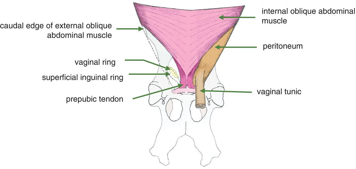
Figure 1.11 Ventral view of inguinal canal of the pig. The left side of the diagram shows the superimposition of the superficial inguinal ring almost directly upon the vaginal ring in this species.

Figure 1.12 Lateral view of the inguinal canal of the horse. A window has been cut from the pelvic part of the tendon of the external abdominal oblique muscle immediately adjoining the superficial inguinal ring. This exposes that part of the internal oblique abdominal muscle which forms the medial (deep) wall of the inguinal canal.
The inguinal canal is a potential space extending between the superficial and deep inguinal rings. The canal does not have a surrounding wall. The external opening (superficial inguinal ring) is a slit in the aponeurosis thereby dividing it into two parts, an abdominal part (cranially) and a pelvic part (caudally).
Species variations: The deep inguinal ring is different in the pigfrom that in the horse due to the different extent to which the internal oblique abdominal muscle inserts caudally. In the horsethe deep inguinal ring is small, being bordered caudally by the inguinal ligament and cranially by the caudal edge of the internal oblique muscle ( Figure 1.12) In the pigthe deep inguinal ring is larger, bordered caudally by the inguinal ligament, cranially by the caudal edge of the internal abdominal oblique muscle and medially by the lateral border of the rectus abdominal muscle and the prepubic tendon ( Figure 1.11). In the other domestic animals, the anatomy of the inguinal ring is between the horse and the pig but tends to be closer to the latter.
In the male foetus of all species an outpouching of parietal peritoneum, the vaginal process(Figure 16.6), enters the inguinal canal. The gubernaculum(see Section 16.9) develops from mesenchyme partly within the inguinal canal and joins the testes to the scrotum. In the adult male the inguinal canal contains the vaginal tunic, the cremaster muscle and the spermatic cord in addition to the external pudendal artery and vein, the inguinal lymph vessels and nerves. In the adult female it is only in the bitch that a rudimentary vaginal process extends through the inguinal canal.
1.7 Hernias
A hernia occurs when an organ or mesentery pushes through an opening in the muscle or tissue that normally holds it in place. Hernias occur most commonly in the abdomen when there is a deficit or weakness in the abdominal wall, but they may also occur at the diaphragm or the perineum. The several sites where hernias may occur are as follows:
1 Inguinal
2 Umbilical
3 Perineal
4 Diaphragmatic
5 Post‐operative.
1.7.1 Inguinal hernia
The vaginal process develops in the embryo as an extension of the parietal peritoneum. Therefore the cavity of the vaginal process is continuous with the peritoneal cavity via the vaginal ring. In the male of all species and the bitch it is possible for abdominal contents (e.g. small intestine or great omentum) to protrude through the vaginal ring and enter the vaginal process. Within the vaginal process the herniated organ or tissue passes through the inguinal canal and may enter the scrotum. An inguinal hernia may or may not be reducible; an irreducible hernia may become strangulated if the blood supply becomes interrupted.
Congenital inguinal hernias are common in pigs, but in sheep they are thought to be a result of trauma. In the horse inheritance has not been proven, but they are more common in certain breeds.
1.7.2 Umbilical hernia
Normally, at birth, the umbilical ring closes and the umbilical blood vessels, the vitelline duct and the allantoic stalk begin to degenerate. If contraction of the umbilical ring does not occur completely it is possible for abdominal contents to enter the aperture and appear as a soft swelling beneath the umbilical scar.
1.7.3 Perineal hernia
The perineum is the part of the body wall that occludes the caudal opening of the pelvic cavity; it surrounds the anus and the urogenital opening. The major portion of the perineum, comprising the levator ani and coccygeus muscles, is called the pelvic diaphragm. A hernia through the pelvic diaphragm can occur as a result of a defect in the musculature. This is a fairly complex anatomical area including the external anal sphincter, superficial gluteal muscle and sacrotuberous ligament.
Perineal hernias occur mainly in older male dogs and certain breeds, e.g. the Boxer, Boston Terrier, Pekingese and crossbreds.
1.7.4 Diaphragmatic hernia
The diaphragm separates the abdominal cavity from the thorax. Sudden increases in pressure in the abdomen can result in tears of the diaphragm with the consequence that abdominal organs can be forced into the thoracic cavity. This situation can occur as the result of a road traffic accident. Congenital hernias of the diaphragm are of rare occurrence in the dog and horse.
1.7.5 Post‐operative hernia
A surgical incision in the abdominal wall is normally closed either with sutures or staples. If there is breakdown along the incision, there is a risk of herniation of abdominal organs. This situation may result from a variety of circumstances, e.g. poor healing of the incision due to inadequate vascularisation, pressure from the weight of the abdominal organs, faulty suturing technique and interference with the wound by the patient.
2 Gastrointestinal Function
2.1 Introduction
In simple life forms, e.g. unicellular organisms such as the amoeba, food particles are ingested by a process of active envelopment with the resulting formation of a food vacuole within the cytoplasm. In higher orders of animal life this process plays no significant part in the process of digestion, although it is retained by macrophagecells in certain cellular activities such as phagocytosis, the ingestion or engulfing of particulate matter. Macrophages are a type of white blood cell found mainly in connective tissue and blood.
The development of the digestive tract involves cell specialisation occurring within the different regions of the tract and in relation to the functions of these different regions. Thus, while certain cells are specialised for absorbing food in a suitably processed form, other cells are primarily protective in function or are associated with transportation and with pre‐ treatment of the food to allow its utilisation. Further specialisation has evolved in relation to the nature of the food, so that marked variations occur in the gastrointestinal anatomy in herbivores as compared to carnivores. In general, however, the tract of higher vertebrates is divided into distinct anatomical regions, namely the buccal cavity, oesophagus, stomach, small intestines, large intestines and rectum. Further subdivisions of these regions are present.
Читать дальшеИнтервал:
Закладка:
Похожие книги на «King's Applied Anatomy of the Abdomen and Pelvis of Domestic Mammals»
Представляем Вашему вниманию похожие книги на «King's Applied Anatomy of the Abdomen and Pelvis of Domestic Mammals» списком для выбора. Мы отобрали схожую по названию и смыслу литературу в надежде предоставить читателям больше вариантов отыскать новые, интересные, ещё непрочитанные произведения.


Обсуждение, отзывы о книге «King's Applied Anatomy of the Abdomen and Pelvis of Domestic Mammals» и просто собственные мнения читателей. Оставьте ваши комментарии, напишите, что Вы думаете о произведении, его смысле или главных героях. Укажите что конкретно понравилось, а что нет, и почему Вы так считаете.