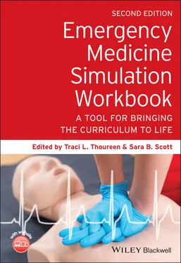
Emergency Medicine Simulation Workbook
Здесь есть возможность читать онлайн «Emergency Medicine Simulation Workbook» — ознакомительный отрывок электронной книги совершенно бесплатно, а после прочтения отрывка купить полную версию. В некоторых случаях можно слушать аудио, скачать через торрент в формате fb2 и присутствует краткое содержание. Жанр: unrecognised, на английском языке. Описание произведения, (предисловие) а так же отзывы посетителей доступны на портале библиотеки ЛибКат.
- Название:Emergency Medicine Simulation Workbook
- Автор:
- Жанр:
- Год:неизвестен
- ISBN:нет данных
- Рейтинг книги:3 / 5. Голосов: 1
-
Избранное:Добавить в избранное
- Отзывы:
-
Ваша оценка:
- 60
- 1
- 2
- 3
- 4
- 5
Emergency Medicine Simulation Workbook: краткое содержание, описание и аннотация
Предлагаем к чтению аннотацию, описание, краткое содержание или предисловие (зависит от того, что написал сам автор книги «Emergency Medicine Simulation Workbook»). Если вы не нашли необходимую информацию о книге — напишите в комментариях, мы постараемся отыскать её.
Emergency Medicine Simulation Workbook
Emergency Medicine Simulation Workbook — читать онлайн ознакомительный отрывок
Ниже представлен текст книги, разбитый по страницам. Система сохранения места последней прочитанной страницы, позволяет с удобством читать онлайн бесплатно книгу «Emergency Medicine Simulation Workbook», без необходимости каждый раз заново искать на чём Вы остановились. Поставьте закладку, и сможете в любой момент перейти на страницу, на которой закончили чтение.
Интервал:
Закладка:
See flow diagram ( Figure 1.15) for scenario changes based on learner actions.
Case Narrative, Continued
For all learners, the case should begin with obtaining a complete history as well as performing a thorough physical exam. The nurse can help the learners to set the patient up on the monitor to obtain vital signs. This should also include obtaining a fingerstick glucose, in light of the patient's slightly altered mental state.
Learners should order initial labs, imaging, IV fluids and medications.
If there is inadequate fluid resuscitation, the patient's hypotension will worsen. Continued failure to appropriately fluid resuscitate will lead to PEA, requiring CPR.
For novice learners, administration of an appropriate fluid bolus (30 cc/kg) will cause the patient's blood pressure to increase to a mean arterial pressure greater than 65 mmHg and his heart rate will slowly lower. With these improvements, the patient's mental status will also improve. Laboratory tests and imaging will then be available for review and appropriate consultation should be made with general surgery/GI, as well as an admitting physician. The case will end after appropriate disposition and consultation have occurred.
For advanced learners, despite initiation of IV fluids and broad‐spectrum antibiotics, the patient will continue to be hypotensive and altered, requiring initiation of vasopressors and intubation. Failure to do so will result in PEA arrest. Once intubation is performed and vasopressors are initiated, the patient will stabilize. Consultation with general surgery/GI and the intensive care admitting physician should then occur. The case will end after appropriate disposition and consultation have occurred.
Instructor Notes
Pathophysiology
Acute (or ascending) cholangitis results from inflammation and infection of the biliary system, often related to a blockage of the bile ducts or hepatic ducts:Biliary obstruction leads to bacterial growth within the bile and subsequent infection.Common causes of blockage include choledocholithiasis, malignancy, strictures, primary sclerosing cholangitis, and AIDS‐related cholangiopathy.
Most frequent pathogens found in acute cholangitis include Escherichia coli, Klebsiella spp., Enterococcus spp., and Enterobacter spp.
Anaerobes such as Bacteroides fragilis and Clostridium perfringens have also been accountable, often in elderly patients or those with prior biliary surgery [1].
Clinical Features
Charcot's triad:Fever, right upper‐quadrant abdominal pain, and jaundice.“Classic” presentation, although it is only around 25% sensitive for acute cholangitis to have all three, 80–90% of patients with acute cholangitis will have fever and/or abdominal pain [2].
Reynold's pentad:Charcot's triad plus hypotension and altered mental state.Severe presentation, but only seen in 5–7% of cases [2].
Diagnosis
Laboratory tests that may support diagnosis:Leukocytosis (with neutrophilic predominance).Transaminitis.Hyperbilirubinemia (conjugated).Elevated alkaline phosphatase.Elevated gamma‐glutamyl transferase level.
Imaging:Ultrasound:Best initial study. Can detect dilation of the common bile duct, gallstones, and other evidence of pathology.CT with IV contrast:Helpful for looking at other potential causes of biliary obstruction.May show complications such as hepatic abscesses.CT findings that may support the diagnosis of cholangitis include dilation of intra‐ or extra‐hepatic biliary ducts, thickening of the ductal walls, presence of gallstones. Magnetic resonance cholangiopancreatography:Sensitive imaging modalityMay not be readily available at all institutions.
Management
Resuscitation:IV fluid administration.Broad‐spectrum IV antibiotics (with Gram‐negative and anaerobic coverage).Vasopressor administration (if needed).
Analgesia.
Specialist consultation (general surgery and/or gastroenterology) for intervention:The type of intervention will depend on etiology:Endoscopic retrograde cholangiopancreatography for biliary drainage, sphincterotomy, stone extraction, biliary stent placement.Open surgical drainage.If a patient is initially seen at a facility without these consult services, transfer to the nearest facility that has these resources should be undertaken [2, 3].
Debriefing Plan
Allow approximately 20–30 minutes for debriefing after this scenario.
Potential Questions for Discussion
What are signs that a patient may have sepsis?
What is the appropriate management of a patient with sepsis?
How can you assess for end‐organ dysfunction in a patient with sepsis?
What are the indications for vasopressor initiation in sepsis?
What is Charcot's triad?
What is Reynold's pentad?
REFERENCES FOR SEPSIS
1 1. Ahmed, M. (2018). Acute cholangitis – an update. World J. Gastrointest. Pathophysiol. 9 (1): 1–7.
2 2. Ely, R., Long, B., and Koyfman, A. (2018). The emergency medicine−focused review of cholangitis. J. Emerg. Med. 54 (1): 64–72.
3 3. Mayumi, T., Okamoto, K., Takada, T. et al. (2018). Tokyo Guidelines 2018: management bundles for acute cholangitis and cholecystitis. J. Hepatobiliary Pancreat. Sci. 25 (1): 96–100.
SIGMOID VOLVULUS
Educational Goals
Learning Objectives
1 Assess a patient presenting with acute abdominal pain, utilizing a focused history and physical exam (MK, PC).
2 Formulate a differential diagnosis for acute abdominal pain (MK).
3 Recognize the signs and symptoms of a possible intestinal obstruction (MK).
4 Demonstrate appropriate utilization of lab tests and imaging studies to evaluate abdominal pain (MK, PC).
5 Recognize an agitated patient and use verbal de‐escalation and negotiation skills (ICS, P).
6 Demonstrate professionalism while treating a patient with behavioral issues (P, ICS).
7 Demonstrate appropriate surgical consultation (P, ICS, SBP).
Critical Actions Checklist
Recognize clinical signs of obstruction (MK).
Obtain appropriate IV access (PC).
Obtain imaging (MK).
Administer analgesic medication and antiemetics for patient's symptoms (PC).
Recognize an abnormal bowel gas pattern (concerning for obstruction) on x‐ray and/or CT (MK).
Maintain a calm and professional composure while communicating with a difficult, disruptive patient (PC, ICS, P).
Consult general surgery for emergent management of volvulus (P, ICS, SBP).
Simulation Set‐Up
| Environment: | ED treatment room. |
| Mannequin: | Adult, male, simulator mannequin moulaged to appear disheveled (e.g. clothes may be slightly dirty and/or torn or used in appearance). |
Props:
Images (see online component for sigmoid volvulus, Scenario 1.3 at https://www.wiley.com/go/thoureen/simulation/workbook2e):Abdominal x‐ray showing evidence of distended sigmoid colon ( Figure 1.16).Radiology report of abdominal x‐ray ( Figure 1.17).CT abdomen/pelvis with IV contrast (static image 1‐cut) ( Figure 1.18).Radiology report of sigmoid volvulus ( Figure 1.19).ECG showing sinus tachycardia ( Figure 1.20).
Laboratory tests (see online component as above):Complete blood count ( Table 1.22).Basic metabolic panel ( Table 1.23).Liver function panel ( Table 1.24).Lipase ( Table 1.25).Lactic acid ( Table 1.26).Troponin ( Table 1.27).Coagulation panel ( Table 1.28).Urinalysis ( Table 1.29).Urine microscopy ( Table 1.30).
Available supplies:
Adult code cart with basic airway supplies.
Читать дальшеИнтервал:
Закладка:
Похожие книги на «Emergency Medicine Simulation Workbook»
Представляем Вашему вниманию похожие книги на «Emergency Medicine Simulation Workbook» списком для выбора. Мы отобрали схожую по названию и смыслу литературу в надежде предоставить читателям больше вариантов отыскать новые, интересные, ещё непрочитанные произведения.
Обсуждение, отзывы о книге «Emergency Medicine Simulation Workbook» и просто собственные мнения читателей. Оставьте ваши комментарии, напишите, что Вы думаете о произведении, его смысле или главных героях. Укажите что конкретно понравилось, а что нет, и почему Вы так считаете.
