Ian Peate - Anatomy and Physiology for Nursing and Healthcare Students at a Glance
Здесь есть возможность читать онлайн «Ian Peate - Anatomy and Physiology for Nursing and Healthcare Students at a Glance» — ознакомительный отрывок электронной книги совершенно бесплатно, а после прочтения отрывка купить полную версию. В некоторых случаях можно слушать аудио, скачать через торрент в формате fb2 и присутствует краткое содержание. Жанр: unrecognised, на английском языке. Описание произведения, (предисловие) а так же отзывы посетителей доступны на портале библиотеки ЛибКат.
- Название:Anatomy and Physiology for Nursing and Healthcare Students at a Glance
- Автор:
- Жанр:
- Год:неизвестен
- ISBN:нет данных
- Рейтинг книги:5 / 5. Голосов: 1
-
Избранное:Добавить в избранное
- Отзывы:
-
Ваша оценка:
Anatomy and Physiology for Nursing and Healthcare Students at a Glance: краткое содержание, описание и аннотация
Предлагаем к чтению аннотацию, описание, краткое содержание или предисловие (зависит от того, что написал сам автор книги «Anatomy and Physiology for Nursing and Healthcare Students at a Glance»). Если вы не нашли необходимую информацию о книге — напишите в комментариях, мы постараемся отыскать её.
Everything you need to know about anatomy and physiology … An ideal introduction and revision guide for anatomy and physiology
www.wiley.com
www.wiley.com/email
All content reviewed by students for students
www.reviewnursingbooks.com
www.wiley.com/buy/9781119757207 at a Glance
at a Glance
At a Glance
Anatomy & Physiology for Nursing & Healthcare Students
Anatomy and Physiology for Nursing and Healthcare Students at a Glance — читать онлайн ознакомительный отрывок
Ниже представлен текст книги, разбитый по страницам. Система сохранения места последней прочитанной страницы, позволяет с удобством читать онлайн бесплатно книгу «Anatomy and Physiology for Nursing and Healthcare Students at a Glance», без необходимости каждый раз заново искать на чём Вы остановились. Поставьте закладку, и сможете в любой момент перейти на страницу, на которой закончили чтение.
Интервал:
Закладка:
Those terms that are used to describe locations and positions reference a person in what is known as the anatomical position. The international standard anatomical position is standing upright as seen in Figure 1.1; whenever referring to anatomical terms, always apply them to the person standing in the anatomical position. By using this as a standard posture for anatomical descriptions, confusion can be avoided even when in reality the person is in some other position.
The position is defined as if the body is standing erect with hips and knees extended, head forward facing, eyes open looking directly forwards with the mouth closed. The arms are by the sides (shoulders adducted), the palms are facing forward (elbows extended and wrists supinated), and the feet together. In this position, the radius and ulna are parallel.
Anatomical terms
It is important to understand and use anatomical terminology when making a description of body parts so there is a shared method of communicating (a common language) with nurses, doctors and other healthcare staff. This is done in order to accurately describe anatomical locations irrespective of their language. Knowing about anatomical terms makes things safer and clearer and will save time.
Anatomical terms (using a specific vocabulary) describe the directions within the body and also the body’s reference planes, cavities and regions ( Figure 1.2). There are a number of occasions when a nurse or other healthcare worker is required to record information in nursing or medical notes with the intention of communicating with others or telling others the exact body part or location. Standard terms for describing human anatomy including the body and its organs are required to do this.
Directional terms
Directional terms describe the positions of structures relative to other structures or locations in the body.
When referring to left and right, reference is being made to the left and right side of the person standing in the anatomical position, not to the left and right side of the observer.
Anterior (also called ventral) refers to the front of the body and posterior (dorsal is also used) to the back of the body. The nipples, for example, are on the anterior (ventral) surface of the body, the buttocks are superior (dorsal).
Superior means above, towards the head, and inferior means below, towards the feet. The umbilicus is superior to the genitalia but inferior to the head.
Proximal and distal are only used to describe two points on the same arm or leg. Proximal means close to where the arm or leg is inserted into the body. Distal means further away from where the arm or leg is inserted into the body. The knee is proximal to the ankle as the knee is closer to where the leg inserts into the body. With regard to the arm, the wrist is distal to the elbow as the wrist is further away from where the arm inserts into the body.
Medial refers to any point that is closer to the midline of the body and lateral means any point further away from the midline. The midline is an imaginary line that separates the body in half vertically. The inner thigh is medial and the outer thigh is lateral.
Planes
To describe the anatomical positions of the internal structures, planes or sections are used ( Figure 1.3). There are four planes.
1 Sagittal
2 Frontal
3 Transverse
4 Oblique
The sagittal, vertical (top to bottom) plane divides the body into left and right sides. It is known as a midsagittal plane when it divides the body down the middle into equal left and right sides. If the divide does not pass exactly midline, this is known as parasagittal. The frontal plane divides the body into anterior (ventral) and posterior (dorsal) portions. The transverse plane divides the body into superior and inferior portions. The oblique plane is a slanted plane (at an angle) passing through the body.
Body cavities
These areas contain internal organs. The two main cavities are the dorsal and ventral cavities ( Figure 1.4). The dorsal cavity (sometimes called caudal) is on the posterior of the body, containing the cranial cavity and spinal cavity. The ventral cavity is on the anterior of the body, divided into the thoracic cavity and abdominopelvic cavity; the diaphragm divides the ventral cavity into two subcavities: thoracic and abdominal ( Table 1.1).
2 Genetics and genomics
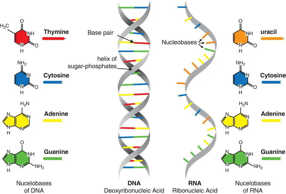
Figure 2.1 DNA and RNA.
Table 2.1 Types of RNA.
| Type of RNA | Description |
|---|---|
| Messenger RNA (mRNA) | Copies portions of genetic code, a process known as transcription, and transports these copies to ribosomes, the cellular factories that facilitate the production of proteins from this code |
| Transfer RNA (tRNA) | Responsible for bringing amino acids, basic protein building blocks, to these protein factories, in response to the coded instructions introduced by the mRNA. This protein‐building process is called translation |
| Ribosomal RNA (rRNA) | The protein builder of the cell, without which protein production would not occur |
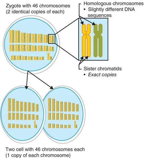
Figure 2.2 Mitosis.

Figure 2.3 Meiosis.
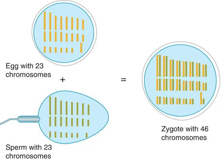
Figure 2.4 Fertilisation.
Genetics is the study of the way particular features or diseases are inherited through genes passed down from one generation to the next. The idea of having a single gene for this or a single gene for that (determining fate) is not a good way of describing the complexity of genes. There are groups of genes that work together, influenced by a variety of environmental and other factors. The genome can be seen as the body’s instruction manual, with a copy of it in almost every healthy cell in the body. The study of the genome and the technologies that are required to analyse and interpret it is known as genomics.
DNA and RNA
Both deoxyribonucleic acid (DNA) and ribonucleic acid (RNA) are made of nucleotides (bases) which are the building blocks, responsible for the storage and reading of genetic information that underpins all life. DNA encodes all genetic information, also acting as a biological store allowing the blueprint of life to be passed between generations. RNA reads and then decodes what is stored. This is a multistep process with specialised RNAs for each step ( Table 2.1).
DNA and RNA are nucleic acids, known as linear polymers, consisting of sugars, phosphates and bases, but there are differences between the two. The differences permit the two molecules to work together, fulfilling essential roles. See Figure 2.1for the differences. The complementary base pairs in DNA are adenine (‘A’), thymine (‘T’), guanine (‘G’) and cytosine (‘C’) and RNA shares adenine (‘A’), guanine (‘G’) and cytosine (‘C’) with DNA, but contains uracil (‘U’) instead of thymine ( Figure 2.1).
Читать дальшеИнтервал:
Закладка:
Похожие книги на «Anatomy and Physiology for Nursing and Healthcare Students at a Glance»
Представляем Вашему вниманию похожие книги на «Anatomy and Physiology for Nursing and Healthcare Students at a Glance» списком для выбора. Мы отобрали схожую по названию и смыслу литературу в надежде предоставить читателям больше вариантов отыскать новые, интересные, ещё непрочитанные произведения.


Обсуждение, отзывы о книге «Anatomy and Physiology for Nursing and Healthcare Students at a Glance» и просто собственные мнения читателей. Оставьте ваши комментарии, напишите, что Вы думаете о произведении, его смысле или главных героях. Укажите что конкретно понравилось, а что нет, и почему Вы так считаете.