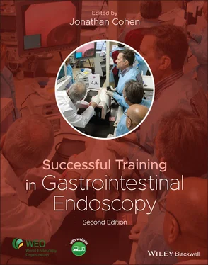
Successful Training in Gastrointestinal Endoscopy
Здесь есть возможность читать онлайн «Successful Training in Gastrointestinal Endoscopy» — ознакомительный отрывок электронной книги совершенно бесплатно, а после прочтения отрывка купить полную версию. В некоторых случаях можно слушать аудио, скачать через торрент в формате fb2 и присутствует краткое содержание. Жанр: unrecognised, на английском языке. Описание произведения, (предисловие) а так же отзывы посетителей доступны на портале библиотеки ЛибКат.
- Название:Successful Training in Gastrointestinal Endoscopy
- Автор:
- Жанр:
- Год:неизвестен
- ISBN:нет данных
- Рейтинг книги:3 / 5. Голосов: 1
-
Избранное:Добавить в избранное
- Отзывы:
-
Ваша оценка:
- 60
- 1
- 2
- 3
- 4
- 5
Successful Training in Gastrointestinal Endoscopy: краткое содержание, описание и аннотация
Предлагаем к чтению аннотацию, описание, краткое содержание или предисловие (зависит от того, что написал сам автор книги «Successful Training in Gastrointestinal Endoscopy»). Если вы не нашли необходимую информацию о книге — напишите в комментариях, мы постараемся отыскать её.
Teaches trainee gastroenterologists the endoscopic skills needed to meet the medical training requirements to practice gastroenterology and helps clinical specialists refresh their skills to pass their recertification Successful Training in Gastrointestinal Endoscopy, Second Edition
Successful Training in Gastrointestinal Endoscopy, Second Edition
Successful Training in Gastrointestinal Endoscopy — читать онлайн ознакомительный отрывок
Ниже представлен текст книги, разбитый по страницам. Система сохранения места последней прочитанной страницы, позволяет с удобством читать онлайн бесплатно книгу «Successful Training in Gastrointestinal Endoscopy», без необходимости каждый раз заново искать на чём Вы остановились. Поставьте закладку, и сможете в любой момент перейти на страницу, на которой закончили чтение.
Интервал:
Закладка:
16 Chapter 25Table 25.1 Characteristics of some the currently used partially or fully cov...Table 25.2 Characteristics of currently used covered stents to minimize recu...Table 25.3 Characteristics of currently available enteral stents.
17 Chapter 28Table 28.1 Suggested standard for abnormal values for endoscopic sphincter o...
18 Chapter 33Table 33.1 Check list for mastery in training to recognize and manage compli...Table 33.2 Risk factors for post‐ERCP pancreatitis in multivariate analyses.
19 Chapter 38Table 38.1 ASGE IT&T Center Interactive, Hands‐On, and Didactic Courses in 2...
20 Chapter 39Table 39.1 Guidelines for assessment of EUS competence *[11].Table 39.2 Guidelines for assessment of competence in ERCP* [11].Table 39.3 Levels of ERCP complexity. (Adapted from ASGE workshop, reference...
List of Illustrations
1 Chapter 1 Figure 1.1 An example of a typical apprenticeship contract in colonial Ameri... Figure 1.2 Roller demonstration model (1971): Homemade model showing alpha l... Figure 1.3 Hair dryer tube model (1972). Figure 1.4 St Mark’s/KeyMed model (1975): Commercially available with semire... Figure 1.5 Electronic targeting model (1975): Tested hand–eye coordination.... Figure 1.6 Endoscopic Pong Game (1977): Tested hand–eye coordination. Figure 1.7 Imperial College/St Mark’s simulator (1980): Limited shaft insert... Figure 1.8 Imperial College/St Mark’s simulator MK2 (1985): Full shaft inser... Figure 1.9 (a) Novel ERCP endotrainer introduced by Joseph Leung, MD that al... Figure 1.10 (a) Artificial tissue colonoscopy “Phantom” simulator, U. of Tüb... Figure 1.11 (a) Compact‐EASIE porcine model hemostasis simulator. (b) Close‐... Figure 1.12 Immersion AccuTouch ®colonoscopy simulator. Figure 1.13 GI Mentor II (Simbionix) colonoscopy virtual reality simulator.... Figure 1.14 Remote teaching of flexible endoscopy from New York to Kyabirwa,...
2 Chapter 2 Figure 2.1 An example of a stepwise or “progressive” model of simulation‐bas...
3 Chapter 4 Figure 4.1 Stages of endoscopy skill acquisition Figure 4.2 “Preparation‐Training‐Wrap‐up” framework outlining the components... Figure 4.3 Set‐up of an endoscopy suite during training to optimize the trai... Figure 4.4 ACT model of performance enhancing feedback: A sk the trainee, C on... Figure 4.5 This image shows a common loop visualized with the assistance of ...
4 Chapter 5 Figure 5.1 White light high‐resolution endoscopy (HRE) image of (a) early er... Figure 5.2 Long‐segment BE is evident on this low‐magnification white light ... Figure 5.3 Low‐magnification white light HRE image of normal gastric antrum ... Figure 5.4 Low‐magnification white light HRE image of normal fundus with two... Figure 5.5 Low‐magnification white light HRE image of normal duodenal bulb. ... Figure 5.6 White light HRE view of normal duodenal folds. The villiform arch... Figure 5.7 White light HRE view of (a) esophageal squamous cell carcinoma (S... Figure 5.8 Recommended grip technique for the endoscope with left index and ... Figure 5.9 (a) Upward tip deflection demonstrated with the thumb pushing the... Figure 5.10 White light HRE view showing erythema of the aryepiglottic folds... Figure 5.11 Angulation and hooking of the endoscope tip to aid with duodenal... Figure 5.12 (a) Mid‐esophageal cancer with luminal obstruction. (b) Subseque... Figure 5.13 Alternative hand position for ESD in which the index and middle ... Figure 5.14 Sample case from GI Mentor ™.
5 Chapter 6 Figure 6.1 Colon anatomy. This illustration demonstrates the anatomy of the ... Figure 6.2 Rectal anatomy. Just inside the anal sphincter muscles, the denta... Figure 6.3 Endoscopic view of the rectum. This endoscopic view shows the sem... Figure 6.4 Endoscopic view of the transverse colon. The transverse colon is ... Figure 6.5 Endoscopic view of the hepatic flexure. At the splenic and hepati... Figure 6.6 Endoscopic view of the cecum. This view of the cecum demonstrates... Figure 6.7 Endoscope options. Four different endoscopes can be used for lowe... Figure 6.8 How to hold the scope. These images demonstrate the proper manner... Figure 6.9 Scope dials. The colonoscope's dials are shown here. The large in... Figure 6.10 Scope valves. The top “red” valve activates the scope's suction ... Figure 6.11 Trap in suction circuit. When a polyp is removed with a snare, a... Figure 6.12 Rectal intubation techniques. This illustration demonstrates the... Figure 6.13 Torque to change from horizontal to vertical. The two images dep... Figure 6.14 One‐handed technique. With the one‐handed advancement technique,... Figure 6.15 Two‐handed technique. With the two‐handed technique, the right h... Figure 6.16 Lumen identification. When the lumen cannot be readily identifie... Figure 6.17 Retroflex views in rectum. Retroflexion in the rectum allows for... Figure 6.18 Some examples of key colonic abnormalities that trainees should ... Figure 6.19 Snare polypectomy. When a snare is required to remove a polyp, t... Figure 6.20 Looping causing perforation. In this sigmoid loop, the scope is ... Figure 6.21 Force vector. In this illustration, the tip of the scope is defl... Figure 6.22 Sigmoid loop. As the scope makes multiple turns in the sigmoid c... Figure 6.23 Alpha‐loop. One of the most common types of sigmoid loop formati... Figure 6.24 N‐loop. The N‐loop is also a common type of loop formation in th... Figure 6.25 Reverse alpha‐loop. A reverse alpha‐loop follows a similar confi... Figure 6.26 Transverse colon loop. Like the sigmoid, the transverse colon is... Figure 6.27 Acute turn. When attempting to navigate an acute turn, novices w... Figure 6.28 Torque to open folds. When less acute turns are encountered, the... Figure 6.29 Terminal ileum intubation. To intubate the ileocecal valve, the ... Figure 6.30 Incorrect TI maneuver. Like the acute turns, novice endoscopists... Figure 6.31 A trainee using a virtual reality colonoscopy simulator. Figure 6.32 A static mechanical model, the colonoscopy Erlangen active simul... Figure 6.33 Ex vivo models. In these images, harvested animal models are lai... Figure 6.34 An endoscopist practices on an ex vivo bovine colon model. Figure 6.35 Learning curves. Mayo's colonoscopy skills assessment tool (MCSA...
6 Chapter 7Figure 7.1 (a) Computerized EUS simulator (GI Mentor II, Simbionix Inc., Cle...Figure 7.2 EUS phantom images.Figure 7.3 (a) EASIE‐R EUS simulator. (b) Depicts a close‐up of the Easie‐R ...Figure 7.4 Depicts an EUS image produced from the EASIE‐R simulator.
7 Chapter 8Figure 8.1 Using one finger to control the air and suction button allows eas...Figure 8.2 Holding the ERCP scope with two fingers on the air and suction bu...Figure 8.3 A combination of 12 different scope maneuvers to position the cat...Figure 8.4 Tip of the papillotome can be used to change the orientation of t...Figure 8.5 An inflated balloon inside the bile duct can be used as a pivot t...Figure 8.6 Image of the Neo‐papilla model (a) and its cartoon schematic (b)....Figure 8.7 (a) EMS (ERCP mechanical simulator) allows in vitro practices wit...Figure 8.8 The Italian ERCP simulator model.
8 Chapter 9Figure 9.1 Small bowel capsule. Image courtesy of Olympus America Inc. and M...Figure 9.2 Upper gastrointestinal capsule. Medtronic, Inc.Figure 9.3 Colon capsule. Medtronic, Inc.Figure 9.4 Capsule endoscopy equipment (desktop computer, printer, recording...Figure 9.5 Jelly Bean test.Figure 9.6 PillCam™ patency capsule. Medtronic, Inc.Figure 9.7 AdvanCE ®device. Advanced Devices.Figure 9.8 Esophageal varices. Medtronic, Inc.Figure 9.9 Barrett’s esophagus. Medtronic, Inc.Figure 9.10 Suspected blood indicator (SBI). Medtronic, Inc.Figure 9.11 Study review in dual mode. Medtronic, Inc.Figure 9.12 Surface ulceration.Figure 9.13 Central umbilication.Figure 9.14 Lobulated mucosa.Figure 9.15 Bridging folds.
Читать дальшеИнтервал:
Закладка:
Похожие книги на «Successful Training in Gastrointestinal Endoscopy»
Представляем Вашему вниманию похожие книги на «Successful Training in Gastrointestinal Endoscopy» списком для выбора. Мы отобрали схожую по названию и смыслу литературу в надежде предоставить читателям больше вариантов отыскать новые, интересные, ещё непрочитанные произведения.
Обсуждение, отзывы о книге «Successful Training in Gastrointestinal Endoscopy» и просто собственные мнения читателей. Оставьте ваши комментарии, напишите, что Вы думаете о произведении, его смысле или главных героях. Укажите что конкретно понравилось, а что нет, и почему Вы так считаете.


