Paul M. Speight - Shear's Cysts of the Oral and Maxillofacial Regions
Здесь есть возможность читать онлайн «Paul M. Speight - Shear's Cysts of the Oral and Maxillofacial Regions» — ознакомительный отрывок электронной книги совершенно бесплатно, а после прочтения отрывка купить полную версию. В некоторых случаях можно слушать аудио, скачать через торрент в формате fb2 и присутствует краткое содержание. Жанр: unrecognised, на английском языке. Описание произведения, (предисловие) а так же отзывы посетителей доступны на портале библиотеки ЛибКат.
- Название:Shear's Cysts of the Oral and Maxillofacial Regions
- Автор:
- Жанр:
- Год:неизвестен
- ISBN:нет данных
- Рейтинг книги:5 / 5. Голосов: 1
-
Избранное:Добавить в избранное
- Отзывы:
-
Ваша оценка:
Shear's Cysts of the Oral and Maxillofacial Regions: краткое содержание, описание и аннотация
Предлагаем к чтению аннотацию, описание, краткое содержание или предисловие (зависит от того, что написал сам автор книги «Shear's Cysts of the Oral and Maxillofacial Regions»). Если вы не нашли необходимую информацию о книге — напишите в комментариях, мы постараемся отыскать её.
Shear’s Cysts of the Oral and Maxillofacial Regions
Shear’s Cysts of the Oral and Maxillofacial Regions Fifth Edition
Shear's Cysts of the Oral and Maxillofacial Regions — читать онлайн ознакомительный отрывок
Ниже представлен текст книги, разбитый по страницам. Система сохранения места последней прочитанной страницы, позволяет с удобством читать онлайн бесплатно книгу «Shear's Cysts of the Oral and Maxillofacial Regions», без необходимости каждый раз заново искать на чём Вы остановились. Поставьте закладку, и сможете в любой момент перейти на страницу, на которой закончили чтение.
Интервал:
Закладка:
The relationship between the glandular odontogenic cyst and neoplasia has been speculative and is based largely on its histological similarity to intraosseous mucoepidermoid carcinoma. This is discussed in detail in Chapter 10. This histological similarity can make it very difficult to reach a definitive diagnosis and may result in misdiagnosis of lesions. A number of papers have reported lesions with histological features of a glandular odontogenic cyst that have recurred as mucoepidermoid carcinomas, but some of these are poorly illustrated and in others the diagnostic criteria are not clear. Nevertheless, this has led to suggestions that the glandular odontogenic cyst may be a precursor lesion, or a ‘benign variant’ of mucoepidermoid carcinoma. The debate is further fuelled by the observation that the glandular odontogenic cyst has a high recurrence rate, leading some authors to label it ‘biologically aggressive’ (Greer et al. 2018 ). Mucoepidermoid carcinoma is characterised by specific rearrangements of the MAML2 gene and large studies have shown that glandular odontogenic cysts do not show this change, suggesting that the two lesions are distinct and that the cyst is not a precursor to the carcinoma (Bishop et al. 2014 ). More recently, two studies have reported lesions with the features of glandular odontogenic cyst that have recurred as MAML2 positive mucoepidermoid carcinomas, and have suggested that this is further evidence that mucoepidermoid carcinomas can arise from glandular odontogenic cysts (Greer et al. 2018 ; Nagasaki et al. 2018 ). In both cases, an alternative explanation is that the primary lesions were also mucoepidermoid carcinomas, but were either misdiagnosed or were histologically indistinguishable from cysts. Taken together, however, these studies raise a number of questions and provide sufficient evidence to justify further research into the relationship between glandular odontogenic cyst and mucoepidermoid carcinoma. The two possibilities to be explored are that glandular odontogenic cyst is a benign variant or precursor of mucoepidermoid carcinoma or, more likely, that in some cases the two lesions are histologically indistinguishable. Until these issues are clarified, care must be taken to use strict criteria for diagnosis and histological assessment must be accompanied by careful assessment of the clinical and radiological features (see Boxes 10.1and 10.2, Tables 10.3 and 10.4).
An Approach to Diagnosis of Cysts of the Jaws
An accurate diagnosis of a cyst requires knowledge, common sense, and a logical approach to the assimilation of all the information available about each lesion. With few exceptions, it is not possible to reach a correct diagnosis without considering the clinical, radiological, and histological features. This is especially relevant to histopathologists, who may be called upon to make a diagnosis on a small biopsy. To do so without considering the clinical and radiological features often leads to diagnostic errors and incorrect management (Barrett et al. 2017 ). In each chapter of this book we present all the features of each cyst type in context and discuss the differential diagnoses and potential errors that might occur if only small biopsies are examined. Throughout we advise on the importance of correlating clinical, radiological, and histological features and suggest that a diagnosis of an intraosseous lesion should never be made without considering the radiology. We also recommend that a diagnosis should never be made on a small biopsy if there is any uncertainty – when in doubt, the pathologist should request a further biopsy. In this chapter, we briefly discuss an approach to making a correct diagnosis and summarise the key features that can assist in making a diagnosis of cysts of the maxillofacial regions ( Tables 2.2– 2.4). With regard to the odontogenic cysts, an accurate diagnosis is facilitated by an understanding of tooth development and the pathogenesis of each cyst type (see ‘Pathogenesis of Cysts’ and Table 2.1)
In most cases, the clinical features of cysts are not specific, since the most common presenting feature is a swelling with few other symptoms. The surgeon therefore must rely on additional features to assist in making a provisional diagnosis. For example, a swelling associated with an absent tooth may suggest a dentigerous cyst, and a small, bluish‐coloured swelling on the lower lip of a child is almost certainly a mucocele. Ultimately, however, a final diagnosis is usually made on histological examination, but before this both the clinician and the pathologist should examine the radiographs or imaging.
Radiology of Cysts of the Jaws
The characteristic radiological feature of all cysts of the jaws is a well‐demarcated radiolucency with a well‐defined and often corticated margin. Further features that assist in diagnosis include the shape and size of the lesion and the site, but in most cases it is the relationship to the teeth that provides the best indication of the type of cyst. A conventional plane radiograph is usually sufficient to determine the extent and relationships of jaw cysts, but computed tomography (CT) and magnetic resonance imaging (MRI) are often useful and may be essential for planning surgery of larger lesions. These relationships are discussed and illustrated in each chapter, but here we present an overview of characteristic radiological signs and the basic principles of an approach to interpreting the radiology. Table 2.2shows the cyst types that have characteristic radiological features, provides a cross reference to the figures in each chapter, and summarises the diagnostic utility of each feature.
Table 2.2 Characteristic radiological features that assist in the diagnosis of cysts of the jaws.
| Cyst type | Radiological sign | Diagnostic utility | Figure references |
|---|---|---|---|
| Radicular cyst | A radiolucency at the apex of a tooth | All radicular cysts are located at the opening of the root canal, almost always at the apex. This feature can be considered diagnostic of radicular cyst if the tooth is also non‐vital. The radicular cyst also lies within the lamina dura. If the tooth is vital then other lesions must be considered, but are rare. This may include cemental lesions (cementoblastoma, cemento‐osseus dysplasia) or, in the anterior maxilla, nasopalatine duct cyst | Figures 2.2and 3.4 |
| Paradental cyst | A radiolucency superimposed over the distobuccal aspect of an impacted third molar. The distal follicular space and lamina dura are intact | This feature is diagnostic of paradental cyst. If an intact follicular space cannot be seen, then the radiolucency may be due to a hyperplastic follicle or pericoronitis | Figures 4.2and 4.5 |
| Dentigerous cyst | Radiolucency surrounds the crown of an unerupted tooth | All dentigerous cysts show this feature. However, it is not specific, since it may be seen in about 30% of keratocysts, up to 50% of orthokeratinised odontogenic cysts, and occasionally in calcifying odontogenic cysts | Figures 5.5–5.12, 5.14 (dentigerous cyst), 7.7 (odontogenic keratocyst), 12.3 (orthokeratinised odontogenic cyst) |
| Odontogenic keratocyst | Mesiodistal extension with minimal buccolingual expansion | This appearance is almost pathognomonic for keratocysts in the mandible. Note however that glandular odontogenic cyst may also show this growth pattern (see below). Other cyst types and ameloblastomas show ballooning expansion. The feature is best visualised on computed tomography (CT) scans | Figures 7.6 (odontogenic keratocyst), 10.4, 10.5 (glandular odontogenic cyst, ameloblastoma) |
| A well‐demarcated unilocular radiolucency in the ascending ramus not associated with a tooth | Such a radiolucency is most likely to be an odontogenic keratocyst. If the cyst is associated with an unerupted tooth a dentigerous cyst cannot be excluded, and if it is multilocular an ameloblastoma must be considered. A keratocyst is even more likely if there is little buccolingual expansion (see above) | Figure 7.11 | |
| Lateral periodontal cyst | Well‐defined, round corticated radiolucency lateral to the tooth root. Periodontal space and lamina dura are intact | This feature is characteristic of lateral periodontal cyst. Lesions are rarely greater than 10 mm in diameter. If the lamina dura surrounds the cyst, or cannot be seen, a lateral radicular cyst must be considered. If the radiolucency is larger than 10 mm or is multilocular, an alternative diagnosis must be considered: possibly botryoid odontogenic cyst, keratocyst, or glandular odontogenic cyst | Figure 8.2 |
| Glandular odontogenic cyst | Large multilocular radiolucency crosses the midline of the mandible in a symmetrical pattern | This is not diagnostic, but is typical of the glandular odontogenic cyst. In some reports up to 85% of cases are located in the anterior mandible. Keratocysts and ameloblastoma may be multilocular, but are more often located in the posterior mandible | Figures 10.3 and 10.4 |
| Calcifying odontogenic cyst | A cystic radiolucency associated with irregular calcifications | About 25% of calcifying odontogenic cysts are associated with an odontoma and show irregular radiopacities either in or adjacent to the cyst. Note that simple bone cyst is occasionally associated with calcifications, but these are usually multiple and represent florid cemento‐osseous dysplasia ( Chapter 17) | Figures 11.4 and 11.5 |
| A cystic radiolucency with a peripheral band of calcifications | About 50% of calcifying odontogenic cysts contain dentinoid in the wall or show dystrophic calcification in the lining. A peripheral band of calcification is characteristic and is best seen on CT scans | ||
| Nasopalatine duct cyst | A radiolucency in the midline of the anterior maxilla | Almost diagnostic of nasopalatine duct cyst. Very rarely a radicular cyst may be in the midline. Occasional nasopalatine duct cysts are displaced laterally, in which case a radicular cyst must be considered. Note that the nasopalatine duct cyst is not associated with the periodontal ligament and the lamina dura may be intact | Figures 13.7 and 13.8 |
| A heart‐shaped radiolucency in the midline of the anterior maxilla | This appearance is diagnostic of nasopalatine duct cyst and is seen in about 20% of cases | Figure 13.7 | |
| Nasolabial cyst | An upward or posterior convexity of the inferior margin of the nasal aperture or anterior floor of the nose | Nasolabial cyst is a soft tissue cyst, but it may distort the margin of the nasal aperture. This ‘distorted anchor appearance’ is diagnostic of nasolabial cyst. It is only seen on an anterior occlusal radiograph | Figure 14.3 |
| Simple bone cyst | A scalloped margin at the superior aspect of a mandibular cyst, which rises up and embraces the roots of multiple teeth | This feature is typical and almost diagnostic of simple bone cyst and is seen in 50% or more of cases. It has been described as the tooth roots ‘hanging’ into the cyst cavity ( Chapter 17) | Figure 17.1 |
| A cone‐shaped margin at the anterior aspect of a mandibular cyst | This feature is specific to simple bone cyst. The margins converge at a 45° angle to form a cone. However, it is only seen in about 10% of cases | Figure 17.1 | |
| Stafne bone cavity | A corticated unilocular radiolucency at the angle of the mandible below the inferior dental (ID) canal | 85% of Stafne bone cavities are located in the posterior mandible and are always below the ID canal. This excludes a lesion of odontogenic origin. The feature can be regarded as diagnostic | Figure 17.5 |
| A radiolucency that, in a coronal view, is open on the lingual aspect of the mandible | This feature is best visualised on CT and is diagnostic of Stafne bone cavity | Figure 17.6 |
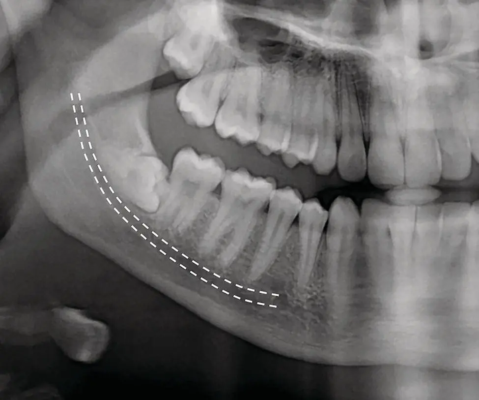
Figure 2.1 In the posterior region of the mandible, the course of the inferior dental (ID) canal (hashed lines) allows the tooth‐bearing areas (the alveolar bone) to be clearly distinguished from the basal bone of the mandible. Radiolucencies below the ID canal are not odontogenic in origin (see text for details).
Читать дальшеИнтервал:
Закладка:
Похожие книги на «Shear's Cysts of the Oral and Maxillofacial Regions»
Представляем Вашему вниманию похожие книги на «Shear's Cysts of the Oral and Maxillofacial Regions» списком для выбора. Мы отобрали схожую по названию и смыслу литературу в надежде предоставить читателям больше вариантов отыскать новые, интересные, ещё непрочитанные произведения.


Обсуждение, отзывы о книге «Shear's Cysts of the Oral and Maxillofacial Regions» и просто собственные мнения читателей. Оставьте ваши комментарии, напишите, что Вы думаете о произведении, его смысле или главных героях. Укажите что конкретно понравилось, а что нет, и почему Вы так считаете.