Digital Dentistry
Здесь есть возможность читать онлайн «Digital Dentistry» — ознакомительный отрывок электронной книги совершенно бесплатно, а после прочтения отрывка купить полную версию. В некоторых случаях можно слушать аудио, скачать через торрент в формате fb2 и присутствует краткое содержание. Жанр: unrecognised, на английском языке. Описание произведения, (предисловие) а так же отзывы посетителей доступны на портале библиотеки ЛибКат.
- Название:Digital Dentistry
- Автор:
- Жанр:
- Год:неизвестен
- ISBN:нет данных
- Рейтинг книги:5 / 5. Голосов: 1
-
Избранное:Добавить в избранное
- Отзывы:
-
Ваша оценка:
Digital Dentistry: краткое содержание, описание и аннотация
Предлагаем к чтению аннотацию, описание, краткое содержание или предисловие (зависит от того, что написал сам автор книги «Digital Dentistry»). Если вы не нашли необходимую информацию о книге — напишите в комментариях, мы постараемся отыскать её.
A guide to all current basic digital imaging and CAD-CAM procedures, with an emphasis on the most popular systems and software programs. An atlas of multidisciplinary cases that were treated with digital dentistry, from diagnosis and treatment planning to execution and follow-up, in order of complexity Assessment of the scientific basis for using digital dentistry in each category A presentation of clinical cases to support the use of digital methodologies in all relevant scenarios An exploration of the role of digital dentistry in dental public health, preventive dentistry, and dental education Ideal for dental clinicians—general practitioners and specialists—as well as all other dental professionals, such as dental technologists, dental hygienists, and dental students,
is an essential tool and reference work to help dental practitioners streamline and update their practice with the most up-to-date technologies.
Digital Dentistry — читать онлайн ознакомительный отрывок
Ниже представлен текст книги, разбитый по страницам. Система сохранения места последней прочитанной страницы, позволяет с удобством читать онлайн бесплатно книгу «Digital Dentistry», без необходимости каждый раз заново искать на чём Вы остановились. Поставьте закладку, и сможете в любой момент перейти на страницу, на которой закончили чтение.
Интервал:
Закладка:
SUMMARY
This chapter will discuss all the terms and definitions that the dental professional needs to know to understand the procedures discussed in the following chapters. Such definitions include abbreviations and general concepts of digital imaging and digital workflow. The chapter also presents a history of the use of CAD‐CAM in dentistry in the last two decades, and the basic knowledge required plus ideas and alternatives to start with digital dentistry.
1.1 Definitions
Digital dentistry is the term used to describe the different modalities of dental treatment workflow that are mostly performed with the use of digital technologies. Several digital methods have been incorporated to dental practice to replace conventional methods and techniques in order to enhance treatment planning and predictability of execution. Nowadays, digital dentistry is considered a whole field of study within dentistry. As with any other field of study, digital dentistry involves a learning curve to be mastered and used in the clinical routine. Ultimately, the dental professional is responsible for using existing digital tools appropriately for patient treatment. In other words, the basic theories of dentistry are still the same and should be very well known by the professional, who will be able to use these new digital tools to enhance predictability in executing the treatment plan.
In order to become familiar with digital dentistry and take advantage of its benefits, it is required to learn a series of important concepts and abbreviations. The most important of these are discussed below.
1.1.1 Three‐Dimensional Imaging
Conventional two‐dimensional (2D) imaging modalities usually have several limitations such as image distortion, magnification, superimposition of anatomical structures, and lack of three‐dimensional (3D) information for diagnosis and planning. In this context, 3D imaging modalities such as cone beam computed tomography (CBCT), intraoral and facial scanning systems provide 3D digital images for dentistry [1–3]. CBCT imaging allows for visualization and assessment of bone structures with high diagnostic accuracy and precision. For CBCT images, the professional needs to understand image acquisition parameters, since the quality of the image affects the quality of the work in digital dentistry. There are several CBCT acquisition parameters, such as field of view size (FOV), peak kilovoltage (kVp), milliamperage (mA), and voxel size. Each of these parameters has an influence on CBCT quality [2–5].
Intraoral and facial scanning can capture 3D patient images that can be used for digital treatment planning systems ( Figure 1.1). The software will then develop a digital representation of the 3D object surfaces available, which will be automatically converted into 3D images composed by wireframe models.
Any 3D images can be rendered and edited in the 3D space, before being converted and saved in a specific file format [5]. As discussed in the next chapter, three file formats are commonly used in digital dentistry: OBJ, STL, and PLY. These files are based on the geometric reconstruction of objects by vectors, triangles or polygons, considering their positioning in a 3D space. After all data is ready, it is possible to store the shape of a model and other details such as color or texture.
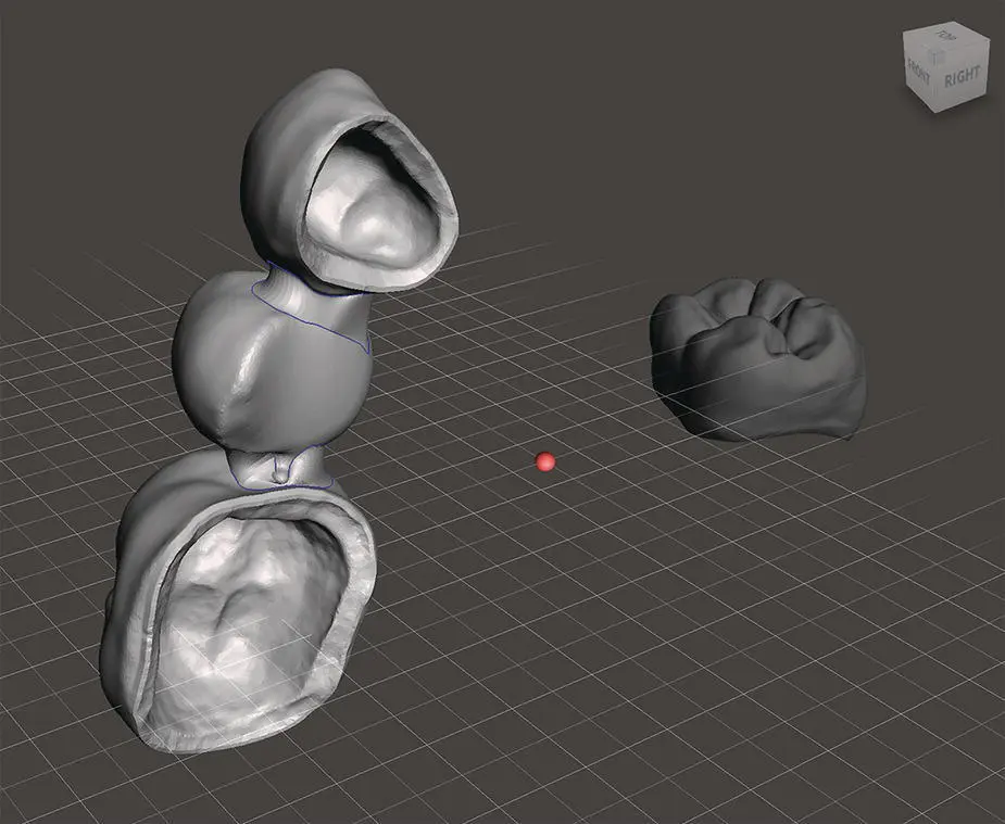
Figure 1.1 Three‐dimensional objects imported in different coordinates of the 3D space (screen capture of MeshMixer software, Autodesk). Note that the fixed bridge is closer to the screen than the molar crown. The dynamic grid is used to orientate the spatial disposition of the 3D objects.
Three‐dimensional images can be manipulated in various ways, depending on the characteristics of the software. For example, with DICOM and STL files, using the CAD software one can plan and perform digital surgery of dental implants and wax‐up of future prostheses. After digital planning, the implant surgery guide, temporary crowns, and definitive crowns can be printed with additive manufacturing devices or milled by subtractive manufacturing devices [5, 6].
1.1.2 Coordinates and Planes
All 3D images are created or rendered in a virtual space of coordinates and planes. Any objects that are digitally designed within the 3D coordinates can be fully edited in the virtual space, before being manufactured. The coordinate system is a method of assigning numbers to points. In three dimensions, three numbers are required to specify a point. Plain 2D images have numbers related to only two coordinates (x and y). The coordinate that represents the third dimension is usually an axis called z. The z‐axis is perpendicular to both the x‐axis and the y‐axis ( Figure 1.2).
The coordinates and the respective planes provide references for the location, size, and volume of the 3D images. All 3D objects have their coordinates fixed in a virtual plane of the imaging software. It is important to make sure that multiple 3D objects to be manipulated or aligned are positioned in the same spatial coordinates, which can be used as spatial references. Therefore, 3D files from different imaging methods should be in the same 3D coordinates in order to be superimposed or combined with the aim of creating a virtual patient, as explained further in this chapter.
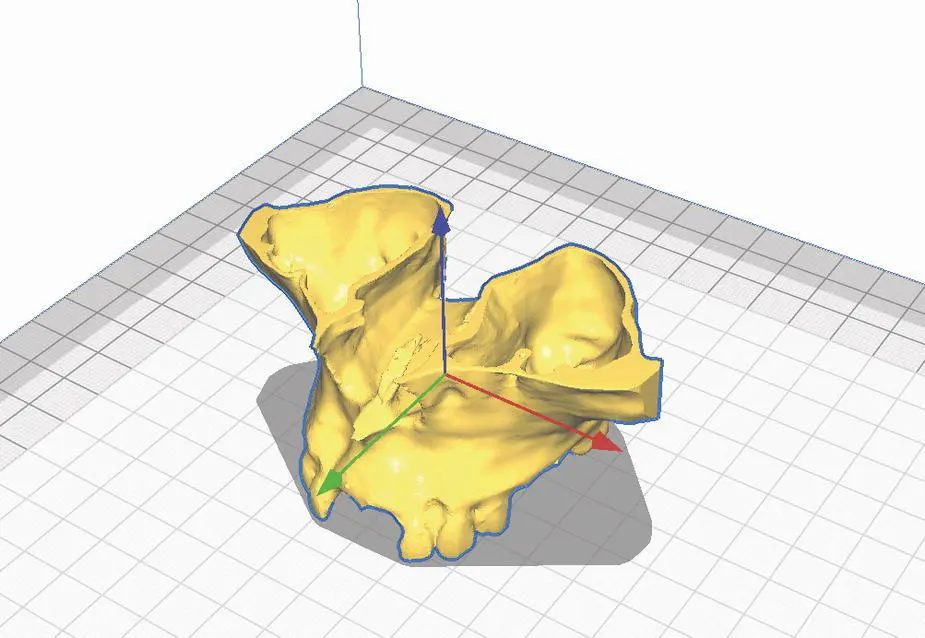
Figure 1.2 A 3D object (reconstructed model of a maxillary CBCT scan) is positioned in the 3D space of a software (Ultimaker Cura) to be 3D printed. Note the three axes depicted by the software in different colors (x‐axis in red, y‐axis in green, z‐axis in blue).
1.1.3 Computer‐Aided Design and Computer‐Aided Manufacturing (CAD‐CAM)
The term computer‐aided/assisted design is usually abbreviated as CAD. The methods used for image acquisition (CBCT, scanning imaging, photographs) and manipulation (software programs) can be included in CAD. On the other hand, computer‐aided/assisted manufacturing (CAM) includes processes such as 3D printers (additive manufacturing) and milling devices (subtractive manufacturing). CAD‐CAM technologies are currently used in biomedical engineering, clinical medicine, customized medical implants, tissue engineering, dentistry, artificial joint manufacture, and robotic surgery. Furthermore, the use of CAD‐CAM technologies has been increasing in various fields of study of medicine and dentistry [5, 6]. Among the main devices that can be digitally designed and manufactured are different types of dental restorations and prostheses, surgical guides, occlusal splints, dental casts, and orthodontic aligners [5, 7]. Details of the main clinical applications of CAD‐CAM in dentistry are further addressed in the next chapters.
1.1.4 Mesh
The term mesh is used to describe the surface of a 3D object composed of triangular or polygon faces. A mesh object does not have any actual curvature. Instead, the appearance of curvatures in a 3D image composed of meshes is obtained by increasing the number of surfaces. The most common file format of these 3D images is the STL file [5], which will be discussed in detail in the next chapter.
1.1.5 Image‐Guided Treatment
Since 3D patient scans are taken prior to dental treatment, CAD‐CAM technology can be used for the fabrication of surgical guides, preparation guides, and maxillofacial surgical templates. Most of these applications require 3D hard and soft tissue images generated by CBCT and optical scanning image modalities, respectively. Based on such images, CAD‐CAM guides can be designed and manufactured to orientate directions of drilling procedures and incisions [5].
Читать дальшеИнтервал:
Закладка:
Похожие книги на «Digital Dentistry»
Представляем Вашему вниманию похожие книги на «Digital Dentistry» списком для выбора. Мы отобрали схожую по названию и смыслу литературу в надежде предоставить читателям больше вариантов отыскать новые, интересные, ещё непрочитанные произведения.


Обсуждение, отзывы о книге «Digital Dentistry» и просто собственные мнения читателей. Оставьте ваши комментарии, напишите, что Вы думаете о произведении, его смысле или главных героях. Укажите что конкретно понравилось, а что нет, и почему Вы так считаете.