McLeod Ian - Swimming Anatomy
Здесь есть возможность читать онлайн «McLeod Ian - Swimming Anatomy» весь текст электронной книги совершенно бесплатно (целиком полную версию без сокращений). В некоторых случаях можно слушать аудио, скачать через торрент в формате fb2 и присутствует краткое содержание. Год выпуска: 2009, ISBN: 2009, Жанр: Природа и животные, на английском языке. Описание произведения, (предисловие) а так же отзывы посетителей доступны на портале библиотеки ЛибКат.
- Название:Swimming Anatomy
- Автор:
- Жанр:
- Год:2009
- ISBN:978-0736075718
- Рейтинг книги:5 / 5. Голосов: 1
-
Избранное:Добавить в избранное
- Отзывы:
-
Ваша оценка:
Swimming Anatomy: краткое содержание, описание и аннотация
Предлагаем к чтению аннотацию, описание, краткое содержание или предисловие (зависит от того, что написал сам автор книги «Swimming Anatomy»). Если вы не нашли необходимую информацию о книге — напишите в комментариях, мы постараемся отыскать её.
Swimming Anatomy — читать онлайн бесплатно полную книгу (весь текст) целиком
Ниже представлен текст книги, разбитый по страницам. Система сохранения места последней прочитанной страницы, позволяет с удобством читать онлайн бесплатно книгу «Swimming Anatomy», без необходимости каждый раз заново искать на чём Вы остановились. Поставьте закладку, и сможете в любой момент перейти на страницу, на которой закончили чтение.
Интервал:
Закладка:
Muscles Involved
Primary:Biceps brachii
Secondary:Brachialis, forearm and finger flexors
Swimming Focus
This exercise is useful if you are having difficulty maintaining your form with the barbell or dumbbell biceps curls or if you want to isolate the biceps brachii and brachialis. As the name implies, the primary purpose of this exercise is to concentrate on the curling motion and, in turn, to strengthen the elbow flexors. The key is to maintain the elbow in a stabilized position against the inner thigh and perform the exercise in a slow, controlled manner.
CHAPTER 3
SHOULDERS
The shoulder girdle is important because it serves as the link between the arms and the trunk. It is the main rotation point about which all the arm movements take place during each of the four strokes. The shoulder girdle is composed of three bones: the clavicle (collarbone), scapula (shoulder blade), and humerus. Three joints make up the shoulder girdle: the sternoclavicular joint, which is the junction between the sternum (breastbone) and clavicle; the acromioclavicular joint, which is made up of the scapula and clavicle; and the glenohumeral joint, which is composed of the humerus and scapula. This chapter focuses on the movements that take place at the glenohumeral joint, which in layman’s terms is the shoulder joint, and the movements of the scapula. The shoulder joint is one of the most flexible joints in the human body, as demonstrated by our ability to place our hands anywhere in our field of vision. This wide range of motion is possible because of the combination of six movements that occur at the shoulder girdle. Flexion involves raising the arm forward away from the body, as if you were raising your hand to answer a question. Extension, the reverse motion, involves lowering the hand from a flexed position. Moving your hand away from your body by raising it to the side is called abduction, and bringing your hand back toward the midline of your body is called adduction. The final two movements are rotational. External rotation involves rotation of the hand from the midline of the body in an outward motion. Internal rotation entails rotating your hand inward, as if you were bringing it in to rub your belly.
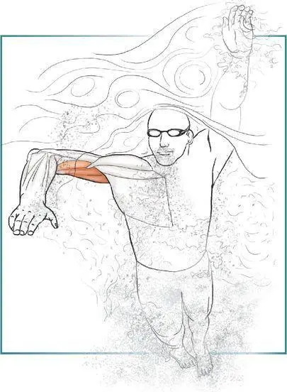
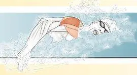
The muscles about the shoulder girdle can be classified into four groups: scapular pivoters, shoulder protectors, humeral positioners, and humeral propellers; an easy way to remember the four groups is to keep in mind the four Ps. The scapular pivoters are the trapezius, rhomboid major, rhomboid minor, serratus anterior, and pectoralis minor. As the name implies, these muscles are responsible for the upward and downward pivoting motion of the shoulder blade. They also account for the shoulder blade movements of elevation and depression and the movements of retraction and protraction. Upward rotation of the scapula is easily visualized if you stand behind a swimmer and watch him or her raise the arms to the sides up over the head. Elevation is simply the movement that occurs when you shrug your shoulders. Retraction is the movement performed when you pinch the shoulder blades together. A combination of these movements in unison with movement at the shoulder joint allows the wide variety of overhead movements that we are capable of performing. To observe the importance of these combined movements, place a hand on another person’s shoulder blade. While you hold it in place, ask the person to lift a hand overhead. Notice the varied movements of the shoulder blade as the arm is moved through a variety of positions.
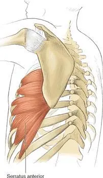
The trapezius is a large triangular muscle that attaches along the midline of the body to numerous points along the spine, starting at the base of the skull and ending at the bottom of the rib cage. From its attachment, the trapezius tapers outward to insert onto points on the clavicle and scapula. The trapezius can be divided into upper, middle, and lower portions. The upward portion is responsible for elevating and upwardly rotating the scapula. The middle portion aids in retraction, and the lower portion contributes to depression and downward rotation. The rhomboid major and minor run from the inner border of the shoulder blade to attachments on the spine. They work in conjunction with the middle portion of the trapezius to pinch the shoulder blades back. The serratus anterior is also attached along the inner border of the shoulder blade, but instead of running toward the midline, it runs in between the shoulder blade and the rib cage to attach along the outer surface of the first nine ribs (figure 3.1). Its two primary responsibilities are to assist in upward rotation of the scapula and to hold the scapula flat against the rib cage. Finally, the pectoralis minor is a small muscle on the front part of the rib cage that goes from an attachment on ribs 2 through 3 to a landmark on the superior aspect of the shoulder blade called the coracoid process. The pectoralis minor aids the lower fibers of the trapezius in depressing the scapula.
Figure 3.1Serratus anterior.
The scapular pivoters have three main areas of influence on the swimming athlete. First, proper upward rotation of the scapula is vital to allowing the swimmer to reach far out in front of the body when entering the hand into the water. The more elongated the swimmer can be, the more efficient the stroke will become. The second role is best described using the analogy that the shoulder blade and the scapular pivoters are like the foundation of a house. Building a spectacular house is foolish if the foundation is eventually going to crumble and fall apart. The same goes for the shoulder girdle and the scapula. If the scapular pivoters are weak, the rest of the kinetic chain making up the arms will eventually deteriorate and the risk of injury will increase. As discussed in chapter 2, when performing upper-extremity exercises, particularly those that target the shoulder joints, you should set the shoulder blades into a stable position. See the sidebar on page 13 for an explanation of how to set the shoulder blades. Lastly, strengthening the posterior scapular pivoters (trapezius, rhomboids, and serratus anterior) helps to overcome the forward rounded-shoulder posture commonly seen in swimmers because of overdevelopment of the latissimus dorsi.
The shoulder protector group, also known as the rotator cuff, is made up of the supraspinatus, infraspinatus, teres minor, and subscapularis (figure 3.2). The supraspinatus lies along the top part of the shoulder blade and attaches to the head of the humerus. The primary role of the supraspinatus is to help initiate overhead movements of the arm. The infraspinatus and teres minor arise from the back part of the scapula and attach next to the supraspinatus on the head of the humerus. The infraspinatus and teres minor act to rotate the shoulder externally. The subscapularis muscle runs along the front part of the shoulder, and like the other rotator cuff muscles, it originates on the scapula and inserts onto the head of the humerus. As the name implies, the primary action of the rotator cuff muscle group is to perform rotational movements at the shoulder joint. Because of the smaller sizes of these muscles, their contribution to the propulsive forces generated while swimming are relatively small; they do, however, have an important role in aiding in the recovery phase of all the strokes. Another vitally important role is their “cuff” function, which stabilizes the shoulder joint. When considering the role of the rotator cuff in stabilizing the shoulder joint, remember that the shoulder is a ball-and-socket joint that resembles a golf ball sitting on a tee. The rotator cuff muscles act as dynamic stabilizers by creating opposing forces that keep the ball centered on the tee. In some instances an imbalance can arise amongst the rotator cuff muscles, which inhibits their stabilizing mechanism and in turn increases the risk of injury. The shoulder joint sacrifices stability in favor of mobility and therefore depends on the rotator cuff muscle group to act as stabilizers and protectors.
Читать дальшеИнтервал:
Закладка:
Похожие книги на «Swimming Anatomy»
Представляем Вашему вниманию похожие книги на «Swimming Anatomy» списком для выбора. Мы отобрали схожую по названию и смыслу литературу в надежде предоставить читателям больше вариантов отыскать новые, интересные, ещё непрочитанные произведения.


Обсуждение, отзывы о книге «Swimming Anatomy» и просто собственные мнения читателей. Оставьте ваши комментарии, напишите, что Вы думаете о произведении, его смысле или главных героях. Укажите что конкретно понравилось, а что нет, и почему Вы так считаете.