Guy Deutscher - Through the Language Glass, Why the World Looks Different in Other Languages
Здесь есть возможность читать онлайн «Guy Deutscher - Through the Language Glass, Why the World Looks Different in Other Languages» весь текст электронной книги совершенно бесплатно (целиком полную версию без сокращений). В некоторых случаях можно слушать аудио, скачать через торрент в формате fb2 и присутствует краткое содержание. Жанр: Языкознание, на английском языке. Описание произведения, (предисловие) а так же отзывы посетителей доступны на портале библиотеки ЛибКат.
- Название:Through the Language Glass, Why the World Looks Different in Other Languages
- Автор:
- Жанр:
- Год:неизвестен
- ISBN:нет данных
- Рейтинг книги:5 / 5. Голосов: 1
-
Избранное:Добавить в избранное
- Отзывы:
-
Ваша оценка:
Through the Language Glass, Why the World Looks Different in Other Languages: краткое содержание, описание и аннотация
Предлагаем к чтению аннотацию, описание, краткое содержание или предисловие (зависит от того, что написал сам автор книги «Through the Language Glass, Why the World Looks Different in Other Languages»). Если вы не нашли необходимую информацию о книге — напишите в комментариях, мы постараемся отыскать её.
Linguistics has long shied away from claiming any link between a language and the culture of its speakers: too much simplistic (even bigoted) chatter about the romance of Italian and the goose-stepping orderliness of German has made serious thinkers wary of the entire subject. But now, acclaimed linguist Guy Deutscher has dared to reopen the issue. Can culture influence language-and vice versa? Can different languages lead their speakers to different thoughts? Could our experience of the world depend on whether our language has a word for "blue"?
Challenging the consensus that the fundaments of language are hard-wired in our genes and thus universal, Deutscher argues that the answer to all these questions is-yes. In thrilling fashion, he takes us from Homer to Darwin, from Yale to the Amazon, from how to name the rainbow to why Russian water-a "she"-becomes a "he" once you dip a tea bag into her, demonstrating that language does in fact reflect culture in ways that are anything but trivial. Audacious, delightful, and field-changing, Through the Language Glass is a classic of intellectual discovery.
Through the Language Glass, Why the World Looks Different in Other Languages — читать онлайн бесплатно полную книгу (весь текст) целиком
Ниже представлен текст книги, разбитый по страницам. Система сохранения места последней прочитанной страницы, позволяет с удобством читать онлайн бесплатно книгу «Through the Language Glass, Why the World Looks Different in Other Languages», без необходимости каждый раз заново искать на чём Вы остановились. Поставьте закладку, и сможете в любой момент перейти на страницу, на которой закончили чтение.
Интервал:
Закладка:
Until the nineteenth century, scientists tried to understand this phenomenon of “color matching” through some physical properties of light itself. But in 1801 the English physicist Thomas Young suggested in a famous lecture that the explanation lies not in the properties of light but rather in the anatomy of the human eye. Young developed the “trichromatic” theory of vision: he argued that there are only three kinds of receptors in the eye, each particularly sensitive to light in a particular area of the spectrum. Our subjective sensation of continuous color is thus produced when the brain compares the responses from these three different types of receptors. Young’s theory was refined in the 1850s by James Clerk Maxwell and in the 1860s by Hermann von Helmholtz and is still the basis for what is known today about the functioning of the retina.
Color vision is based on three kinds of light-absorbing pigment molecules that are contained within cells of the retina called cones. These three types of cells are known as long-wave, middle-wave, and short-wave cones. The cones absorb photons and send on a signal about the number of photons they absorb per unit of time. The short-wave cones have their peak sensitivity around 425 nm-that is, on the border between violet and blue. This does not mean that these cones absorb photons only at 425 nm. As can be seen from the diagram below (and in color in figure 12), the short-wave cones absorb light at a range of wavelengths, from violet to blue and even some parts of green. But their sensitivity to light decreases as the wavelength moves away from the peak at 425 nm. So when monochromatic green light at 520 nm reaches the short-wave cones, a much smaller percentage of the photons are absorbed compared to light at 425 nm.
The second type of receptors, the middle-wave cones, have their peak sensitivity at yellowish green, around 530 nm. And again, they are sensitive (to a decreasing degree) to a range of wavelengths from blue to orange. Finally, the long-wave cones have their peak sensitivity quite close to the middle-wave cones, in greenish yellow, at 565 nm.
The cones themselves do not “know” what wavelength of light they are absorbing. Each cone by itself is color-blind. The only thing the cone registers is the overall intensity of light that it has absorbed. Thus, a short-wave cone cannot tell whether it is absorbing low-intensity violet light (at 440 nm) or high-intensity green light at (500 nm). And the middle-wave cone cannot tell the difference between light at 550 nm and light in the same intensity at 510 nm.
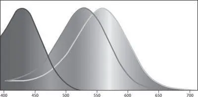
The (normalized) sensitivity of the short-wave, middle-wave, and long-wave cones as a function of wavelength.
The brain works out what color it is seeing by comparing the rates at which photons are absorbed in the three different classes of cones. But there are infinitely many different spectral distributions that could give exactly the same ratios, and we cannot distinguish between them. For example, a monochromatic yellow light at wavelength 580 nm creates exactly the same absorption ratio between the cones as a combination of red light at 620 nm and green light at 540 nm, as mentioned earlier. And there are an infinite number of other such “metameric colors,” different spectral distributions that produce the same absorption ratios between the three types of cones and thus look the same to the human eye.
It is important to realize, therefore, that our range of color sensations is determined not directly by the range of monochromatic lights in the spectrum but rather by the range of possibilities of varying the ratios between the three types of cones. Our “color space” is three-dimensional, and it contains sensations that do not correspond to any colors of the rainbow. Our sensation of pink, for example, is created from an absorption ratio that corresponds not to any monochromatic light but rather to a combination of red and blue lights.
As the light fades at night, a different system of vision comes into play. The cones are not sensitive enough to perceive light in very low intensity, but there are other receptors, called rods, that are so sensitive they can register the absorption of even a single photon! The rods are most sensitive to bluish green light at around 500 nm. Our low-light vision, however, is color-blind. This is not because the light itself “forgets” its wavelength at night but simply because there is just one type of rod. As the brain has nothing with which to compare the responses from the single type of rod, no color sensation can be produced.
There are about six million cones in total in the retina, but the three types are not found in nearly equal numbers: there are relatively few short-wave (violet) cones, more than ten times as many middle-wave (green) cones, and even more long-wave cones. The far greater numbers of middle-wave and long-wave cones means that the eye is more efficient in absorbing light at the long-wave half of the spectrum (yellow and red) than at the short-wave half, so it takes lesser intensity of yellow light to be detected by the eye than blue or violet light. In fact, our day vision has a maximum sensitivity to light of 555 nm, at yellow-green. It is this idiosyncrasy of our anatomy that makes yellow appear brighter to us than blue or violet, rather than any inherent properties of the light itself, since blue light is not in itself less intense than yellow light. (In fact, wavelength and energy are inversely related: the long-wave red light has the lowest energy, yellow light has higher energy than red, but green and blue have higher energy than yellow. The invisible ultraviolet light has even higher energy, enough in fact to damage the skin.)
There is also a different type of unevenness in our sensitivity to colors: our ability to discriminate between fine differences in wavelength is not uniform across the spectrum. We are especially sensitive to wavelength differences in the yellow-green area, and the reason again lies in the accidents of our anatomy. Because the middle-wave (green) and long-wave (yellowish green) receptors are very close in their peak sensitivities, even very small variations in wavelength in the yellow-green area translate into significant changes in the ratios of light absorbed by the two neighboring cones. Under optimal conditions, a normal person can discriminate between yellow hues differing in wavelength by just a single nanometer. But in the blue and violet area of the spectrum, our ability to discriminate between different wavelengths is less than a third of that. And with red hues near the edge of the spectrum, we are even less sensitive to wavelength differences than in the blues.
These two types of unevenness in our sensitivity to color-the feeling of varying brightness and the varying ability to discriminate fine differences in wavelength-make our color space asymmetric. And as mentioned in this footnote, this asymmetry makes certain divisions of the color space better than others in increasing similarity within concepts and decreasing it across concepts.
When one of the three types of cones fails, this reduces color discrimination to two dimensions instead of three, and the condition is thus called dichromacy. The most frequent type of dichromacy is commonly called red-green blindness. It affects about 8 percent of men and 0.45 percent of women, who lack one of the two neighboring types of cones (long-wave or middle-wave). Little is known about the actual color sensations of people with color blindness, because one cannot simply “translate” the sensations of dichromats directly to those of trichromats. A few reports have been collected from the rare people with a red-green defect in one eye and normal vision in the other. Using their normal eye as a reference, such people say that their color-blind eye has the sensation of yellow and blue. But since the neural wiring associated with the normal eye might not be normal in their cases, even the interpretation of such reports is not straightforward.
Читать дальшеИнтервал:
Закладка:
Похожие книги на «Through the Language Glass, Why the World Looks Different in Other Languages»
Представляем Вашему вниманию похожие книги на «Through the Language Glass, Why the World Looks Different in Other Languages» списком для выбора. Мы отобрали схожую по названию и смыслу литературу в надежде предоставить читателям больше вариантов отыскать новые, интересные, ещё непрочитанные произведения.


Обсуждение, отзывы о книге «Through the Language Glass, Why the World Looks Different in Other Languages» и просто собственные мнения читателей. Оставьте ваши комментарии, напишите, что Вы думаете о произведении, его смысле или главных героях. Укажите что конкретно понравилось, а что нет, и почему Вы так считаете.